. 1997 Nov 15;17(22):8695-701.
doi: 10.1523/JNEUROSCI.17-22-08695.1997.

Peroxide modulation of slow onset potentiation in rat hippocampus

Affiliations

Peroxide modulation of slow onset potentiation in rat hippocampus

J M Auerbach et al. J Neurosci. .

Abstract

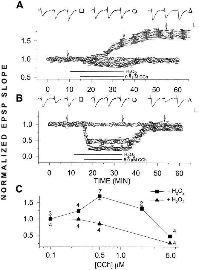
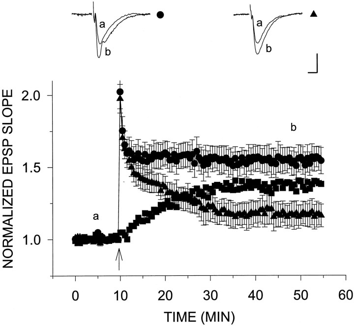
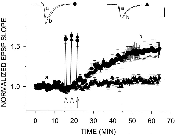
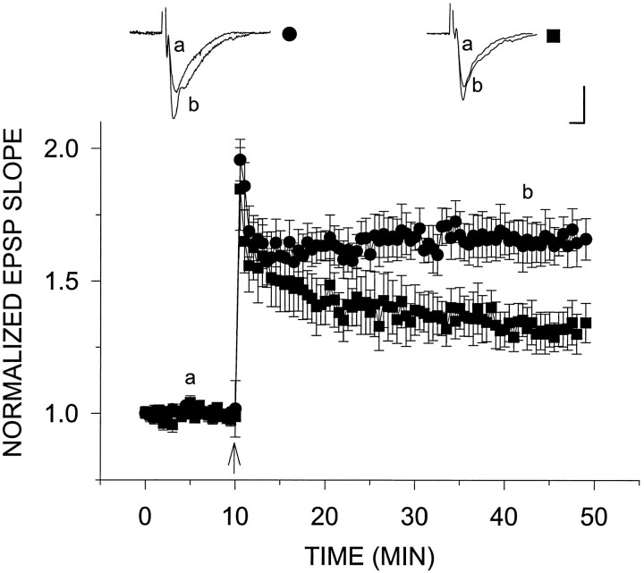
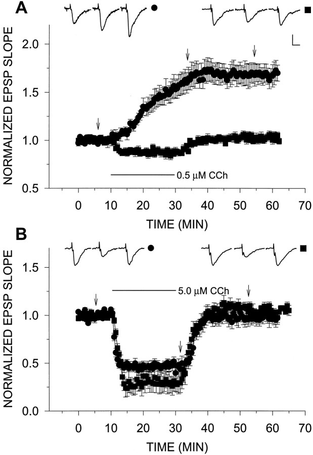
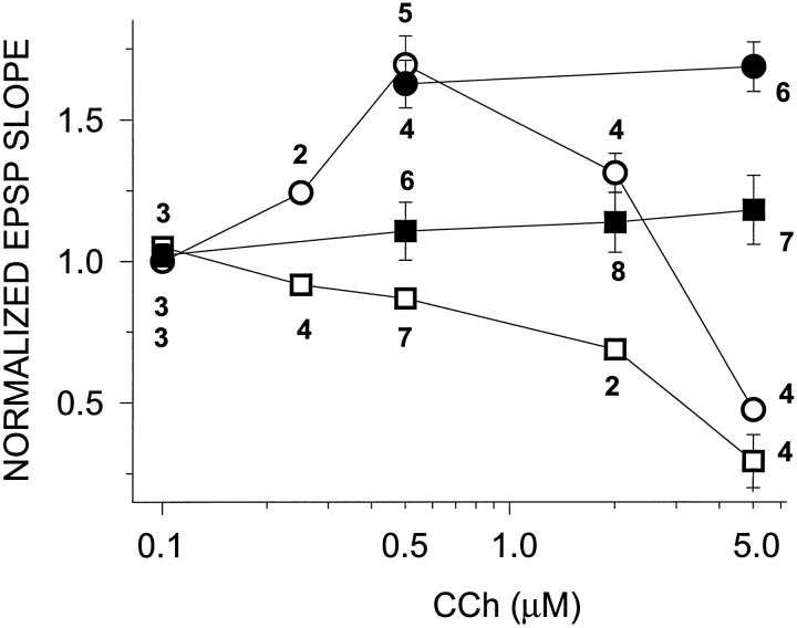
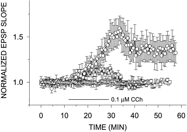
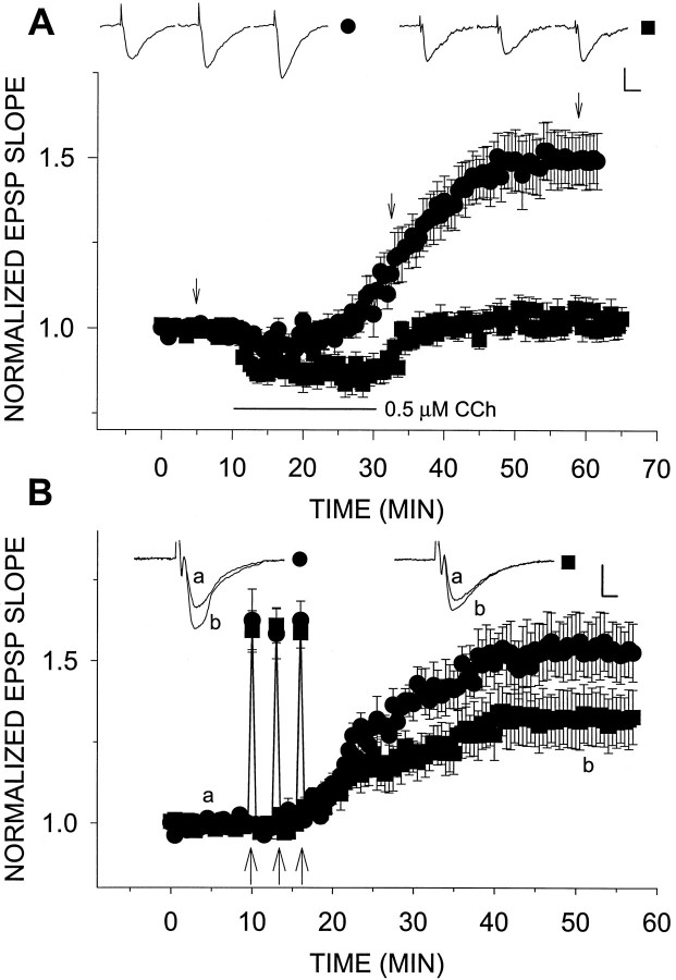
Exposure of rat hippocampal slices to low concentrations of the muscarinic agonist carbachol (CCh) has been shown to produce a slow onset long-term potentiation (LTP) of reactivity to afferent stimulation in CA1 neurons. Although this potentiation shares a number of properties with tetanic LTP, muscarinic LTP (LTPm) is independent of activation of the NMDA receptor. We now demonstrate that low levels of hydrogen peroxide (H2O2) cause hippocampal slices to lose the ability to express LTPm. This powerful effect of H2O2 is selective in that it does not affect the reactivity of hippocampal neurons to higher concentrations of CCh. In fact, H2O2 also blocks induction of a slow onset, non-NMDA-dependent tetanic LTP (NN-LTP). The functional relevance of this action of H2O2 is exemplified by the fact that the hippocampus of aged rats, which produces higher levels of endogenous H2O2 than that of young rats, lacks LTPm and expresses a markedly reduced NN-LTP. In aged rats, the lack of LTPm contrasts with an apparently normal muscarinic suppression of the EPSP slope induced by higher concentrations of CCh. When hippocampal slices from aged animals are treated with catalase, an enzyme that breaks down H2O2, LTPm is restored, and NN-LTP is enhanced. Thus, our study proposes a unique and novel age-dependent peroxide regulation of LTPm in the brain and provides a link between the cholinergic system, aging, and memory functions.


Figures

Fig. 1.
Fig. 1.
A, H2O2selectively blocks LTPm. H2O2 (29 μm) applied for 25 min had no observable effect (n = 4) (○). However, when applied 5 min before and together with CCh for an additional 20 min, H2O2 blocked the induction of LTPm(n = 4) (▵). This can be compared with the control case in which 0.5 μm CCh alone was added to the slices for 20 min, resulting in a potentiation of 69.3 ± 10.2% above baseline (n = 5) (□). B,H2O2 did not block the depression of the EPSP slope induced by 5.0 μm CCh. As above, H2O2 alone had no observable effect (n = 4) (○). CCh (5.0 μm) alone reduced the EPSP slope values to 47.4 ± 3.3% of baseline (n = 4) (□), whereas addition of 29 μm H2O2 with 5.0 μmCCh increased the CCh-induced depression, reducing the EPSP slope to 27.2 ± 4.4% of baseline values (n = 4) (▵). C, Dose–response relations for increasing concentrations of CCh in H2O2 clearly shows that although LTPm was abolished, the depressing effects of CCh were present. In this and the following graphs, representative EPSPs are presented above the grouped data. Each EPSP triplet is from one treatment, as identified by the symbol to theright of the triplet. Each individual EPSP within the triplet was sampled at a time corresponding to thearrows or lower case letters in the graph. Calibration, 0.5 mV, 5 msec. Error bars represent SEM, when larger than the symbol. The bars under the graphs designate the duration of drug application.
Fig. 2.
Fig. 2.
H2O2 reduces the slow component of tetanic LTP. A single tetanus was applied at twice the test intensity 10 min after establishment of stable baseline (arrowhead) to slices from young rats in the presence (▴) and absence (•) of H2O2. H2O2 had no effect on the amount of post-tetanic potentiation immediately after the tetanus. However, H2O2 caused an increased decay in this potentiation, and in its presence the EPSP slope leveled off at 17.4 ± 8.5% above baseline (n = 8) (▴) compared with levels 54.5 ± 8.9% above baseline (n = 12) (•) without H2O2. Subtracting tetanic LTP in the presence of H2O2 from that in its absence yields the middle curve in the graph (▪). Calibration, 0.5 mV, 5 msec.
Fig. 3.
Fig. 3.
H2O2 blocks NN-LTP. On induction of NN-LTP by three consecutive 200 Hz, 1 sec tetani (arrows), the EPSP slope reached a level of 43.3 ± 7.3% above baseline (n = 4) (•). The same induction protocol for NN-LTP in the presence of 29 μmH2O2 caused only a very slight increase in the EPSP slope (6.2 ± 3.4%; n = 4) (▴). Theinsets above the plots are representative EPSPs sampled at the times indicated by letters(a) and (b) in the graph. Calibration, 0.5 mV, 5 msec.
Fig. 6.
Fig. 6.
Impairment of tetanic LTP in slices from aged animals. A single tetanus was applied at twice the test intensity 10 min after establishment of stable baseline (arrowhead) to slices from aged (▪) versus young (•) rats. The amount of post-tetanic potentiation in both groups of slices was similar. Immediately after the tetanus, EPSP slopes from aged slices reached levels 84.7 ± 15.6% (n = 6) (▪) above baseline compared with levels 95.6 ± 7.8% above baseline in young slices (n = 7) (•). However, 30 min after tetanus, plateau potentiation levels reached in aged slices (34.3 ± 7.4%) were substantially less than those in young slices (65.9 ± 7.8%). Calibration, 0.5 mV, 5 msec.
Fig. 4.
Fig. 4.
Slices from aged animals lack LTPm but express cholinergic depression of EPSP slope. A,Addition of 0.5 μm CCh to slices from aged rats resulted in a slight initial depression of the EPSP slope, which recovered to baseline levels once the drug was removed from the medium (n = 7) (▪). This was compared with LTPm in young slices shown here in which 0.5 μm CCh potentiated the EPSP slope to 69.3 ± 10.2% above baseline (n = 5) (•). B, CCh (5.0 μm) reduced the EPSP slope in aged slices to 29.4 ± 9.3% of baseline values (n = 4) (▪) compared with a reduction of 47.4 ± 3.3% in young slices (n = 4) (•). The insets above the plots are representative EPSPs from single experiments sampled at times indicated by the arrows. Calibration, 0.5 mV, 5 msec.
Fig. 5.
Fig. 5.
Dose–response curve of effects of CCh on slices from young (○, •) and old (□, ▪) rats. CCh had a biphasic concentration effect on young slices, i.e., potentiation at low concentrations and depression at high concentrations. Old slices showed only the depression at high CCh concentrations (empty symbols). The potentiating effects of CCh were completely lacking at all concentrations tested. Slices were also tested in the presence of 4-DAMP and picrotoxin (filled symbols). Whereas this treatment revealed LTPminduction at high CCh concentration (5.0 μm) in young slices (68.6 ± 8.7%) (•), only a slight potentiation was seen in aged slices (18.1 ± 12.2%) (▪). The numbersunderneath the symbols in the graph correspond to the number of slices tested for each condition.
Fig. 8.
Fig. 8.
Catalase lowers the threshold for LTPminduction in slices from young rats. Slices treated with 25 U/ml catalase showed a slight potentiation (12.1 ± 3.9% above baseline) of the EPSP slope after exposure to 0.1 μm CCh (n = 6) (□). Slices treated with 100 U/ml showed a stable, long-lasting potentiation 35.3 ± 14.3% above baseline (n = 4) (○). Catalase (100 U/ml) alone added to young slices in the superfusion 10 min after the establishment of a steady baseline had no effect on evoked response (n= 3) (▵).
Fig. 7.
Fig. 7.
Catalase restores LTP in aged slices.A, LTPm is returned to aged slices by treatment with catalase. Slices were incubated in 100 U/ml catalase 1 hr before and continuously throughout the recording session. Under these conditions, 0.5 μm CCh induced LTPm to a level 48.6 ± 8.0% above baseline (n = 8) (•). This was compared with the lack of effect of 0.5 μm CCh in aged slices in the absence of catalase (n = 7) (▪). B, NN-LTP was elevated by catalase-treated aged slices. The treatment was the same as in A. The potentiation in aged slices enabled by catalase increased from 32.2 ± 8.4% (n = 8) (▪) to 58.9 ± 8.9% (n = 6) (•).

References

    1. Aniksztejn L, Ben-Ari Y. Novel form of long-term potentiation produced by a K+ channel blocker in the hippocampus. Nature. 1991;349:67–69. - PubMed
    1. Auerbach JM, Segal M. A novel cholinergic induction of long-term potentiation in rat hippocampus. J Neurophysiol. 1994;72:2034–2040. - PubMed
    1. Auerbach JM, Segal M. Muscarinic receptors mediating depression and long-term potentiation in rat hippocampus. J Physiol (Lond) 1996;492:479–493. - PMC - PubMed
    1. Barnes CA. Normal aging: regionally specific changes in hippocampal synaptic transmission. Trends Neurosci. 1994;17:13–18. - PubMed
    1. Bartus RT, Reginald L, Dean RL, Beer B, Lippa AS. The cholinergic hypothesis of geriatric memory dysfunction. Science. 1982;217:408–417. - PubMed

LinkOut - more resources