. 1997 Nov 15;17(22):8721-8.
doi: 10.1523/JNEUROSCI.17-22-08721.1997.

Nociceptin inhibits T-type Ca2+ channel current in rat sensory neurons by a G-protein-independent mechanism

Affiliations

Nociceptin inhibits T-type Ca2+ channel current in rat sensory neurons by a G-protein-independent mechanism

F A Abdulla et al. J Neurosci. .

Abstract

Nociceptin (orphanin FQ) is a novel, opioid-like, heptadecapeptide that is an endogenous ligand for the opioid receptor-like (ORL1) receptor. Unlike classical opioids, nociceptin can produce hyperalgesia when injected intracerebroventricularly into mice. Despite this, nociceptin has been reported to decrease transmitter release, activate an inwardly rectifying K+ conductance, and suppress high-voltage-activated Ca2+ channel conductances (HVA gCa) in much the same way as micro-, delta-, and kappa-opioids. We report an action of nociceptin that is not shared by morphine: the suppression of low-voltage-activated, transient calcium (barium) current (IBa,T) in acutely dissociated rat dorsal root ganglion (DRG) neurons (EC50 = 100 nM). This effect was reflected as inhibition of bursts of action potentials that can be evoked in "medium-sized" DRG neurons. Experiments with GTP-gamma-S (100 microM), GDP-beta-S (2 mM), or aluminum fluoride (AlF3) (100 microM) in the patch pipette failed to provide evidence for G-protein involvement in nociceptin-induced IBa,T suppression. By contrast, both morphine and nociceptin suppressed HVA gCa, and the latter response was affected by intracellular GTP-gamma-S, GDP-beta-S, and AlF3 in ways that confirmed G-protein involvement. The selective effect of nociceptin on IBa,T may therefore be relevant to understanding why its behavioral actions differ from those of other opioids. This G-protein-independent effect of the action of nociceptin may reflect a new general mechanism of action for opioid peptides within the nervous system.


Figures

Fig. 6.
Fig. 6.
Biophysical aspects of nociceptin-induced T-current suppression. A, Slow speed chart record to show rate of onset and the effect of nociceptin with and without repeated activation of gBa,T. Top record is current, and bottom record is voltage. Deflections result from brief (30 msec) voltage commands to −40 mV from the holding potential of −90 mV applied once every 2.5 sec (0.4 Hz). Nociceptin (1 μm) was first applied (atleft) while gBa,T was repeatedly activated. The current deflections attain a smaller steady-state amplitude 3 min after initiating nociceptin superfusion. Nociceptin was reapplied (at right) in the absence ofgBa,T activation. Voltage commands were recommenced 3 min after initiating nociceptin superfusion, andIBa,T responses were attenuated to the same extent as those recorded when the conductance had been activated repetitively in the presence of nociceptin. B, Normalized steady-state inactivation plots forgBa,T acquired before and during application of 100 nm nociceptin. Cell was held at a series of prepulse potentials (+5 mV increments from −115 mV) for 200 msec before activating gBa,T at −40 mV. Voltage protocol is shown as inset.C1,C2, Original data records used to produce the data in B. A split-clock protocol was used so that 100 msec calibration refers to prepulse and 25 msec calibration refers to recordings of IBa,T.
Fig. 1.
Fig. 1.
Blockade of IBa,T by nociceptin. A1, A2, Comparison of the effects of nociceptin and morphine onIBa,T in the same neuron. Voltage record (step to −40 mV from a holding potential of −90 mV) omitted for clarity. Large capacity current transients accompanying voltage steps have been attenuated or removed by erasing three or more data points (clock speed for data acquisition was 0.05 msec/point).A1, Superimposed records to show almost complete suppression of IBa,T by 1 μmnociceptin. A2, In the same cell, 1 μm morphine suppresses the current by <10%.B, Superimposed records obtained from another cell to show suppression of IBa,T tails by 1 μm nociceptin. Tails recorded at −60 mV after brief 30 msec step to −40mV from a holding potential of −90 mV (voltage record omitted for clarity). C, I–Vrelationships for IBa recorded in the absence and presence of 1 μm nociceptin.Asterisk marks characteristic shoulder onI–V relationship that results fromIBa,T. Note attenuation of current at −40 mV (IBa,T) and −10 mV (attenuation of HVA IBa) by nociceptin.D, I–V relationships forIBa recorded in the absence and presence of 1 μm morphine. Note lack of attenuation of current at potentials more negative than −40 mV (IBa,T, marked byasterisk) and attenuation of HVAIBa recorded at potentials positive to −20 mV. E, Log-concentration-response relationship for nociceptin. Numbers on graph indicate numbers of cells tested with each concentration; error bars, which indicate SEM, are smaller than data markers in some cases. The concentration of nociceptin required for half-maximum suppression of IBa,T(EC50) is 100 nm. F, Data from E replotted as a Hill plot [Log10R/RRm vs Log10 nociceptin concentration ÷ EC50; where Ris amplitude of response and Rm is the maximum response that can be obtained;KD, the dissociation equilibrium constant for nociceptin and its receptor is assumed to equal the EC50 (100 nm)]. Linear fit to data points yields a line with a gradient of 1 (Hill coefficient of 1.0).
Fig. 2.
Fig. 2.
Effects of nociceptin and morphine on HVAgBa. A1,B1, Currents recorded in response to a step from −90 to −10 mV in the presence and absence of 1 μmnociceptin or 1 μm morphine. Note suppression of current and slowed rate of activation in the presence of agonists.A2, B2, Removal of suppression of HVA gBa induced by 1 μm nociceptin or 1 μm morphine with depolarizing prepulses to +100 mV. Current responses to depolarizing commands are off scale; 2 nA/20 msec calibration inA1 refers also to records inB1, and 2 nA/40 msec calibration inA2 refers also to records inB2. Records in A1and A2 from the same cell and records inB1 and B2 from another cell.
Fig. 3.
Fig. 3.
Pharmacology of nociceptin attenuation of T-current. Superimposed data records show (A1) suppression ofIBa,T by 1 μm nociceptin, (A2) a second response to nociceptin recorded in the same cell in the presence of 1 μmnaloxone, (B1) suppression ofIBa,T in another cell by 1 μmnociceptin, and (B2) attenuation of nociceptin-induced inhibition in the second cell by 1 μmnalbzoh. IBa,T evoked by a step to −40 mV from holding potential of −90 mV (voltage record omitted for clarity).
Fig. 4.
Fig. 4.
Evidence against G-protein involvement in the effect of nociceptin on T-currents.A1, Superimposed data records to show suppression of IBa,T by 100 nmnociceptin. A2, Superimposed records from A1 normalized and replotted. Note that the two records superimpose exactly, and the presence of nociceptin does not slow the onset or the rate of inactivation of the current.B1, Graph of time course of an experiment done with a pipette containing 100 μmGTP-γ-S. Points represent amplitudes ofIBa,T or HVA IBaevoked by a double-pulse protocol applied once every 20 sec. BothIBa,T and HVA IBaare suppressed by 1 μm nociceptin. AlthoughIBa,T returns to control amplitude after nociceptin washout, HVA IBa remains suppressed throughout the course of the experiment. The cell had been left to stabilize with intracellularly applied GTP-γ-S for 15 min before the application of nociceptin.B2, Original superimposed data record collected before, during, and after application of nociceptin in the experiment plotted in B1. The cell was stepped from −90 to −40 mV to activategBa,T and then to −10 mV to activate HVAgBa. Nociceptin invokes reversible suppression of the former and irreversible suppression of the latter.
Fig. 5.
Fig. 5.
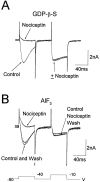
Further evidence against G-protein involvement in the effect of nociceptin on T-currents. A, Effect of 1 μm nociceptin on IBa,T and HVAIBa in the same cell studied with 2 mm GDP-β-S in the pipette. The cell had been allowed to equilibrate with intracellularly applied GDP-β-S for 15 min before the application of nociceptin. Superimposed records acquired before and during nociceptin application show thatIBa,T was suppressed when HVAIBa was unaffected. B, An experiment similar to that of A but using AlF3 (100 μm) instead of GDP-β-S in the pipette. The cell had been allowed to equilibrate with intracellularly applied AlF3 for 20 min before the application of 1 μm nociceptin. Superimposed records acquired before, during, and after nociceptin application show thatIBa,T was reversibly suppressed when HVAIBa was unaffected. Voltage protocol inB applies also to experiment illustrated inA.
Fig. 7.
Fig. 7.
Effects of nociceptin, morphine, and Ni2+ on APs, bursting activity, and afterdepolarizations. All records were obtained from the same cell. APs were evoked from a 5 msec/0.3 nA depolarizing current command applied via the recording electrode. Superimposed records of bursts of APs were recorded before and during superfusion of (A) 1 μm nociceptin, (B) 1 μm morphine, and (C) 100 μm Ni2+. Note that afterdepolarization and burst of spikes are attenuated by nociceptin and Ni2+ but not by morphine. Calibration (40 mV/40 msec) in B and current trace in C apply to all records.

References

    1. Abdulla FA, Smith PA. Ectopic α2-adrenoceptors couple to N-type Ca2+ channels in axotomized rat sensory neurons. J Neurosci. 1997;17:1633–1641. - PMC - PubMed
    1. Bean BP. Neurotransmitter inhibition of neuronal calcium currents by changes in channel voltage dependence. Nature. 1989;340:153–156. - PubMed
    1. Bigay J, Deterre P, Pfister C, Chabre M. Fluoride complexes of aluminium or bretylium act on G-proteins as reversibly bound analogues of the γ phosphate of GTP. EMBO J. 1987;6:2907–2913. - PMC - PubMed
    1. Brauneis U, Oz M, Peoples RW, Weight FF, Zhang I. Differential sensitivity of recombinant N-methyl d-aspartate receptor subunits to inhibition by dynorphin. J Pharmacol Exp Ther. 1996;279:1063–1068. - PubMed
    1. Burch RM, Axelrod J. Dissociation of bradykinin-induced prostaglandin formation from phosphatidylinositol turnover in Swiss 3T3 fibroblasts: evidence for G-protein regulation of phospholipase A2. Proc Natl Acad Sci USA. 1987;84:6374–6378. - PMC - PubMed

Publication types

MeSH terms

LinkOut - more resources