. 1997 Nov 15;17(22):8778-91.
doi: 10.1523/JNEUROSCI.17-22-08778.1997.

Targeted overexpression of the neurite growth-associated protein B-50/GAP-43 in cerebellar Purkinje cells induces sprouting after axotomy but not axon regeneration into growth-permissive transplants

Affiliations

Targeted overexpression of the neurite growth-associated protein B-50/GAP-43 in cerebellar Purkinje cells induces sprouting after axotomy but not axon regeneration into growth-permissive transplants

A Buffo et al. J Neurosci. .

Abstract

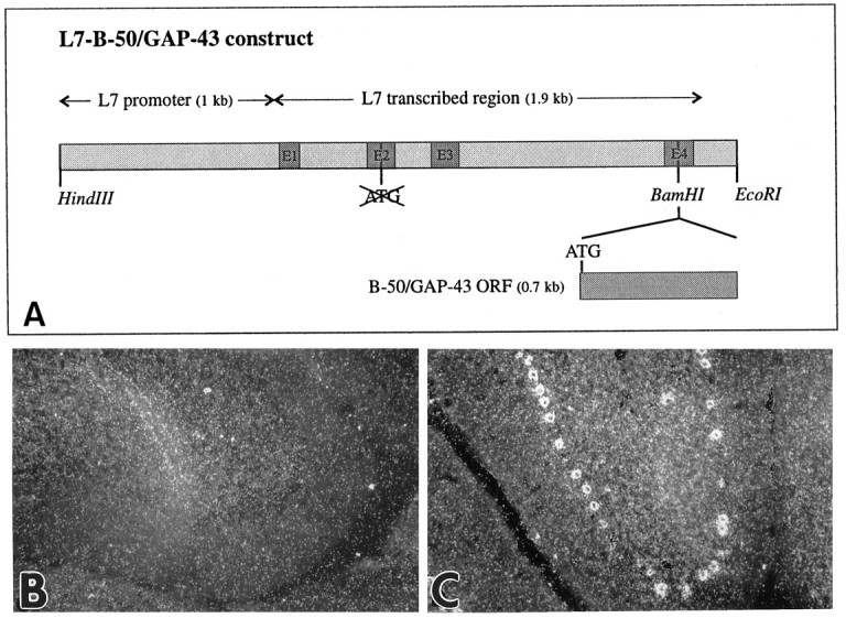
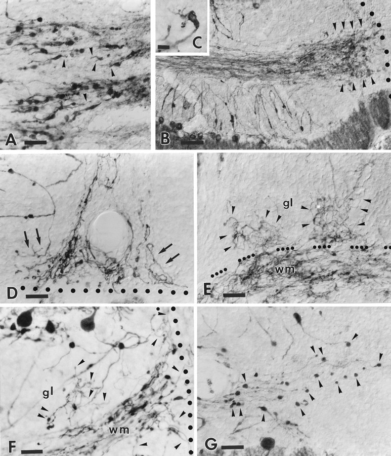
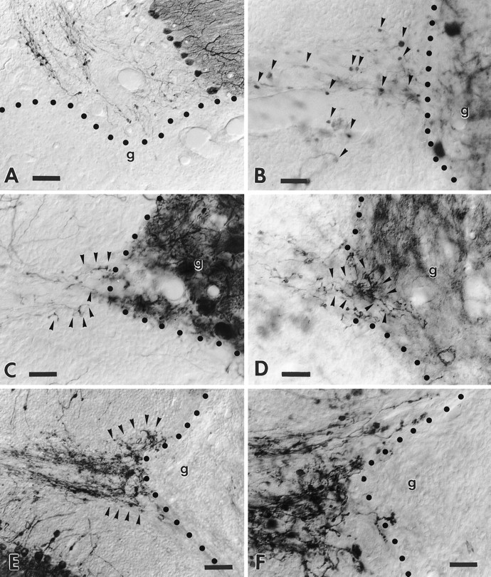
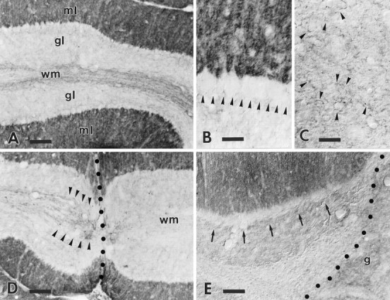
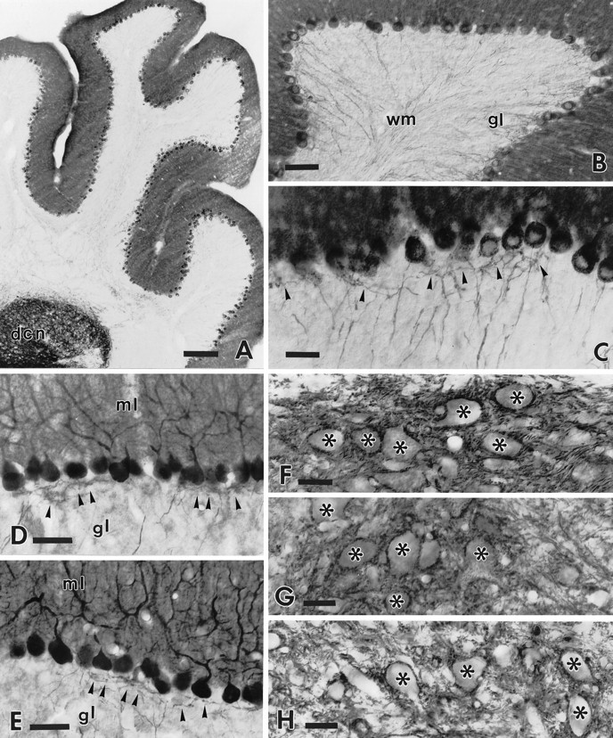
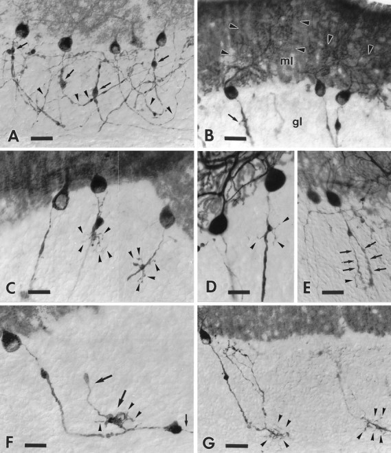
B-50/GAP-43 is a nervous tissue-specific protein, the expression of which is associated with axon growth and regeneration. Its overexpression in transgenic mice produces spontaneous axonal sprouting and enhances induced remodeling in several neuron populations (; ). We examined the capacity of this protein to increase the regenerative potential of injured adult central axons, by inducing targeted B-50/GAP-43 overexpression in Purkinje cells, which normally show poor regenerative capabilities. Thus, transgenic mice were produced in which B-50/GAP-43 overexpression was driven by the Purkinje cell-specific L7 promoter. Uninjured transgenic Purkinje cells displayed normal morphology, indicating that transgene expression does not modify the normal phenotype of these neurons. By contrast, after axotomy numerous transgenic Purkinje cells exhibited profuse sprouting along the axon and at its severed end. Nevertheless, despite these growth phenomena, which never occurred in wild-type mice, the severed transgenic axons were not able to regenerate, either spontaneously or into embryonic neural or Schwann cell grafts placed into the lesion site. Finally, although only a moderate Purkinje cell loss occurred in wild-type cerebella after axotomy, a considerable number of injured transgenic neurons degenerated, but they could be partially rescued by the different transplants placed into the lesion site. Thus, B-50/GAP-43 overexpression substantially modifies Purkinje cell response to axotomy, by inducing growth processes and decreasing their resistance to injury. However, the presence of this protein is not sufficient to enable these neurons to accomplish a full program of axon regeneration.


Figures

Fig. 1.
Fig. 1.
Directed expression of B-50/GAP-43 in cerebellar Purkinje cells of transgenic mice. A, Diagram of the construct used to generate L7-B-50/GAP-43 transgenic mice. The B-50/GAP-43 open reading frame (ORF) was inserted in the unique BamHI site of a vector containing 1 kb of the L7 promoter, the four exons and three introns, and 200 bp downstream of the TGA of the L7 gene. In this vector the endogenous ATGs of the L7 gene had been deleted, and the only translational start signal (ATG) was introduced with the B-50/GAP-43 ORF in the fourth exon of the L7 gene. Details of the cloning of this construct are given in Materials and Methods. B, C, Dark-field micrographs ofin situ hybridization of cerebella of wild-type (B) and transgenic (C) mice from line 1658 (L1658) with a radiolabeled B-50/GAP-43-antisense riboprobe. Cerebellar Purkinje cells do not normally express B-50/GAP-43 mRNA in adulthood (B). In contrast, the B-50/GAP-43 mRNA is abundantly present in these cells in transgenic mice (C).
Fig. 7.
Fig. 7.
Purkinje cell loss in the injured wild-type and transgenic cerebella. A transected transgenic cerebellar lobule, immunolabeled by anti-L7 antibodies and counterstained by thionine, is displayed in A. Arrowheads point to areas where Purkinje cells have degenerated. Note that all surviving neurons are immunolabeled and no immunonegative thionine-stained Purkinje cell perikarya remain. B shows three representative camera lucida reconstructions of lobuli IV–V from transgenic mouse cerebella. Two months after axotomy (center) the number of Purkinje cells is considerably decreased compared with control (left). By contrast, when a graft (shaded area on the right: axotomy/graft) is placed in the lesion track, the injured cells are partially rescued. Quantitative estimations of the number of Purkinje cells/millimeter of Purkinje cell layer are shown in the histogram C. Intact wild-type and transgenic mice have similar values, indicating that no spontaneous cell loss occurs in transgenic animals. By contrast, after axotomy the number of Purkinje cells/millimeter is reduced in both animal sets, although a statistically more severe cell loss is observed in transgenic animals. Finally, when a graft is placed into the lesion site, both wild-type and transgenic mice show similar values that are not statistically different from that obtained from axotomized wild-type cerebella, indicating that axotomy-induced Purkinje cell loss in transgenic mice can be prevented by graft-derived trophic support. Scale bars: A, 40 μm; B, 200 μm.
Fig. 5.
Fig. 5.
Morphological modifications of the distal portion of the transgenic Purkinje cell axons after axotomy. Ashows the transected tips of transgenic Purkinje axons 24 hr after injury (lesion site is beyond the right edge of the picture).Arrowheads indicate fine processes that emanate from the terminal clubs. One month after injury (B) a dense plexus of newly formed sprouts (arrowheads) has developed in the vicinity of the lesion track (dotted line). The inset C shows one such transected axon ending in a terminal club from which several sprouts originate. The structural features and tortuous courses taken by these terminal sprouts (arrows) can be better appreciated inD. Note that despite the profuse growth the newly formed processes do not elongate across the injury site (dotted line). Another terminal plexus is shown in E. In this case the newly formed processes are extended in the white matter (wm) and also (as indicated byarrowheads) in the adjacent granular layer (gl) (dots point to the granular layer–white matter border). Anti-calbindin immunostaining in transgenic cerebella (F) also depicts numerous sprouts (arrowheads) abutting the lesion site (dotted line) or elongating in the nearby granular layer (gl). By contrast, in wild-type animals (G) (anti-L7 immunostaining) the transected axons remain close to the lesion site (just beyond the right edge of the picture), but they terminate with round-shaped terminal clubs (some are indicated by arrowheads). Survival times: 24 hr (A); 30 d (B, C, E, G); 60 d (D, F). Scale bars: A, D, E, F, G, 30 μm; B, 60 μm; C, 8 μm.
Fig. 6.
Fig. 6.
Transgenic Purkinje cell axons are unable to regenerate into growth-permissive transplants. A shows a transected folium from a transgenic cerebellum facing a large embryonic neocortical transplant. Note the anti-L7-immunolabeled axons that end at the graft–host interface. The high-magnification picture (B) shows anti-calbindin-immunolabeled wild-type Purkinje cell axons that terminate with round-shaped terminal clubs (arrowheads) close to the edge of an embryonic cerebellar transplant, highlighted by the presence of grafted Purkinje cells. By contrast, the micrograph C shows a similar situation in a transgenic animal also stained by anti-calbindin antibodies: several thin sprouts (arrowheads) emanate from the transected host axons. Anti-B-50/GAP-43 immunolabeling of the adjacent section (D) shows several sprouts (arrowheads) that elongate for a short distance into the transplant. Note, however, that these sprouts do not show the morphology of terminal varicose branches. The micrographE displays a prominent plexus (arrowheads) of anti-B-50/GAP-43-immunolabeled sprouts abutting a Schwann cell graft. The higher-magnification pictureF shows the typical morphology of the newly formed sprouts that abruptly stop at the graft–host border. In all picturesg indicates the graft, whereas the dotted line highlights the graft–host interface. Survival times: 30 d (A); 60 d (B–F). Scale bars: A, E, 60 μm; B, C, D, F, 30 μm.
Fig. 2.
Fig. 2.
B-50/GAP-43 expression in intact and injured wild-type cerebella. The survey micrograph A shows the anti-B-50/GAP-43 labeling pattern in the wild-type cerebellum. The molecular layer (ml) shows an intense punctate labeling, whereas the granular layer (gl) is almost completely unlabeled. The row of virtually immunonegative Purkinje cell somata at the granular–molecular layer interface is highlighted by the contrast with the strongly labeled molecular layer. Note the presence of several dimly labeled axons running along the axial white matter (wm) of this folium. The higher magnification in B shows the absence of staining in Purkinje cell perikarya (arrowheads). Labeling in the deep nuclei (C) is restricted to sparse varicose branches (some are indicated by arrowheads) displaying a faint immunoreactivity. The labeling pattern in the cortex is substantially unaltered in the transected folia (D): Purkinje cells remain immunonegative. Note that all immunoreactive axons (arrowheads) stop abruptly at the lesion site (dotted line), and no labeled profiles remain in the white matter (wm) on the other side of the injury. A similar picture is observed when the transected folia face a transplant (E), a cerebellar graft (g) in this case: arrows point to the immunonegative Purkinje cell somata (dotted lineindicates the graft–host border). Survival times: 21 d (D), 60 d (E). Scale bars: A, D, 90 μm; B, C, 30 μm;E, 60 μm.
Fig. 3.
Fig. 3.
Cerebellar and Purkinje cell phenotype in the adult intact transgenic cerebella. The low-power micrograph (A) shows the general morphology of an intact transgenic cerebellum stained by anti-B-50/GAP-43 antibodies. The labeling pattern is substantially similar to that of wild-type mice except for the strong staining of Purkinje cell perikarya and neurites. Note also the intense staining of the deep cerebellar nuclei (dcn) attributable to the dense terminal meshwork of Purkinje cell axons. Anti-B-50/GAP-43 immunolabeling of Purkinje cells is shown in B. Note the typical morphology and course of immunoreactive Purkinje cell axons across the granular layer (gl) toward the white matter (wm). The higher magnification picture (C) shows the thin axons emerging from the basal pole of Purkinje cell perikarya and the fine infraganglionic recurrent terminal plexus (arrowheads). D andE show anti-L7 immunolabeling of the cerebellar cortex in wild-type (D) and transgenic (E) mice. Transgenic Purkinje cells display the typical structure and orientation of dendritic trees in the molecular layer (ml). In addition, the infraganglionic recurrent axonal plexuses (indicated by arrowheads in both pictures) in the granular layer (gl) show a similar morphology and extension. MicrographsF–H show the terminal distribution of Purkinje axons in the deep cerebellar nuclei in wild-type (F; anti-L7 labeling) and transgenic (G; anti-L7 labeling) mice;H shows anti-B-50/GAP-43 staining. An almost identical labeling pattern is observed in both wild-type and transgenic animals with the typical clustering of Purkinje axon terminals surrounding the perikarya of unlabeled deep nuclear neurons (indicated byasterisks). Note also the very similar pattern of immunoreactivity obtained by anti-L7 and anti-B-50/GAP-43 antibodies in transgenic cerebella (G, H). Scale bars:A, 200 μm; B, 60 μm; D, E, 50 μm; C, F–H, 30 μm.
Fig. 4.
Fig. 4.
Morphological modifications of the initial portion of the transgenic Purkinje cell axons after axotomy. Ashows the general morphology of anti-B-50/GAP-43-immunostained transected Purkinje axons in the cerebellar cortex. Note the prominent torpedoes (arrows) and the thickened arciform fibers (arrowheads). The dendritic labeling (arrowheads) shown by some injured Purkinje cells is shown in B; arrow points to a fine sprout emitted by a thickened Purkinje axon in the granular layer (gl); ml, molecular layer. The compound micrograph (C) shows two different optical sections from an anti-B-50/GAP-43-immunolabeled transgenic cerebellum. Arrowheads point to several sprouts bearing rare varicosities, budding from the torpedoes of the injured axons. Anti-L7-immunolabeled transgenic Purkinje cells are shown inD and E. Arrowheads inD indicate thin sprouts emerging from a tiny torpedo, whereas arrows in E point to longer and thicker, newly formed processes, one of which emits a fine ramification (arrowhead). F shows an injured Purkinje axon stained by anti-B-50/GAP-43 antibodies. The thickened portion of this neurite ends with a large club from which a thinned corticofugal branch (small arrow) emanates. The large arrows point to a thick recurrent branch of this axon that also bears a large varicosity studded with several short sprouts (arrowheads). Two additional transected Purkinje axons, stained by anti-B-50/GAP-43 antibodies, are shown in G. Note the numerous thin sprouts (arrowheads) emerging from the initial portion of the corticofugal branch. Survival times: 30 d (A, D, G); 14 d (B); 60 d (C, E, F). Scale bars: A, B, E, G, 30 μm; C, D, 20 μm; F, 25 μm.

References

    1. Aigner L, Arber S, Kapfhammer JP, Laux T, Schneider C, Botteri F, Brenner H-R, Caroni P. Overexpression of the neural growth-associated protein GAP-43 induces nerve sprouting in the adult nervous system of transgenic mice. Cell. 1995;83:269–278. - PubMed
    1. Barron KD. Neuronal responses to axotomy: consequences and possibilities for rescue from permanent atrophy or cell death. In: Seil FJ, editor. Neural regeneration and transplantation. Alan Liss; New York: 1989. pp. 79–99.
    1. Baürle J, Grover BG, Grüsser-Cornehls U. Plasticity of GABAergic terminals in Deiters nucleus of Weaver mutant and normal mice: a quantitative light microscopic study. Brain Res. 1992;591:305–318. - PubMed
    1. Baürle J, Oestreicher AB, Gispen WH, Grüsser-Cornehls U. Lesion-specific pattern of immunocytochemical distribution of growth associated protein B-50 (GAP-43) in the cerebellum of weaver and PCD-mutant mice: lack of B-50 involvement in neuroplasticity of Purkinje cell terminals? J Neurosci Res. 1994;38:327–335. - PubMed
    1. Benowitz LI, Routtenberg A. GAP-43: an intrinsic determinant of neuronal development and plasticity. Trends Neurosci. 1997;20:84–91. - PubMed

Publication types

LinkOut - more resources