. 1997 Nov 15;17(22):8804-16.
doi: 10.1523/JNEUROSCI.17-22-08804.1997.

Excitotoxicity in the enteric nervous system

Affiliations

Excitotoxicity in the enteric nervous system

A L Kirchgessner et al. J Neurosci. .

Abstract

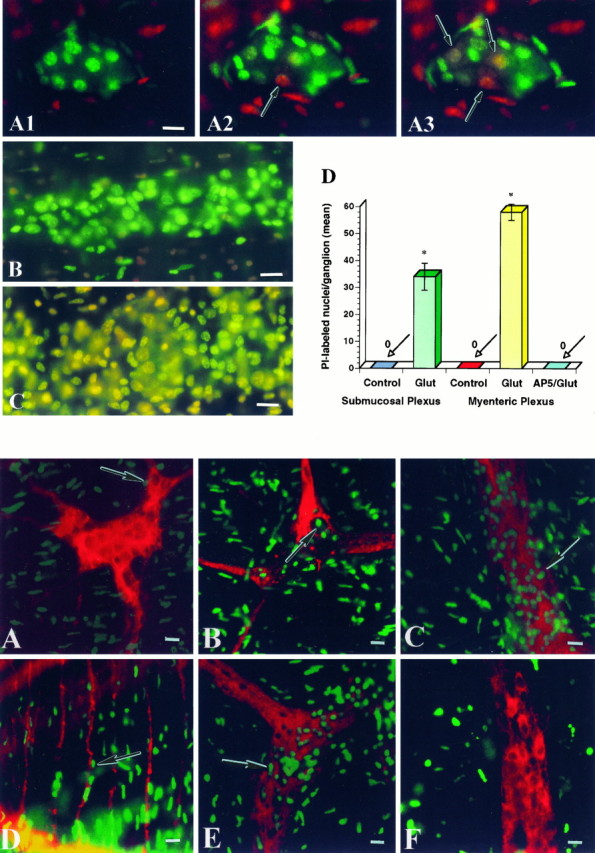
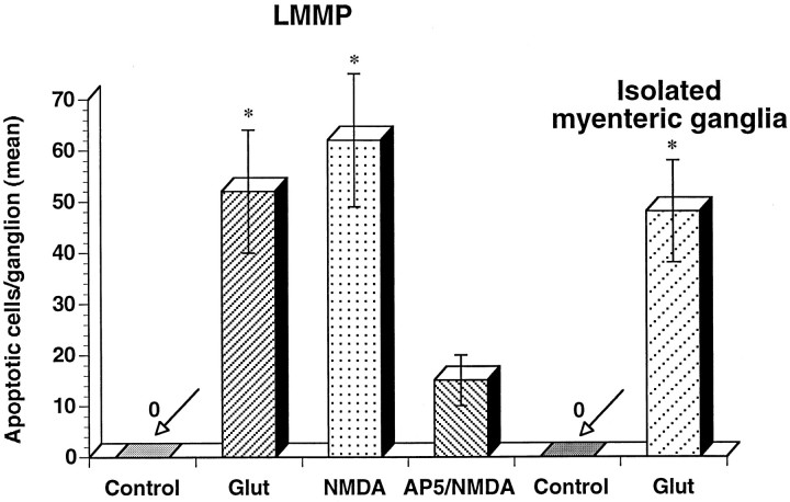
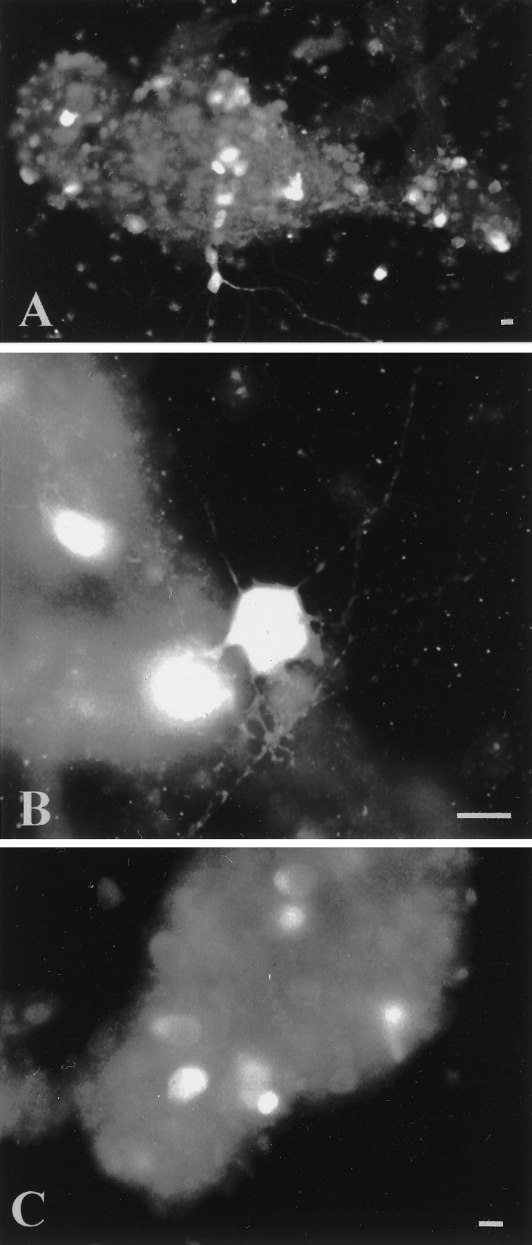
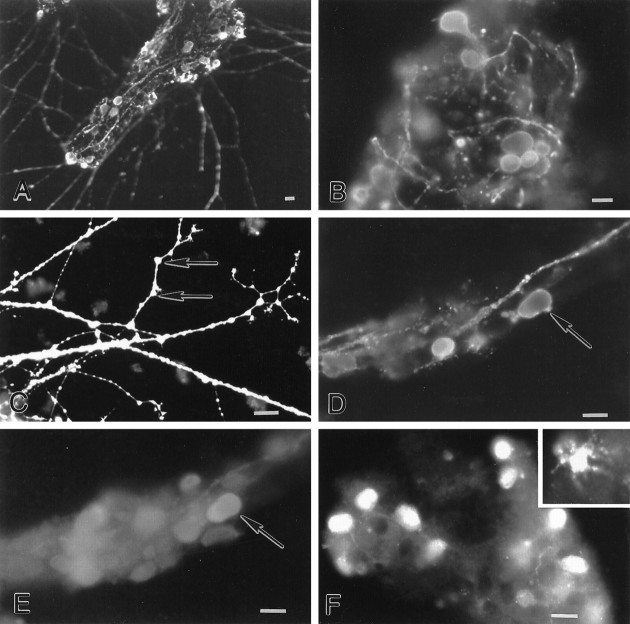
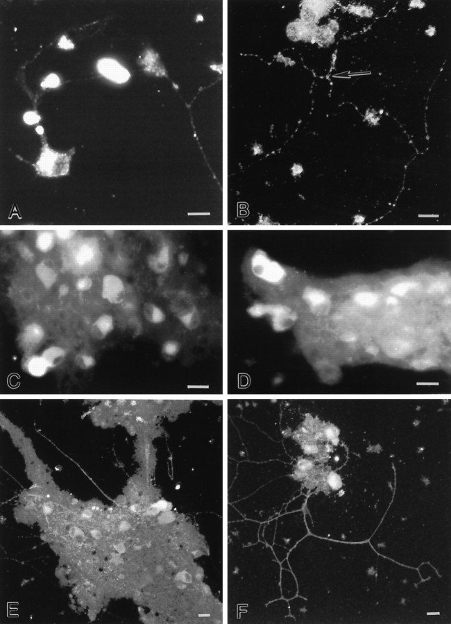
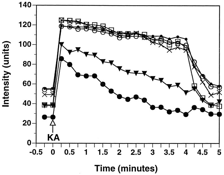
Glutamate, the major excitatory neurotransmitter in the CNS, is also an excitatory neurotransmitter in the enteric nervous system (ENS). We tested the hypothesis that excessive exposure to glutamate, or related agonists, produces neurotoxicity in enteric neurons. Prolonged stimulation of enteric ganglia by glutamate caused necrosis and apoptosis in enteric neurons. Acute and delayed cell deaths were observed. Glutamate neurotoxicity was mimicked by NMDA and blocked by the NMDA antagonist D-2-amino-5-phosphonopentanoate. Excitotoxicity was more pronounced in cultured enteric ganglia than in intact preparations of bowel, presumably because of a reduction in glutamate uptake. Glutamate-immunoreactive neurons were found in cultured myenteric ganglia, and a subset of enteric neurons expressed NMDA (NR1, NR2A/B), AMPA (GluR1, GluR2/3), and kainate (GluR5/6/7) receptor subunits. Glutamate receptors were clustered on enteric neurites. Stimulation of cultured enteric neurons by kainic acid led to the swelling of somas and the growth of varicosities ("blebs") on neurites. Blebs formed close to neurite intersections and were enriched in mitochondria, as revealed by rhodamine 123 staining. Kainic acid also produced a loss of mitochondrial membrane potential in cultured enteric neurons at sites where blebs tended to form. These observations demonstrate, for the first time, excitotoxicity in the ENS and suggest that overactivation of enteric glutamate receptors may contribute to the intestinal damage produced by anoxia, ischemia, and excitotoxins present in food.


Figures

Fig. 1.
Fig. 1.
Top. Glutamate induces necrosis in enteric neurons. A, Whole mounts of submucosa were incubated with SYTO-13 to load enteric cells. Preparations were exposed to glutamate in the presence of PI for 60 min. Neurons that died by necrosis progressively exhibited a fluorescence shift fromgreen to yellow to redbecause PI gained entry into the nucleus 1, Control. SYTO-13-labeled nuclei (green) are present within a ganglion. 2, After a 10 min exposure to glutamate (3 mm). 3, After a 30 min exposure to glutamate (3 mm). Three neurons took up PI (arrows).B, Myenteric ganglion after a 30 min exposure to glutamate. Only SYTO-13-labeled cells are present within the ganglion; however, PI-labeled cells are present outside the ganglion.C, Myenteric ganglion exposed to glutamate (60 min) and subsequently reincubated in culture medium for 24 hr. Many SYTO-13-labeled nuclei within the ganglion have taken up PI (shift toward yellow fluorescence). D, Necrosis of enteric cells stimulated by glutamate. Whole mounts of enteric ganglia (n = 10) were exposed to glutamate (Glut; 60 min) and then incubated in culture medium for 24 hr. A significant number of cells underwent necrosis in both enteric plexuses. Necrotic cells include neurons and probably glia. The NMDA antagonist d-2-amino-5-phosphonopentanoate (AP-5) blocked glutamate-induced necrosis. *p < 0.001. Scale bars: A–C, 30 μm.
Fig. 3.
Fig. 3.
Apoptosis of enteric neurons stimulated by glutamate or NMDA. Neuronal apoptosis is produced by exposure to glutamate (or NMDA) in whole-mount preparations of LMMP (n = 10) and in cultured myenteric ganglia (n = 8). Apoptosis is attenuated by AP-5.
Fig. 4.
Fig. 4.
Characterization of cultured myenteric neurons.A, B, Calretinin-immunoreactive neurons are numerous in cultured myenteric ganglia. Neurons are characterized by short dendrites (B). C, Calbindin-immunoreactive neurons are also found in cultured myenteric ganglia; however, they have a relatively smooth appearance. Scale bars, 30 μm.
Fig. 5.
Fig. 5.
Glutamate-immunoreactive neurons are found in cultured myenteric ganglia. A–C, Glutamate immunoreactivity is found in a subset of cell somas and is abundant in varicose processes. Glutamate immunoreactivity appears to be present on spines (C; arrows). D,E, A subset of glutamate-immunoreactive neurons (D; arrow) contains calbindin immunoreactivity (E; arrow).F, A subset of cultured myenteric neurons expresses EAAC1 immunoreactivity. The majority of EAAC1-immunoreactive cells are smooth in shape; however, “star-shaped” EAAC1-immunoreactive cells are also observed (inset). Scale bars, 30 μm.
Fig. 6.
Fig. 6.
Ionotropic glutamate receptor immunoreactivity is found in cultured myenteric ganglia. A–C, A subset of cultured myenteric neurons express NR1 immunoreactivity. Punctate immunoreactivity is found in the cytoplasm. Moreover, clusters of NR1 immunoreactivity are found along neurites (A,B), near neurite intersections (B;arrow). D, A subset of cultured myenteric neurons express GluR1 immunoreactivity. E,F, A subset of cultured myenteric neurons express GluR2/3 immunoreactivity. Immunostaining is found in the soma and neurites. B, E, F, Confocal photomicrographs. Scale bars, 30 μm.
Fig. 7.
Fig. 7.
Top. Glutamate induces necrosis in cultured myenteric neurons. Cultured myenteric ganglia were incubated with FDA to label viable enteric cells. Cultures were exposed to glutamate in the presence of PI. Neurons that died by necrosis progressively exhibited a fluorescence shift from green toyellow to red because PI gained entry into the nucleus. A, Control. Numerous FDA-labeled cells (green) are present within a ganglion.B, After a 10 min exposure to glutamate (3 mm). C, After a 30 min exposure to glutamate (3 mm). Many FDA-labeled nuclei within the ganglion have taken up PI (shift toward red fluorescence). Scale bars, 30 μm.
Fig. 10.
Fig. 10.
The results depicted in Figure9F–H are shown graphically. R123 intensity in six boutons (arrows in Fig. 9F) was quantified, background-subtracted, and plotted as a function of time. Application of kainic acid (KA) results in an immediate increase in R123 fluorescence, consistent with a loss of mitochondrial potential; however, as exposure to KA continues, R123 intensity decreases with eventual loss of the dye.

References

    1. Ankarcrona M, Dypbukt JM, Bonfoco E, Zhivotovsky B, Orrenius S, Lipton SA, Nicotera P. Glutamate-induced neuronal death: a succession of necrosis or apoptosis depending on mitochondrial function. Neuron. 1995;15:961–973. - PubMed
    1. Bindokas PV, Miller RJ. Excitotoxic degeneration is initiated at non-random sites in cultured rat cerebellar neurons. J Neurosci. 1995;15:6999–7011. - PMC - PubMed
    1. Brorson JR, Manzolillo PA, Miller RJ. Ca2+ entry via AMPA/KA receptors and excitotoxicity in cultured cerebellar Purkinje cells. J Neurosci. 1994;14:187–197. - PMC - PubMed
    1. Burns GA, Stephens KE. Expression of mRNA for the N-methyl-d-aspartate (NMDAR1) receptor and vasoactive intestinal polypeptide (VIP) co-exist in enteric neurons of the rat. J Auton Nerv Syst. 1995;55:207–210. - PubMed
    1. Burns GA, Stephens KE, Benson JA. Expression of mRNA for the N-methyl-d-aspartate (NMDAR1) receptor by the enteric neurons of the rat. Neurosci Lett. 1994;170:87–90. - PubMed

Publication types

MeSH terms

LinkOut - more resources