. 1997 Nov 15;17(22):8828-41.
doi: 10.1523/JNEUROSCI.17-22-08828.1997.

The olivocerebellar projection mediates ibogaine-induced degeneration of Purkinje cells: a model of indirect, trans-synaptic excitotoxicity

Affiliations

The olivocerebellar projection mediates ibogaine-induced degeneration of Purkinje cells: a model of indirect, trans-synaptic excitotoxicity

E O'Hearn et al. J Neurosci. .

Abstract

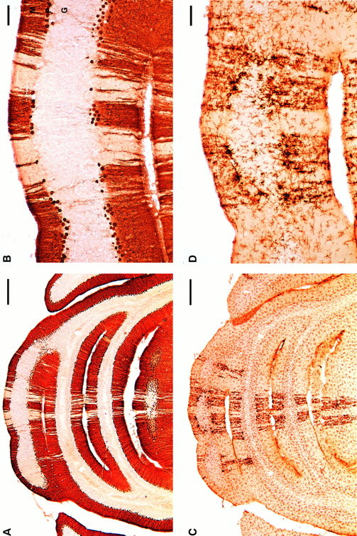
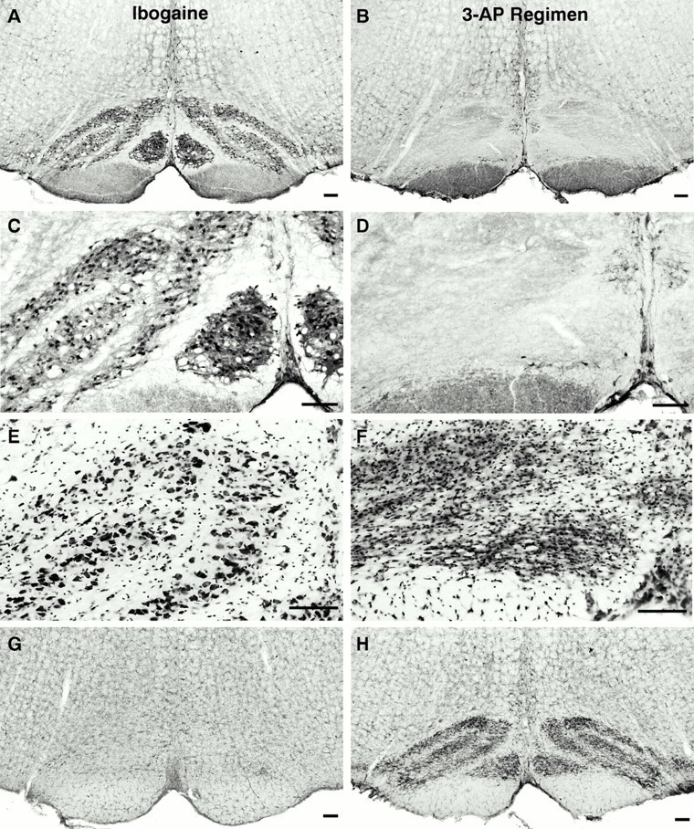
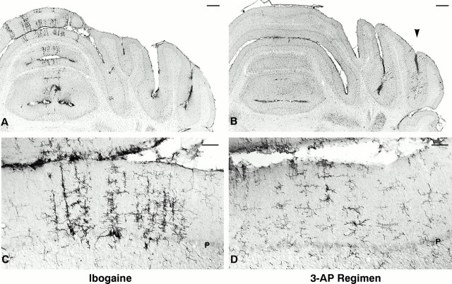
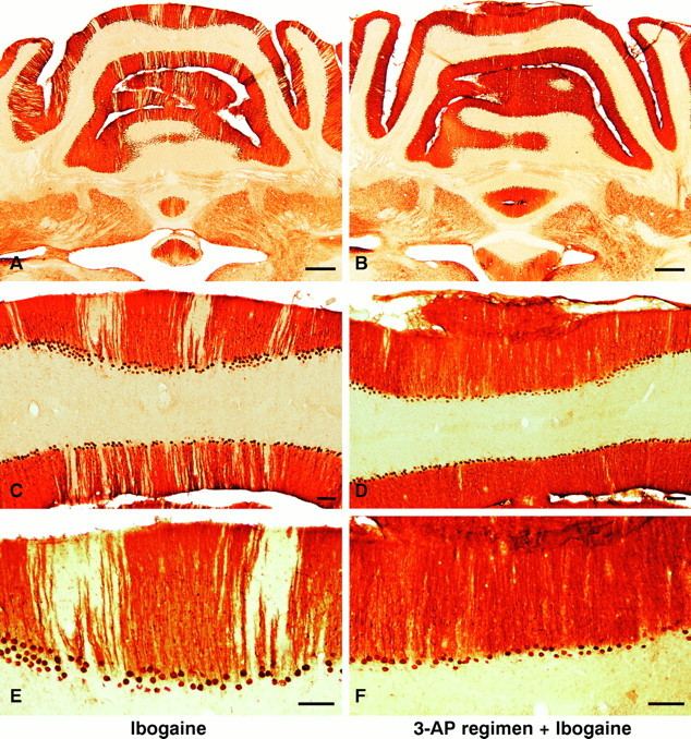
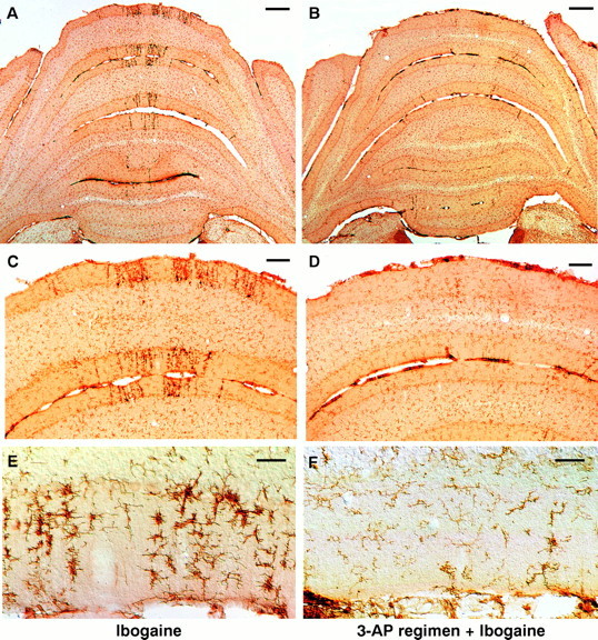
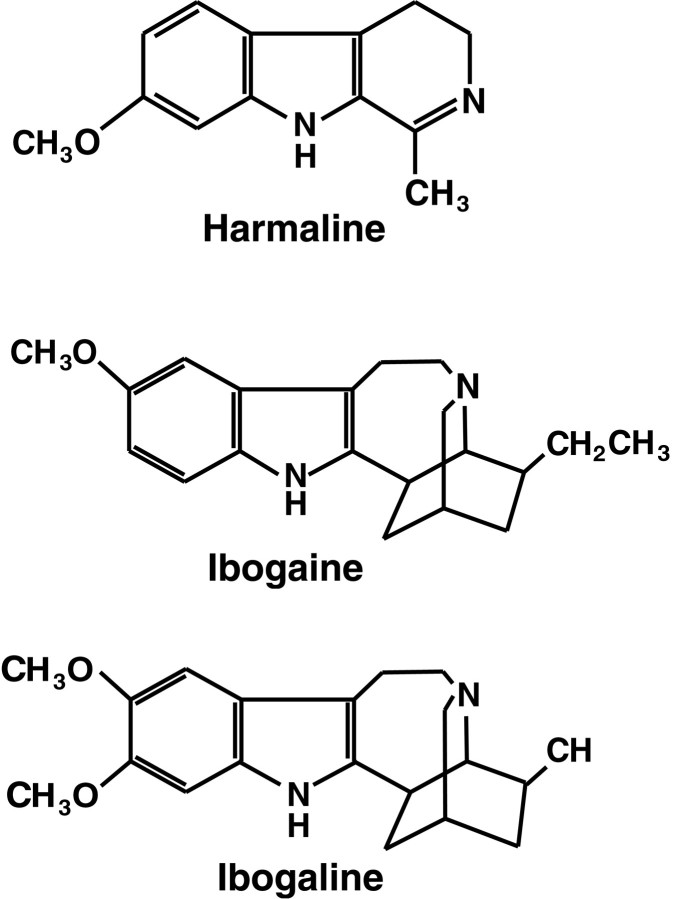
Ibogaine, an indole alkaloid that causes hallucinations, tremor, and ataxia, produces cerebellar neurotoxicity in rats, manifested by degeneration of Purkinje cells aligned in narrow parasagittal bands that are coextensive with activated glial cells. Harmaline, a closely related alkaloid that excites inferior olivary neurons, causes the same pattern of Purkinje cell degeneration, providing a clue to the mechanism of toxicity. We have proposed that ibogaine, like harmaline, excites neurons in the inferior olive, leading to sustained release of glutamate at climbing fiber synapses on Purkinje cells. The objective of this study was to test the hypothesis that increased climbing fiber activity induced by ibogaine mediates excitotoxic Purkinje cell degeneration. The inferior olive was pharmacologically ablated in rats by a neurotoxic drug regimen using 3-acetylpyridine, and cerebellar damage attributed to subsequent administration of ibogaine was analyzed using immunocytochemical markers for neurons and glial cells. The results show that ibogaine administered after inferior olive ablation produced little or no Purkinje cell degeneration or glial activation. That a lesion of the inferior olive almost completely prevents the neurotoxicity demonstrates that ibogaine is not directly toxic to Purkinje cells, but that the toxicity is indirect and dependent on integrity of the olivocerebellar projection. We postulate that ibogaine-induced activation of inferior olivary neurons leads to release of glutamate simultaneously at hundreds of climbing fiber terminals distributed widely over the surface of each Purkinje cell. The unique circuitry of the olivocerebellar projection provides this system with maximum synaptic security, a feature that confers on Purkinje cells a high degree of vulnerability to excitotoxic injury.


Figures

Fig. 1.
Fig. 1.
Ibogaine causes degeneration of Purkinje cells and activation of microglia in discrete radial bands of cerebellar cortex.A, B, Purkinje cells of cerebellar vermis at low (A) and high (B) magnification 7 d after receiving ibogaine (100 mg/kg once). Unstained gaps in the Purkinje cell and molecular layers indicate regions in which Purkinje cells have degenerated (Cam-kin II immunoreactivity, coronal sections). C, D, Clusters of activated microglial cells form darkly stained radial stripes within the cerebellar vermis, in sections adjacent to those showing Purkinje cells. The stripes containing activated microglia are approximately coextensive with regions of Purkinje cell loss (compare densely stainedstripes in C with pale zones in A). The largest and most activated microglia are located in the Purkinje cell layer, where they are presumably phagocytizing a Purkinje cell body (D). Resting microglia are the small, lightly stained cells with fine processes in C andD that are widely distributed throughout all layers of cerebellar cortex and white matter. Microglia are immunoreactive with OX42, which recognizes the complement receptor 3B. Activated microglia are more intensely immunoreactive and have larger processes and cell bodies (D). M, Molecular layer;P, Purkinje cell layer; G, granule cell layer. Scale bars: A, C, 500 μm; B, D, 100 μm.
Fig. 2.
Fig. 2.
Most neurons in the inferior olivary nucleus degenerate after administration of the 3-AP regimen used in this study. In animals treated with ibogaine alone, inferior olive neurons remain intact and exhibit normal morphology (A, C); in contrast, profound loss of neurons is evident (B, D) in the inferior olive of rats that received the 3-AP regimen (3-acetylpyridine, harmaline, and nicotinamide; 13 d survival).A–D, Inferior olivary nucleus: Cam-kin II immunoreactivity at low (A, B) and high (C, D) magnification. E, Large neuronal cell bodies are shown with Nissl stain of rats (E), whereas smaller profiles are glial cells. F, After the 3-AP regimen, neuronal profiles in the inferior olive are absent, and this nucleus has become densely populated with small glial cell bodies. The inferior olive is gliotic because of proliferation of astrocytes and microglia that were activated in response to degeneration of inferior olive neurons. Using a marker for microglial cells (G, H), only lightly stained, resting microglia are observed in the inferior olive from an ibogaine-treated rat (G); in contrast, after the 3-AP regimen, densely packed activated microglia occupy the site of the former inferior olivary nucleus (H) and demarcate the different subregions that were present in this nucleus. Cytochemical markers and stains: A–D, Cam-kin II immunocytochemistry for inferior olive neurons; E, F, Nissl stain of inferior olive; G, H, to identify microglia, the inferior olive is stained with antibody (OX42) that recognizes the complement receptor 3B. This receptor is expressed at moderate levels by quiescent microglia and is greatly increased in activated microglia in response to neuronal injury or degeneration. Scale bars:A–H, 100 μm.
Fig. 3.
Fig. 3.
Activated microglia exhibit a different distribution and morphology in ibogaine- versus 3-AP-treated animals. After ibogaine administration (100 mg/kg; 7 d survival), activated microglia form radial bands located primarily in the vermis (A); the most intensely activated microglia (C) are found at the depth of Purkinje cell bodies (which have degenerated, as observed in adjacent sections). After the 3-AP regimen (13 d survival), occasional activated microglia are observed far laterally in the hemispheres (B, arrowhead), primarily in the ansiform lobule, crus I, and less commonly in the vermis; these activated cells are found mainly in the molecular layer and tended not to be adjacent to Purkinje cell bodies (D). Enlarged, darkly immunoreactive cells are activated microglia. More delicate cellular profiles (C, D) with finer processes, smaller cell bodies, and less intense immunostaining are quiescent microglia. In ibogaine-treated animals, activated microglia (C) are larger and more darkly immunoreactive than those seen after the 3-AP regimen (D), suggesting that 3-AP may cause a smaller degree of neuronal insult. Coronal sections immunostained with antibody OX42 for complement receptor 3B. Scale bars: A, B, 500 μm; C, D, 50 μm. P, Purkinje cell layer.
Fig. 4.
Fig. 4.
A–F, Neuroprotection: ablation of the inferior olive with 3-AP prevents or greatly attenuates Purkinje cell degeneration induced by ibogaine. Left column(A, C, E), Treatment with ibogaine alone produces radial bands of Purkinje cell loss manifested by pale, unstained gaps in the molecular and Purkinje cell layers. Loss of Purkinje cells is most prominent in the vermis but is also present in the paravermis and simplex lobule. Right column (B, D, F), Animals that received the 3-AP regimen followed by ibogaine 6 d later demonstrate marked neuroprotection against Purkinje cell degeneration. The nearly continuous immunostaining of Purkinje cell bodies and of their dendrites in the molecular layer (B, D, F) indicates that there is little or no ibogaine-induced degeneration of Purkinje cells after olive ablation. Infrequently in rats that received the 3-AP regimen plus ibogaine, a single Purkinje cell may have degenerated (see thin gapin neuronal staining of molecular and Purkinje cell layers inupper right corner of D). Photomicrographs show Purkinje cells in coronal sections immunostained with antiserum to Cam-kin II. Ibogaine dose, 100 mg/kg once; in all cases, survival was 7 d after ibogaine administration. Scale bars:A, B, 500 μm; C–F, 100 μm.
Fig. 5.
Fig. 5.
Ablation of the inferior olive with the 3-AP regimen profoundly attenuates subsequent microglial activation induced by ibogaine. Left column (A, C, E), Treatment with ibogaine alone produces radial bands of darkly stained, activated microglia, primarily in the molecular and Purkinje cell layers of the cerebellar vermis. In most cases, the radial bands of microglia are coextensive with bands of degenerating Purkinje cells (Fig. 1). Right column (B, D, F), Animals that received the 3-AP regimen followed by ibogaine 6 d later demonstrate few signs of microglial activation. After the 3-AP regimen, ibogaine no longer produces more than an occasional radial stripe of activated microglia. The great majority of cells seen are resting microglia that are faintly stained and have delicate processes. Activated microglia are infrequent in the rats that received the 3-AP regimen plus ibogaine. Photomicrographs of coronal sections show microglial cells immunostained with antiserum OX42 for complement receptor 3B. Drug doses: A, C, E, ibogaine (100 mg/kg);B, D, F, 3-AP regimen given 6 d before ibogaine (100 mg/kg). In all cases, survival was 7 d after ibogaine administration. Scale bars: A, B, 500 μm; C, D, 100 μm; E, F, 50 μm.
Fig. 6.
Fig. 6.
Chemical structures of the indole alkaloids harmaline, ibogaine, and ibogaline. Harmaline, ibogaine, and ibogaline share an indole nucleus and have nearly identical types of behavioral effects. Zetler et al. (1972, reported that the positions and number of methoxy groups (left) greatly influence tremorigenic potency, whereas the additional ring structures (right) have little effect on drug action within this group of compounds. For comparative potencies of tremor induction, seeZetler et al. (1972, .

References

    1. Adams JH, Duchen LW. Greenfield’s neuropathology. Oxford UP; New York: 1992.
    1. Anderson WA, Flumerfelt BA. A light and electron microscopic study of the effects of 3-acetylpyridine intoxication on the inferior olivary complex and cerebellar cortex. J Comp Neurol. 1980;190:157–174. - PubMed
    1. Anderson WA, Flumerfelt BA. Sensitivity of rat inferior olivary neurons to 3-acetylpyridine. Dev Brain Res. 1984;12:285–291. - PubMed
    1. Azizi SA, Woodward DJ. Inferior olivary nuclear complex of the rat: morphology and comments on the principles of organization within the olivocerebellar system. J Comp Neurol. 1987;263:467–484. - PubMed
    1. Balaban CD. Central neurotoxic effects of intraperitoneally administered 3-acetylpyridine, harmaline and niacinamide in Sprague-Dawley and Long-Evans rats: a critical review of central 3-acetylpyridine neurotoxicity. Brain Res. 1985;356:21–42. - PubMed

Publication types

MeSH terms

LinkOut - more resources