. 1997 Oct;8(10):1845-62.
doi: 10.1091/mbc.8.10.1845.

Endoplasmic reticulum stress-induced mRNA splicing permits synthesis of transcription factor Hac1p/Ern4p that activates the unfolded protein response

Affiliations
Free PMC article

Endoplasmic reticulum stress-induced mRNA splicing permits synthesis of transcription factor Hac1p/Ern4p that activates the unfolded protein response

T Kawahara et al. Mol Biol Cell. 1997 Oct.
Free PMC article

Abstract

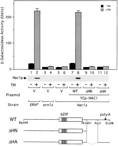
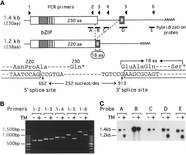
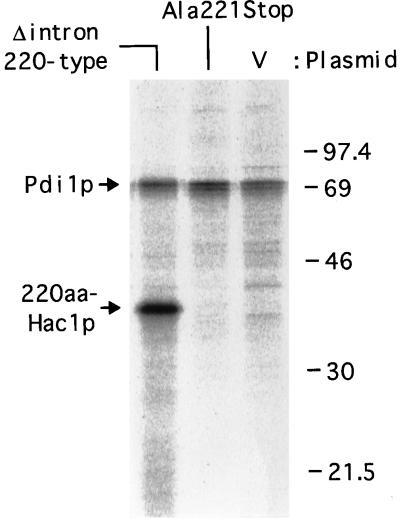
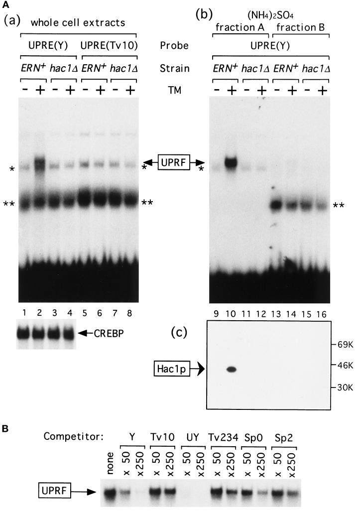
An intracellular signaling from the endoplasmic reticulum (ER) to the nucleus, called the unfolded protein response (UPR), is activated when unfolded proteins are accumulated in the ER under a variety of stress conditions ("ER stress"). We and others recently identified Hac1p/Ern4p as a transcription factor responsible for the UPR in Saccharomyces cerevisiae. It was further reported that Hac1p (238 aa) is detected only in ER-stressed cells, and its expression is mediated by unconventional splicing of HAC1 precursor mRNA. The splicing replaces the C-terminal portion of Hac1p; it was proposed that precursor mRNA is also translated but the putative product of 230 aa is rapidly degraded by the ubiquitin-proteasome pathway. We have identified and characterized the same regulated splicing and confirmed its essential features. Contrary to the above proposal, however, we find that the 238-aa product of mature mRNA and the 230-aa-type protein tested are highly unstable with little of no difference in stability. Furthermore, we demonstrate that the absence of Hac1p in unstressed cells is due to the lack of translation of precursor mRNA. We conclude that Hac1p is synthesized as the result of ER stress-induced mRNA splicing, leading to activation of the UPR.


Figures

Figure 3
Figure 3
Induction of 1.2-kb HAC1 mRNA correlates with the UPR. (A) The ERN+ (KMY1105) and ern1Δ (KMY1115) strains were transformed with a vector alone (V) or a Ern1p expression plasmid (a single-copy or multicopy plasmid indicated by Cen or 2 μm, respectively). Transformants were grown at 30°C in SC(−Ura, Leu) medium to midlogarithmic phase, and aliquots were incubated for 1 h in the presence (+) or absence (−) of tunicamycin (TM). Total RNAs were extracted and analyzed by Northern blot hybridization using DNA probes specific for HAC1, KAR2, or yeast actin ACT1. Positions of the 1.4-kb and 1.2-kb HAC1 mRNAs are indicated. In addition to these two mRNA species, tunicamycin treatment produced a faint band of 0.7 kb, that was not analyzed in this report. (B) The ERN+ and ern1Δ strains with the sec53 background in which the UPRE-CYC1-lacZ reporter gene had been integrated into the respective chromosome (KMY2105 and KMY2115, respectively) were grown at the permissive temperature of 23°C in SC(−Ura) medium to midlogarithmic phase, and aliquots were incubated for an additional hour at 23°C or at the seminonpermissive temperature of 30°C. Total RNAs were extracted and analyzed as in A. (C) The ERN+, SEC+ strain (KMY1105) was treated with tunicamycin at 30°C for the times indicated as in A, and RNA was similarly analyzed by Northern blot hybridization. Relative radioactivities of each band (HAC1, KAR2, and PDI1) were determined using BioImaging Analyzer BAS-2000 (Fuji Photo Film), corrected for ACT1 values, and plotted after normalization to the 0 time values.
Figure 2
Figure 2
Induction of Hac1p requires 3′UTR of HAC1 mRNA. The ERN+, ern1Δ, and hac1Δ strains with the UPRE-CYC1-lacZ reporter gene integrated into the respective chromosome (KMY1105, 1115, and 1145, respectively) were transformed with a vector alone (V) or a single-copy expression plasmid carrying the wild-type (WT) or a mutant (ΔHN or ΔHA) HAC1 gene, whose structures are schematically drawn at the bottom. bZIP and polyA denote the bZIP region and the polyadenylation site, respectively. Transformants were grown at 30°C in SC(−Ura, Leu) medium to a midlogarithmic phase, and aliquots were incubated in the presence (hatched bars) or absence (solid bars) of tunicamycin (TM). Samples taken after 3 h were used for β-galactosidase assays, and the activities are presented as the mean ± SD, based on duplicate determinations with three independent transformants. Separate samples taken after 1 h were used for extracting total proteins that were analyzed by immunoblotting with purified anti-Hac1p antibodies.
Figure 4
Figure 4
ER stress-induced mRNA splicing replaces the C-terminal portion of Hac1p. (A) Structures of the 1.4-kb and 1.2-kb HAC1 mRNAs are schematically presented. Locations of primers 1–6 for RT-PCR and DNA probes A-E for Northern blot hybridization are also indicated. The 1.4-kb mRNA can encode a bZIP protein of 230 aa, whereas the 1.2-kb mRNA resulting from ER-stress–induced splicing can encode a bZIP protein of 238 aa. Shown below are the nucleotide sequences around the 5′ and 3′ splice sites. The splicing deletes the internal 252 nt from nt 662 to 913 (the A of the first ATG codon is set as +1), resulting in the replacement of the C-terminal 10 aa with a stretch of 18 aa encoded by the second exon. (B) Total RNAs were extracted from the ERN+ strain grown for 1 h in the presence (+) or absence (−) of tunicamycin (TM). HAC1 cDNA was prepared by RT-PCR using a fixed 5′ primer 1 and various 3′ primers (primers 2–6), and the products were analyzed by 1% agarose gel electrophoresis. The amounts of PCR products did not reflect the amounts of mRNA well due to excessive cycles used. Positions of the 100-bp ladder DNA size markers are indicated. (C) Total RNAs prepared as in B were analyzed by Northern blot hybridization using the various DNA probes indicated in A, which had been labeled with [32P]dCTP by PCR. Positions of the 1.4-kb and 1.2-kb HAC1 mRNAs are indicated.
Figure 6
Figure 6
Constitutive expression of Hac1p of 238, 220, and 230 aa. (A) Structures of the wild-type (WT) and mutant versions of the HAC1 gene are schematically presented. Thin lines denote nucleotide segments deleted from the HAC1 gene. (B and C) The ern1Δ (KMY1115) and hac1Δ (KMY1145) strains were transformed with a vector alone (V) or a single-copy expression plasmid carrying a mutant version (238, 220, or 230) of the HAC1 gene. Transformants were grown at 30°C in SC(−Ura, Leu) medium to midlogarithmic phase, and aliquots were incubated in the presence (hatched bars and lanes of even numbers) or absence (solid bars and lanes of odd numbers) of tunicamycin (TM). Samples taken after 3 h were used for β-galactosidase assays, and the activities are presented as the mean ± SD, based on duplicate determinations with three independent transformants. Separate samples taken after 1 h were used for extracting total proteins or total RNAs that were analyzed by immunoblotting using anti-Hac1p, anti-Kar2p, and anti-Pdi1p antibodies or by Northern blot hybridization using DNA probes specific for HAC1 and ACT1. Hac1p of 238, 220, and 230 aa were also translated in vitro as described in MATERIALS AND METHODS. The positions of molecular mass markers and Hac1p of 238, 220, and 230 aa are indicated. * denotes a nonspecific band. (B, inset) The levels of Kar2p and Pdi1p in total proteins extracted from tunicamycin-untreated hac1Δ cells.
Figure 7
Figure 7
Stability of Hac1p of 238, 220, and 230 aa. (A) Transformants of the hac1Δ strain in which each of the mutant versions of the HAC1 gene described in Figure 6A had been introduced were grown at 30°C in SC(−Ura, Leu) medium to midlogarithmic phase, and aliquots were treated with cycloheximide (20 μg/ml). Immediately after the times indicated, the cultures were poured over ice and the cells were collected by centrifugation. Total proteins were extracted and analyzed by immunoblotting using anti-Hac1p and anti-Kar2p antibodies. For Hac1p of 220 aa, a shorter exposure of the film is also shown below. * denotes a nonspecific band. (B) The hac1Δ strain (KMY1045) was cotransformed with a plasmid carrying the mutant (230-type) HAC1 gene and the LEU2-selectable marker and a plasmid carrying the intron-less (238-type) HAC1 gene and the URA3-selectable marker. The resulting transformant was grown, pulse-labeled for 5 min with [35S]methionine and [35S]cysteine, and then chased for the times indicated as described in MATERIALS AND METHODS. The hac1Δ strain carrying either the 230-type or 238-type mutant HAC1 gene was pulse-labeled for 5 min and shown for comparison. Hac1p and Pdi1p (internal control) were immunoprecipitated and subjected to SDS-PAGE (12% gel). The positions of molecular mass markers, Pdi1p, and Hac1p of 238 aa and 230 aa are indicated. Radioactivities of each band were determined by using a BioImaging Analyzer BAS-2000 and corrected for Pdi1p values. Data from two separate experiments (indicated by circles and triangles) are plotted after normalization to the 0 time value of 230aa-Hac1p.
Figure 10
Figure 10
Synthesis rates of 220aa-Hac1p encoded by intron-less and intron-containing mRNAs. The hac1Δ strain (KMY1145) was transformed with a vector alone (V) or a mutant version (Δintron 220-type or Ala221Stop) of the HAC1 gene. Each transformant was grown and pulse-labeled for 5 min with [35S]methionine and [35S]cysteine as described in MATERIALS AND METHODS. Hac1p and Pdi1p were immunoprecipitated and subjected to SDS-PAGE (12% gel). Radioactive bands were visualized by using a BioImaging Analyzer BAS-2000. The positions of molecular mass markers, 220aa-Hac1p, and Pdi1p are indicated in kilodaltons.
Figure 9
Figure 9
Production of Hac1p only from mature mRNA. At the top, the nucleotide sequence of the wild-type (WT) HAC1 gene around the 5′ splice site and its deduced amino acid sequence are shown. In a mutant HAC1 gene designated WT(XbaI), a XbaI site (underlined) was created at nt 640 between the two MfeI sites in YCp-HAC1WT that changed aa 214 and 215 from Leu-Asp to Ser-Arg. Δintron(XbaI) contains a XbaI site in YCp-HAC1Δintron similarly. The Ala221Stop mutant contains a stop codon TAG (double underlined) instead of the codon for Ala221. The Ala221Stop′ mutant contains a 2-nt (TA) insertion between the codons for Pro220 and Ala221 that creates a stop codon (double underlined) immediately after Pro220 but maintains nucleotide sequences at the 5′ splice site from the position −2 (AGCCG—). The Asn216Stop mutant contains a stop codon TAG (double underlined) instead of the codon for Asn216. The hac1Δ strain (KMY1145) was transformed with a vector alone (V) or each of these constructs as indicated. Transformants were grown at 30°C in SC(−Ura, Leu) medium to midlogarithmic phase, and aliquots were incubated for 3 h in the presence (hatched bars) or absence (solid bars) of tunicamycin (TM). β-Galactosidase activities in cell extracts were determined, and are presented as the mean ± SD, based on duplicate determinations with three independent transformants. Separate samples taken after 1 h were used for extracting total proteins or total RNAs that were analyzed by immunoblotting using anti-Hac1p antibodies or by Northern blot hybridization using DNA probes specific for HAC1 and ACT1. Hac1p of 238 and 220 aa were also translated in vitro. * denotes a nonspecific band.
Figure 1
Figure 1
Induction of Hac1p by tunicamycin. (A) Whole cell extracts, (NH4)2SO4 fraction A, and (NH4)2SO4 fraction B were prepared from the ERN+ (KMY1105) and hac1Δ (KMY1145) strains that had been grown in YPD medium to a midlogarithmic phase and incubated for 1 h with (+) or without (−) tunicamycin (TM). (a) Forty micrograms of proteins in each of whole cell extracts were mixed with 0.3 ng of 32P-labeled wild-type UPRE [designated UPRE(Y), 9060 cpm], a point mutant of UPRE [designated UPRE(Tv10), 9390 cpm], or CRE (7590 cpm). (b) Forty micrograms of proteins in each of (NH4)2SO4 fraction A or fraction B were mixed with 0.2 ng (9130 cpm) of 32P-labeled UPRE(Y). Protein-bound probes were separated from free probes in a 5% nondenaturing gel. The specific binding to UPRE(Y) is marked as UPRF. * and ** denote nonspecific bindings that were also detected when 32P-labeled CRE was used as a probe. Only specific binding to CRE was shown below and marked as CREBP. (c) Fifty micrograms of proteins in (NH4)2SO4 fraction A or fraction B were subjected to SDS-PAGE (12% gel) and immunoblotted with purified anti-Hac1p antibodies. The positions of molecular mass markers are indicated. (B) The specific binding between 0.3 ng (7870 cpm) of 32P-labeled UPRE(Y) and UPRF in the (NH4)2SO4 fraction A (20 μg of proteins) from tunicamycin-treated ERN+ strain was competed by 50- or 250-fold molar excess of unlabeled UPRE(Y) or various mutant forms of UPRE, whose activity in mediating the UPR was well characterized previously (Mori et al., 1996). Only specific binding is shown. Tv10, a point mutant of Y described in the text, is virtually inactive in mediating the UPR in vivo. UY contains a palindrome of 7 bp that is more active than Y. Tv234 contains three transversions upstream of the palindromic sequence. Sp0 and Sp2 are mutant versions of UPRE in which the half-sites in the palindromic sequence are separated by spacers of 0 and 2 nt, respectively, instead of 1 nt in Y. Tv234, Sp0, and Sp2 exhibit very weak activities in vivo. Neither 5′ nor 3′ termini of competitor oligonucleotides were filled in, and possible formation of concatemers may explain the large molar excess of unlabeled UPRE-Y required for competition under these conditions.
Figure 5
Figure 5
Expression of cDNA corresponding to 1.2-kb HAC1 mRNA lacking the intron constitutively activates the UPR. (A) The hac1Δ strain (KMY1145) was transformed with a single-copy expression plasmid (YCp-HAC1) carrying the wild-type (WT) or intron-less (Δintron) HAC1 gene. Transformants were grown at 30°C in SC(−Ura, Leu) medium to midlogarithmic phase, and aliquots were incubated for 1 h in the presence (+) or absence (−) of tunicamycin (TM). Total proteins and total RNAs were extracted and analyzed by immunoblotting using anti-Hac1p antibodies and by Northern blot hybridization using DNA probes specific for HAC1, KAR2, LHS1/SSI1/CER1, SCJ1, PDI1, EUG1, FKB2, and ACT1, respectively. (B) The ERN+, ern1Δ, and hac1Δ strains (KMY1105, 1115, and 1145, respectively) were transformed with a vector alone (V), YCp-HAC1WT, or YCp-HAC1Δintron. Transformants were grown at 30°C in SC(−Ura, Leu) medium to midlogarithmic phase, and aliquots were incubated for 3 h in the presence (hatched bars) or absence (solid bars) of tunicamycin (TM). β-Galactosidase activities in cell extracts were determined and are presented as the mean ± SD, based on duplicate determinations with three independent transformants.
Figure 8
Figure 8
Effects of various ubc mutations on the UPR. (A) Various strains indicated on the abscissa were transformed with the UPRE-CYC1-lacZ reporter gene carried on a single-copy vector (pSCZ-Y, hatched bars) or a multicopy vector (pMCZ-Y, stippled bars) and transformants were grown at 30°C in SC(−Ura) medium. The amounts of β-galactosidase expressed without tunicamycin treatment were determined with midlogarithmic-phase cells and are presented as the mean ± SD, based on duplicate determinations with three independent transformants after normalization to the values for the UBC+ strain of MATα. (B) The UBC+, ubc4Δ, ubc5Δ, and ubc7Δ strains carrying pMCZ-Y were grown at 30°C in SC(−Ura) medium to midlogarithmic phase, and aliquots were incubated in the presence (+) or absence (−) of tunicamycin (TM). Samples taken after 1 h were used for extracting total RNAs that were analyzed by Northern blot hybridization using DNA probes specific for UBC4, HAC1, and ACT1. Total proteins were extracted from the UBC+, ubc4Δ, ubc5Δ, and ubc7Δ strains that had been grown in YPD medium to midlogarithmic phase, then incubated for 1 h in the presence (+) or absence (−) of tunicamycin, and analyzed by immunoblotting using anti-Hac1p, anti-Kar2p, and anti-Pdi1p antibodies. (C) The ERN1 and HAC1 loci in the ubc5Δ and ubc7Δ strains were disrupted as described previously (Mori et al., 1996). Various strains indicated on the abscissa were transformed with pMCZ-Y. Transformants were grown at 30°C in SC(−Ura) medium to midlogarithmic phase, and aliquots were incubated in the presence (hatched bars) or absence (solid bars) of tunicamycin. Samples taken after 3 h were used for β-galactosidase assays, and the activities are presented as the mean ± SD, based on duplicate determinations with three independent transformants.

References

    1. Baxter BK, James P, Evans T, Craig EA. SSI1 encodes a novel Hsp70 of the Saccharomyces cerevisiae endoplasmic reticulum. Mol Cell Biol. 1996;16:6444–6456. - PMC - PubMed
    1. Brodsky JL, McCracken AA. ER-associated and proteasome-mediated protein degradation: how two topologically restricted events came together. Trends Cell Biol. 1997;7:151–156. - PubMed
    1. Chen P, Johnson P, Sommer T, Jentsch S, Hochstrasser M. Multiple ubiquitin-conjugating enzymes participate in the in vivo degradation of the yeast MATα2 repressor. Cell. 1993;74:357–69. - PubMed
    1. Clark MW, Abelson J. The subnuclear localization of tRNA ligase in yeast. J Cell Biol. 1987;105:1515–1526. - PMC - PubMed
    1. Cox JS, Shamu CE, Walter P. Transcriptional induction of genes encoding endoplasmic reticulum resident proteins requires a transmembrane protein kinase. Cell. 1993;73:1197–1206. - PubMed

Publication types

MeSH terms