. 1997 Oct;8(10):2003-15.
doi: 10.1091/mbc.8.10.2003.

Amphiphysin heterodimers: potential role in clathrin-mediated endocytosis

Affiliations
Free PMC article

Amphiphysin heterodimers: potential role in clathrin-mediated endocytosis

P Wigge et al. Mol Biol Cell. 1997 Oct.
Free PMC article

Abstract

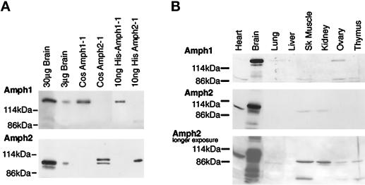
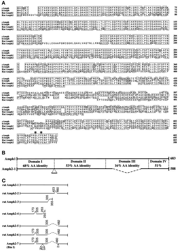
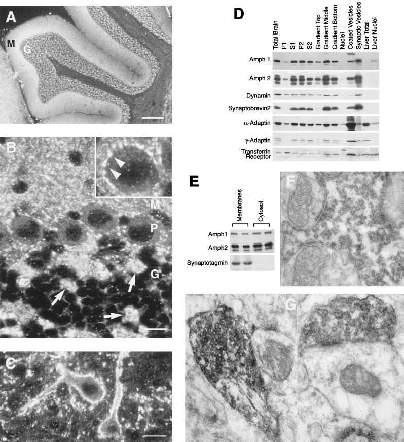
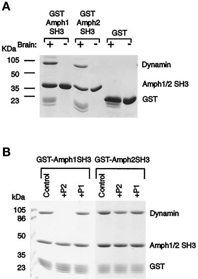
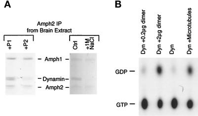
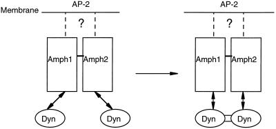
Amphiphysin (Amph) is a src homology 3 domain-containing protein that has been implicated in synaptic vesicle endocytosis as a result of its interaction with dynamin. In a screen for novel members of the amphiphysin family, we identified Amph2, an isoform 49% identical to the previously characterized Amph1 protein. The subcellular distribution of this isoform parallels Amph1, both being enriched in nerve terminals. Like Amph1, a role in endocytosis at the nerve terminal is supported by the rapid dephosphorylation of Amph2 on depolarization. Importantly, the two isoforms can be coimmunoprecipitated from the brain as an equimolar complex, suggesting that the two isoforms act in concert. As determined by cross-linking of brain extracts, the Amph1-Amph2 complex is a 220- to 250-kDa heterodimer. COS cells transfected with either Amph1 or Amph2 show greatly reduced transferrin uptake, but coexpression of the two proteins rescues this defect, supporting a role for the heterodimer in clathrin-mediated endocytosis. Although the src homology 3 domains of both isoforms interact with dynamin, the heterodimer can associate with multiple dynamin molecules in vitro and activates dynamin's GTPase activity. We propose that it is an amphiphysin heterodimer that drives the recruitment of dynamin to clathrin-coated pits in endocytosing nerve terminals.


Figures

Figure 2
Figure 2
Characterization of Amph2 and its antiserum. (A) Amph2 is a 92-kDa protein present in the brain in the same molar ratio as Amph1. Total brain extract, extracts of COS cells transfected with either of the Amphs, and bacterially expressed protein were loaded on SDS-PAGE gels for comparison, followed by immunoblotting with each of the Amph antibodies. (B) Tissue distribution of Amph2 resembles that of Amph1. Brain extract (20 μg) and each of the other tissues (5 μg) were loaded on SDS-PAGE gels and immunoblotted with each antibody as indicated. A longer exposure of Amph2 reveals Amph2 reactivity in other tissues.
Figure 1
Figure 1
Primary structure of rat Amph2. (A) Sequence alignment showing homology of rat Amph2 to Amph1. Boxes show regions of amino acid identity in comparison to rat Amph1. The chick and human Amph sequences are shown for comparison. Amino acids conserved in among all SH3 domains are indicated with an asterisk. These sequences have been submitted to the GenBank database under accession numbers Y13380 (Amph2) and Y13381 (Amph1). (B) Overall comparison of Amph1 and Amph2. Amph is divided into four domains. Domain A is predicted to be α-helical and has a average pI of 9.0. Domain B is proline rich and acidic. Domain C is not conserved and acidic. Domain D is an SH3 module and acidic. (C) Amph2 splice variants. Comparison of Amph2 clones. Spliced introns are indicated by dotted lines. Note that Amph2–3 and Amph2–4 contain Amph2 sequence at their 3′ untranslated regions and, therefore, are unlikely to be cloning artifacts.
Figure 3
Figure 3
Subcellular distribution of Amph2 in rat brain. (A) By using Amph2 antiserum, the strongest staining for Amph2 is in the molecular (M) and granule cell (G) layers of the cerebellum. (B) At higher magnification, the presence of Amph in mossy fiber terminals of the granule cell layer (G) can be clearly seen (arrows). The Purkinje cell bodies (P) were surrounded by numerous small reactive terminals. This is more clearly seen in the inset (arrowheads). (C) Many cells in the pontine nucleus were densely innervated by fine Amph2 containing terminals. Bars: A, 300 μm; B, 14 μm; C, 9 μm. (D) Western blot analysis of subcellular fractions of rat brain. Subcellular fractionation was carried out according to MATERIALS AND METHODS. S1-P2 refer to successive pellets (P) or supernatants (S) of rat brain homogenized in 0.32 M buffered sucrose (see McMahon et al., 1992). The P2 fraction contains crude synaptosomes and was used for Percoll gradient separation into three predominant layers, the middle layer of which is enriched in isolated nerve terminals. Five micrograms of protein were loaded in each lane. (E) Amph1 and Amph2 both exist as membrane-associated and cytoplasmic pools. Synaptosomes were hypotonically lysed, followed by pelleting at 150 000 × g for 30 min to obtain a crude membrane fraction and cytosol, cleared of all synaptic vesicles (as shown by synaptotagmin control). Each fraction was loaded in duplicate, at 10 μg of protein per lane. (F and G) Electron micrographs showing the ultrastructural localization of Amph2 in the rat cerebellum. The large mossy fiber terminals in the granule cell layer were found to be heavily stained with Amph2 reaction product (F), as were climbing fiber terminals in the molecular/Purkinje cell layer (G). Within these structures, Amph2 staining was heavily concentrated on the outer layer of synaptic vesicles.
Figure 4
Figure 4
Amph2 associates with dynamin in vitro. (A) The SH3 domain of Amph2, like Amph1, binds specifically and stoichiometrically to dynamin in brain extracts. GST fusion proteins, and GST as a control, were incubated with brain extract followed by analysis of bound proteins by SDS-PAGE and Coomassie staining. Note the presence of a fainter ∼145-kDa band (probably synaptojanin). (B) The P2 peptide differentially affects dynamin binding to Amph1 and Amph2. GST-tagged SH3 domains were incubated as in A but in the presence or absence of each of the indicated peptides (at a concentration of 200 μM). Bound dynamin was visualized by Coomassie blue staining.
Figure 5
Figure 5
Amph phosphorylation and dephosphorylation. (A) Time course of Amph2 dephosphorylation is more rapid and extensive than Amph1 or DynI. Seconds refer to the time after addition of KCl. Data are averaged from four separate experiments (means ± SEM). (B) Repetitive depolarization/repolarization of synaptosomes induces cyclical changes in the phosphorylation state of the Amphs. After the first stimulation (Stim1), synaptosomes were pelleted for 5 s at 13,000 × g and resuspended in repolarization buffer (HBM containing 4.5 mM KCl), allowed to equilibrate for 1 min at 37°C, before addition of KCl back to 35 mM (Stim2). Dynamin, Amph1, and Amph2 were immunoprecipitated with their respective antiserum. (C) Effect of PKC activator (PMA) and inhibitor (Ro31–8220) on the phosphorylation state of Amph1 and Amph2. Synaptosomes were labeled as described in A and 10 μM of either PMA or Ro31–8220 were added just before harvesting.
Figure 6
Figure 6
Amph1 and Amph2 form heterodimers in vivo. (A) Immunoprecipitation of Amph1 and Amph2 from brain as an equimolar complex. Rat brain extract in buffer A was immunoprecipitated with antiserum to either Amph2 or, as a control, dynamin. Bound proteins were analyzed by Coomassie blue staining. Note the presence of dynamin in both immunoprecipitates (see Figure 8A). (B) Immunoprecipitation of the two isoforms as a complex in COS cells. COS cells were transiently transfected with Amph1, Amph2, or the two together. Extracts of each were separately immunoprecipitated with antibodies against Amph1 or Amph2 and processed for Western blotting. Total proteins were also loaded to show the starting material. (Due to the strength of the Amph2 antibody, total brain (lane 1) appears to have a much higher level of Amph2 compared with Amph1, but as we have shown in Figure 2A, they are present in a 1:1 ratio.) (C) Cross-linking with DTSSP suggest the Amph1–Amph2 oligomer is a heterodimer. Rat brain cytosol (10 mg/ml) was incubated with different concentrations of DTSSP and separated by SDS-PAGE on 7% gels before blotting with the antibodies indicated.
Figure 7
Figure 7
The heterodimer binds tightly to dynamin and increases its GTPase. (A) Effect of peptides and high salt treatment on dynamin binding to the heterodimer. Amph2 immunoprecipitations of brain extract were done in buffer A containing 200 μM peptide P1 or P2 (left) or in different salt concentrations (right; Ctrl, low salt [buffer A]; 1M NaCl, high salt wash) and separated by SDS-PAGE on 7% gels for Coomassie staining. (B) The heterodimer activates dynamin’s GTPase in vitro. Purified dynamin was incubated with [α-32P]GTP in the presence of different effectors. The heterodimer (0.2 μg in lane 1 and 2.0 μg in lane 2) was purified from brain cytosol by Amph2 immunoprecipitation. All reactions were brought to the same dynamin concentration (0.5 μg in 50 μl). Taxol-polymerized microtubules were included as a positive control.
Figure 8
Figure 8
Support for heterodimer formation and function in COS cells. (A) Immunofluorescence micrograph showing a blockade of transferrin uptake (green perinuclear staining) in cells overexpressing Amph1 (stained red) or Myc-Amph2 (stained blue). The mauve cell marked with an arrow is cotransfected with both constructs (and therefore overexpressing both isoforms) and takes up transferrin normally. The transferrin uptake assay was done according to Wigge et al. (1997). (B) Quantitation of the effect. Sixty cells (20 from each category) were measured for transferrin uptake and the mean value was expressed as a percentage of normal transferrin uptake (i.e., that of untransfected cells).
Figure 9
Figure 9
Hypothetical model for the action of the Amph heterodimer in dynamin recruitment. The SH3 domains of both isoforms are accessible in the dimer to interact with separate dynamin molecules, which are brought into close proximity, catalyzing the formation of intermolecular links leading to oligomerization. The heterodimer is likely to be membrane localized through its interaction with AP-2 adaptor complexes (the question mark in the diagram). Amph1 and Amph2 probably dimerize through coiled-coil interactions between their N-terminal α-helical domains (a region weakly conserved with the Rvs proteins). Dissociation of the complex could be brought about by dynamin’s GTPase activity, breaking up the oligomer and returning the separate components to the cytosol.

Similar articles

Cited by

References

    1. Bauer F, Urdaci M, Aigle M, Crouzet M. Alteration of a yeast SH3 protein leads to conditional viability with defects in cytoskeletal and budding patterns. Mol Cell Biol. 1993;13:5070–5084. - PMC - PubMed
    1. Bauerfeind, R., David, C., and De Camilli, P. (1995). Amphiphysin, a nerve-terminal protein with a putative function in synaptic vesicle endocytosis, is dephosphorylated upon stimulation of neurotransmitter release. Mol. Biol. Cell 6 (suppl), 405a.
    1. Bottger A, Spruce BA. Proenkephalin is a nuclear protein responsive to growth arrest and differentiation signals. J Cell Biol. 1995;130:1251–1262. - PMC - PubMed
    1. Butler MH, David C, Ochoa GC, Freyberg Z, Daniell L, Grabs D, Cremona O, De Camilli P. Amphiphysin II (SH3P9; BIN1), a member of the amphiphysin/Rvs family, is concentrated in the cortical cytomatrix of axon initial segments and nodes of ranvier in brain and around T tubules in skeletal muscle. J Cell Biol. 1997;137:1355–1367. - PMC - PubMed
    1. Clark SG, Stern MJ, Horvitz HR. C. elegans cell-signalling gene sem-5 encodes a protein with SH2 and SH3 domains. Nature. 1992;356:340–344. - PubMed

MeSH terms

Associated data