. 1997 Oct;8(10):2017-38.
doi: 10.1091/mbc.8.10.2017.

Nup93, a vertebrate homologue of yeast Nic96p, forms a complex with a novel 205-kDa protein and is required for correct nuclear pore assembly

Affiliations
Free PMC article

Nup93, a vertebrate homologue of yeast Nic96p, forms a complex with a novel 205-kDa protein and is required for correct nuclear pore assembly

P Grandi et al. Mol Biol Cell. 1997 Oct.
Free PMC article

Abstract

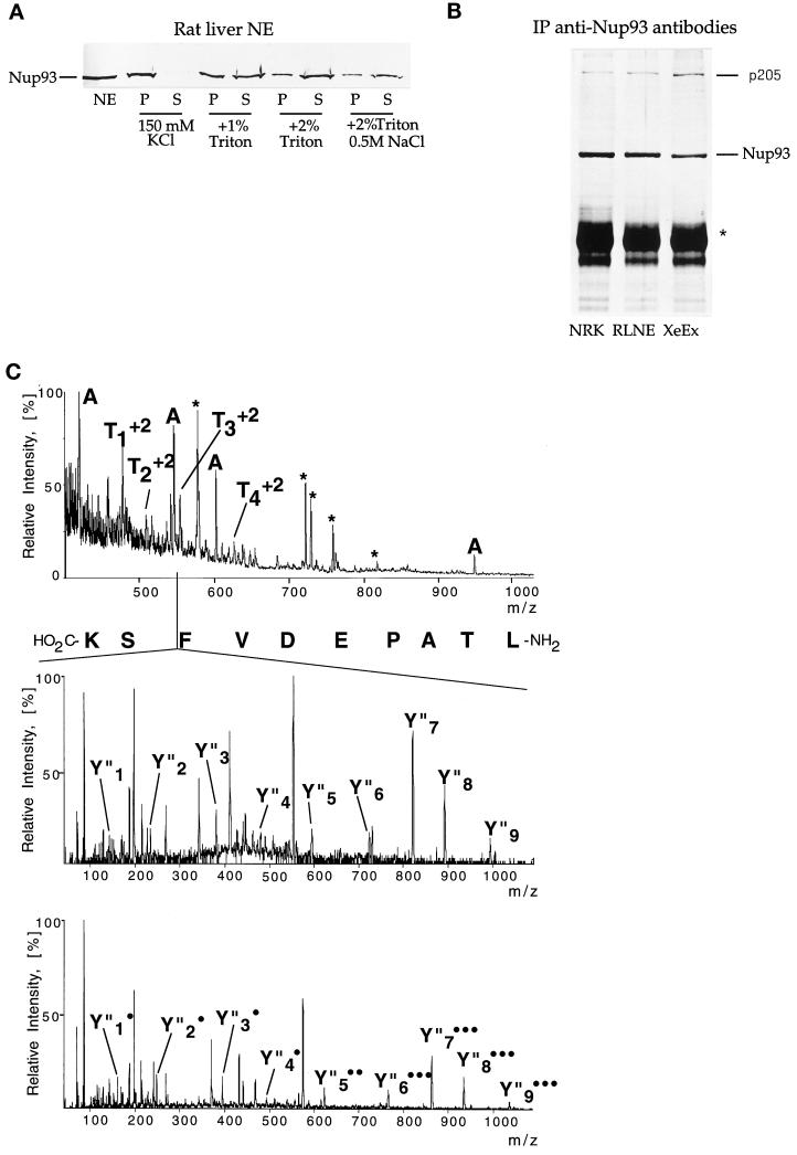
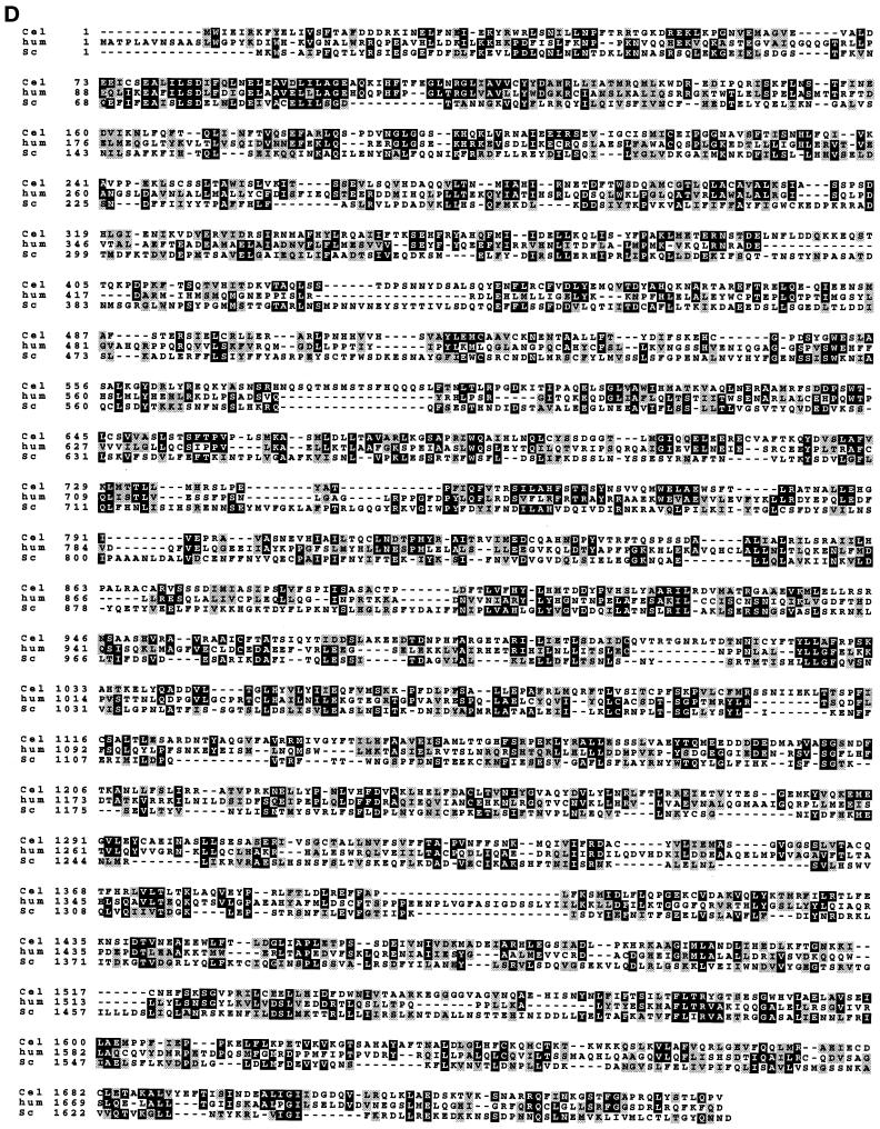
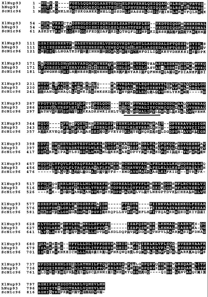
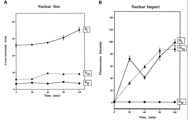
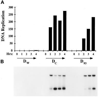
Yeast and vertebrate nuclear pores display significant morphological similarity by electron microscopy, but sequence similarity between the respective proteins has been more difficult to observe. Herein we have identified a vertebrate nucleoporin, Nup93, in both human and Xenopus that has proved to be an evolutionarily related homologue of the yeast nucleoporin Nic96p. Polyclonal antiserum to human Nup93 detects corresponding proteins in human, rat, and Xenopus cells. Immunofluorescence and immunoelectron microscopy localize vertebrate Nup93 at the nuclear basket and at or near the nuclear entry to the gated channel of the pore. Immunoprecipitation from both mammalian and Xenopus cell extracts indicates that a small fraction of Nup93 physically interacts with the nucleoporin p62, just as yeast Nic96p interacts with the yeast p62 homologue. However, a large fraction of vertebrate Nup93 is extracted from pores and is also present in Xenopus egg extracts in complex with a newly discovered 205-kDa protein. Mass spectrometric sequencing of the human 205-kDa protein reveals that this protein is encoded by an open reading frame, KIAAO225, present in the human database. The putative human nucleoporin of 205 kDa has related sequence homologues in Caenorhabditis elegans and Saccharomyces cerevisiae. The analyze the role of the Nup93 complex in the pore, nuclei were assembled that lack the Nup93 complex after immunodepletion of a Xenopus nuclear reconstitution extract. The Nup93-complex-depleted nuclei are clearly defective for correct nuclear pore assembly. From these experiments, we conclude that the vertebrate and yeast pore have significant homology in their functionally important cores and that, with the identification of Nup93 and the 205-kDa protein, we have extended the knowledge of the nearest-neighbor interactions of this core in both yeast and vertebrates.


Figures

Figure 7
Figure 7
Nup93 is depleted from Xenopus egg cytosol by immunodepletion with anti-Nup93 antibodies. To determine the extent of depletion of Nup93 from the Nup93-depleted extract and membrane, different amounts of Dc cytosol were electrophoresed on an 8% polyacrylamide gel in lanes 1–6 (0.01, 0.02, 0.04, 0.1, 0.2, and 0.5 μl, respectively). D93 extract was electrophoresed in lane 8 (1 μl). Examination indicates that the D93 cytosol was depleted of more than 99% of Nup93 when compared with Dc in lane 2 (0.02 μl). Washed membranes (1 μl) contained a small percentage of Nup93, as shown in lane 9, estimated to be 0.2% of the total Nup93 normally present in the egg extract.
Figure 5
Figure 5
Nup93, an evolutionarily conserved nucleoporin, is physically associated with a 205-kDa protein. (A) Western blot decorated with anti-Nup93 antibodies. The blot contains purified NEs and derived soluble (S) and insoluble (P) fractions that were obtained by extracting NEs with the indicated salt and detergent concentrations. Note that 2% Triton X-100 solubilizes the majority of Nup93 (70–80%). (B) Silver staining of immunoprecipitates (IP) obtained with anti-Nup93 antibodies from NRK cells (lane NRK), RLNEs (lane RLNE), and Xenopus egg extracts (lane XeEx). The bands corresponding to Nup93 and p205 are indicated. The star indicates the position of the heavy chain of IgG. (C, top) Mass spectrum of the peptide mixture extracted after in-gel tryptic digest of the band with apparent mass 205 kDa. Peaks that are different from known trypsin autolysis products (marked by an asterisk) were fragmented in turn. Peaks designated by A originate from immunoglobulins, as was shown by sequencing and database searching using peptide sequence tags (Mann and Wilm, 1994). (C, middle and bottom) Sequencing of one of the peptide ions of the 205-kDa protein. The doubly charged ion with m/z 553.8 was isolated and fragmented in the mass spectrometer and tandem mass spectra acquired. Collision fragmentation of tryptic peptides resulted in continuous series of fragments containing the carboxyl terminus of the peptide (Y′′ ions). The methylated form of the same peptide (produced by esterification of the whole peptide mixture) was also fragmented (C, bottom). Esterification with methanol identified the carboxyl-terminal ions by their characteristic 14-Da mass shift for every fragment containing the carboxyl terminus and an additional 14-Da shift for each internal Asp and Glu residue present in the fragment. The number of incorporated methyl groups is designated by • (C, bottom). The actual sequence (LTAPEDVFSK) is retrieved by software-assisted comparison of the two sets of sequence data (derivatized and underivatized) by matching precise mass differences between adjacent peaks in the series of Y′′ ions. Two more peptides, Ta (LLPEQLLK) and Tb (MLALALLDR), were sequenced in the same way. Note that L stands both for leucine and isoleucine because mass spectrometry cannot distinguish between these two isobaric amino acids. Only partial sequence information was obtained for the peptide Td but the spectrum was unambiguously matched to the sequence DLPSADSVQYR in the sequence of the 205-kDa protein using a peptide sequence tag, assembled from the tandem mass spectrometric data. (D) Sequence alignment of evolutionarily conserved 205-kDa proteins: the aligned p205 sequences from human KIAA0225 (hum), C. elegans CEK12D12 (Cel), and S. cerevisiae ORF YJL039c (Sc) are shown to document that the proteins align along their complete length. Amino acids common to two or three of the the genes are shaded in black and structurally related amino acids are shaded in gray.
Figure 5
Figure 5
Nup93, an evolutionarily conserved nucleoporin, is physically associated with a 205-kDa protein. (A) Western blot decorated with anti-Nup93 antibodies. The blot contains purified NEs and derived soluble (S) and insoluble (P) fractions that were obtained by extracting NEs with the indicated salt and detergent concentrations. Note that 2% Triton X-100 solubilizes the majority of Nup93 (70–80%). (B) Silver staining of immunoprecipitates (IP) obtained with anti-Nup93 antibodies from NRK cells (lane NRK), RLNEs (lane RLNE), and Xenopus egg extracts (lane XeEx). The bands corresponding to Nup93 and p205 are indicated. The star indicates the position of the heavy chain of IgG. (C, top) Mass spectrum of the peptide mixture extracted after in-gel tryptic digest of the band with apparent mass 205 kDa. Peaks that are different from known trypsin autolysis products (marked by an asterisk) were fragmented in turn. Peaks designated by A originate from immunoglobulins, as was shown by sequencing and database searching using peptide sequence tags (Mann and Wilm, 1994). (C, middle and bottom) Sequencing of one of the peptide ions of the 205-kDa protein. The doubly charged ion with m/z 553.8 was isolated and fragmented in the mass spectrometer and tandem mass spectra acquired. Collision fragmentation of tryptic peptides resulted in continuous series of fragments containing the carboxyl terminus of the peptide (Y′′ ions). The methylated form of the same peptide (produced by esterification of the whole peptide mixture) was also fragmented (C, bottom). Esterification with methanol identified the carboxyl-terminal ions by their characteristic 14-Da mass shift for every fragment containing the carboxyl terminus and an additional 14-Da shift for each internal Asp and Glu residue present in the fragment. The number of incorporated methyl groups is designated by • (C, bottom). The actual sequence (LTAPEDVFSK) is retrieved by software-assisted comparison of the two sets of sequence data (derivatized and underivatized) by matching precise mass differences between adjacent peaks in the series of Y′′ ions. Two more peptides, Ta (LLPEQLLK) and Tb (MLALALLDR), were sequenced in the same way. Note that L stands both for leucine and isoleucine because mass spectrometry cannot distinguish between these two isobaric amino acids. Only partial sequence information was obtained for the peptide Td but the spectrum was unambiguously matched to the sequence DLPSADSVQYR in the sequence of the 205-kDa protein using a peptide sequence tag, assembled from the tandem mass spectrometric data. (D) Sequence alignment of evolutionarily conserved 205-kDa proteins: the aligned p205 sequences from human KIAA0225 (hum), C. elegans CEK12D12 (Cel), and S. cerevisiae ORF YJL039c (Sc) are shown to document that the proteins align along their complete length. Amino acids common to two or three of the the genes are shaded in black and structurally related amino acids are shaded in gray.
Figure 1
Figure 1
Human and Xenopus Nup93 exhibit significant sequence homology to yeast Nic96p. The deduced amino acid sequence of human and Xenopus Nup93 and yeast Nic96p were aligned using the programs pileup/prettybox. Amino acids common to two or three of the the genes are shaded in black; structurally related amino acids are shaded in gray. A highly conserved short peptide sequence within yeast Nic96p that is essential for cell growth is also indicated. The accession number of the human Nup93 gene is D42085 and of the Xenopus gene is U63919.
Figure 2
Figure 2
Nup93 is present in cells from human to Xenopus and is enriched in RLNE preparations. (A) Western blot analysis of HeLa whole cell homogenate (lane HeLa), Xenopus egg extract (lane Xen.egg), and RLNEs (lane RLNE) labeled with anti-Nup93 antibodies. The bands visible correspond to human and rat liver Nup93 and Xenopus Nup93. (B) Western blot analysis of the different fractions obtained during RLNE purification were labeled with affinity-purified anti-Nup93 (top), rabbit polyclonal anti-p62 (middle), and rabbit polyclonal anti-fibrillarin antibodies (bottom). The bands corresponding to Nup93, p62, and fibrillarin are indicated. Lane NP, nuclear pellet; lane DNAse, DNase washes; lane NaCl, 0.5 M salt wash; lanes NE 1× and 10×, NEs loaded in 1- or 10-fold volume equivalents.
Figure 3
Figure 3
Nup93 is a novel nucleoporin. Indirect immunofluorescence staining of HeLa cells using anti-Nup93 antibodies. The punctuate staining of the NE of a confocal section is overlapping with the staining of NPCs obtained with mAb414.
Figure 4
Figure 4
Localization of Nup93 in RLNES. (A) Cross-section of a single NE and selected examples of NPCs labeled with the polyclonal anti-Nup93 antibody conjugated to 8-nm colloidal gold by preembedding labeling. The rightmost examples (middle and bottom) are NPCs labeled with the mAbs QE5 and RL31 (anti-p62) conjugated to 8-nm colloidal gold, respectively. Cytoplasmic (c) and nuclear (n) sides of the NE are indicated. Bar, 100 nm. (B–D) Quantitative analysis of gold particles associated with the nuclear side of NPCs after preembedding labeling with 5 μg/ml anti-Nup93 antibody. (B) Sixty particles were scored and 20 μg/ml of anti-Nup93 antibody. (C) Fifty-five particles were scored after postembedding labeling with 1 μg/ml anti-Nup93 antibody. (D) Forty particles were scored.
Figure 6
Figure 6
Interaction of Nup93 with p62. Western blots of immunoprecipitations using HeLa cell extracts as a source of Nup93. (A) Immunoprecipitation with anti-Nup93 antibodies. Western blots were decorated with anti-Nup93 antibodies (top), anti-p62 antibodies (middle), and anti-lamin antibodies (bottom). (B) Immunoprecipitation with anti-p62 antibodies. Western blots were decorated with anti-Nup93 antibodies (top) and anti-p62 antibodies (bottom). The bands corresponding to Nup93, p62, and lamins are indicated. Stars mark the position of the heavy chain of the IgG. Lane H, homogenate; lane S, immune supernatant; lane P, immune pellet.
Figure 8
Figure 8
Nup93-complex–depleted nuclei are impaired in nuclear growth but are competent for nuclear import. (A) Nuclear size. Reconstituted nuclei were allowed to form at room temperature for 2 h, and then the karyophilic transport substrate (ss-HSA) was added. At this time, which we called t = 0, nuclei were quantitated for import level and measured for size. Depleted WGA binding protein nuclei (Dw) were done as a negative control for comparison. In A, the cross-sectional area of depleted Nup93 nuclei (D93) is reduced by a factor of 4 compare to mock-depleted nuclei (Dc) over a time period of 4 h. (B) Nuclear import. Nuclear import of the transport substrate appears normal in nuclei lacking Nup93. Transport level was normalized to 16× integration for all time points.
Figure 9
Figure 9
DNA replication in depleted Nup93 nuclei (D93) is delayed. Replication was performed with demembranated sperm chromatin at 1000 sperm/μl in Dw, Dc, and D93 egg lysate. Replication is seen at 2 h in D93 nuclei, whereas it could be seen in 1 h in the Dc nuclei. To quantitate, a rectangular box was drawn for each lane to include the two bands for measurement. The units on the Y-axis are arbitary units given by the Phosphoimager model 445SI.
Figure 10
Figure 10
Anchored nuclei formed in Nup93- complex–depleted Xenopus egg cytosol show greatly decreased staining with mAb414. Egg cytosol was immunodepleted with either anti-Nup93 antibodies (D93) or nonspecific rabbit IgG (Dc). The rim stain of Dc nucleus (a) and the rim stain of a D93 nucleus (b) at 1500× magnification are shown. The mAb414 staining of the D93 nucleus is dimmer and patchy compared with the control. (c) Another D93 nucleus at a higher-intensity integration, which is 4× the integration of a and b. (d) Same nucleus as in c is shown but image is focused on the surface of the nucleus.
Figure 11
Figure 11
Antibodies against Nup98 reveal decreased staining of NPCs with mAb414 and inhibit nuclear growth and DNA replication. (A) mAb414 staining and nuclear growth: addition of 3 μl (4.5 μg) of anti-Nup93 antibodies to a Xenopus nuclear reconstitution extract causes a strong decrease in the immunofluorescence staining of nuclear pore proteins recognized by mAb414 and a reduced sperm nuclei size as seen by Hoechst staining. Control experiments were performed by adding 3 μl (4.5 μg) of unspecific rabbit IgG (unspecific antibodies). Two sperms of each sample are shown. Note that the pictures of the samples incubated with anti-Nup93 antibodies were exposed twice as long as the samples treated with unspecific antibodies. (B) DNA replication: the addition of 1 μl (1.5 μg) and 3 μl (4.5 μg) of anti-Nup93 antibodies (lanes 2 and 3, respectively) but not 3 μl of buffer (lanes 1) or 3 μl (4.5 μg) of unspecific rabbit IgGs (lanes 4) to the nuclear reconstitution extract causes a strong inhibition of newly synthesized (32P-labeled) high molecular weight DNA normally produced upon DNA replication. Equal volumes were taken at time points 0 (t = 0 h), after 1 h (t = 1 h), and after 2 h (t = 2 h) of incubation, digested with proteinase K, and loaded onto a agarose gel.

Similar articles

Cited by

References

    1. Aitchison JD, Rout MP, Marelli M, Blobel G, Wozniak RW. Two novel related yeast nucleoporins Nup170p and Nup157p: complementation with the vertebrate homologue Nup155p and functional interactions with the yeast nuclear pore-membrane protein Pom152p. J Cell Biol. 1995;131:1133–1148. - PMC - PubMed
    1. Akey CW, Radermacher M. Architecture of the Xenopus nuclear pore complex revealed by three-dimensional cryo-electron microscopy. J Cell Biol. 1993;122:1–19. - PMC - PubMed
    1. Allende ML, Amsterdam A, Becker T, Kawakami K, Gaiano N, Hopkins N. Insertional mutagenesis in zebrafish identifies two novel genes, pescadillo and dead eye, essential for embryonic development. Genes Dev. 1996;10:3141–3155. - PubMed
    1. Becker J, Melchior F, Gerke V, Bischoff FR, Ponstingl H, Wittinghofer A. RNA1 encodes a GTPase-activating protein specific for Gsp1p, the Ran-TC4 homologue of Saccharomyces cerevisiae. J Biol Chem. 1995;270:11860–11865. - PubMed
    1. Blobel G, Potter VR. Nuclei from rat liver: isolation method that combines purity with high yield. Science. 1966;154:1662–1665. - PubMed

Publication types

MeSH terms