. 1997 Oct;8(10):2055-75.
doi: 10.1091/mbc.8.10.2055.

Cell-adhesive responses to tenascin-C splice variants involve formation of fascin microspikes

Affiliations
Free PMC article

Cell-adhesive responses to tenascin-C splice variants involve formation of fascin microspikes

D Fischer et al. Mol Biol Cell. 1997 Oct.
Free PMC article

Abstract

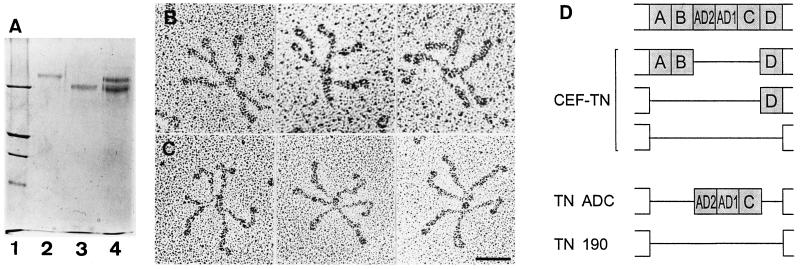
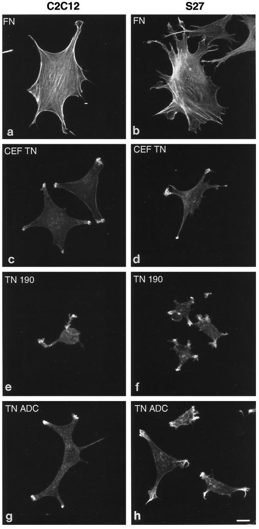
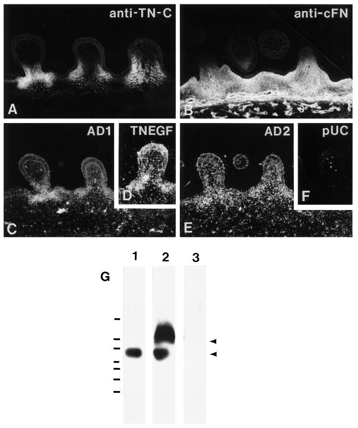
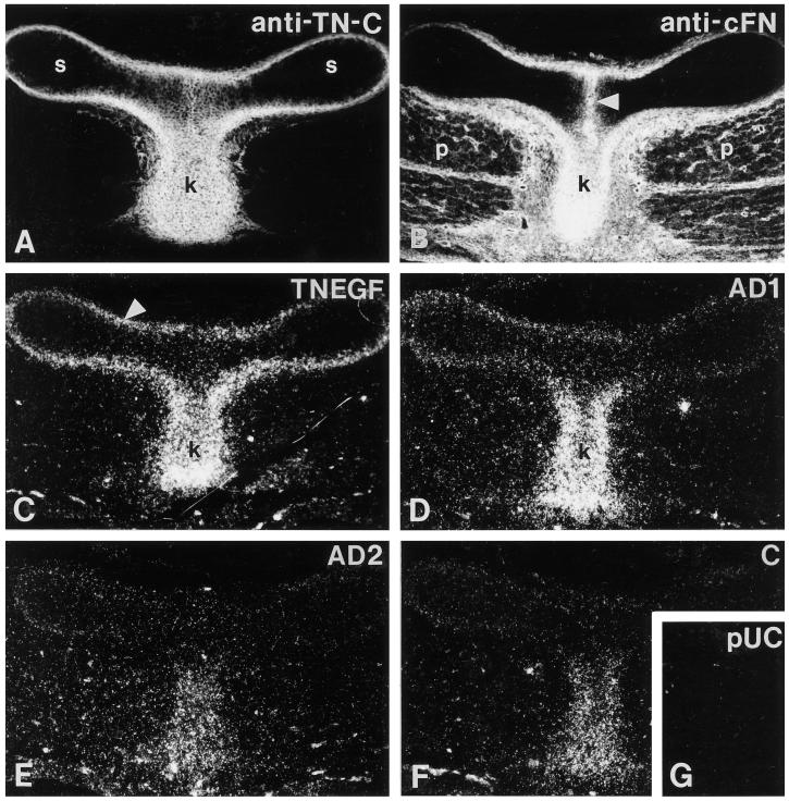
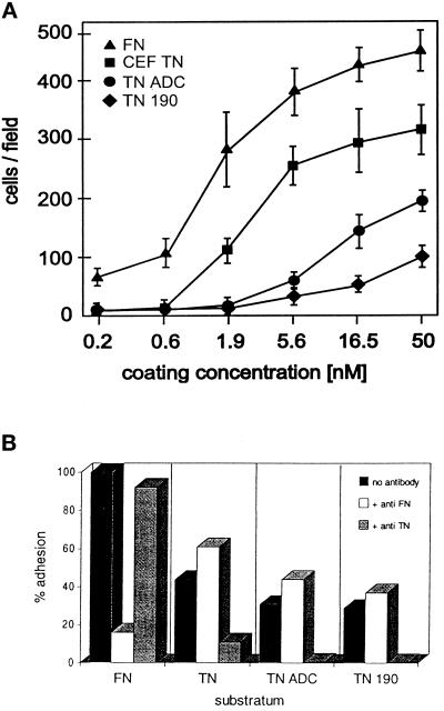
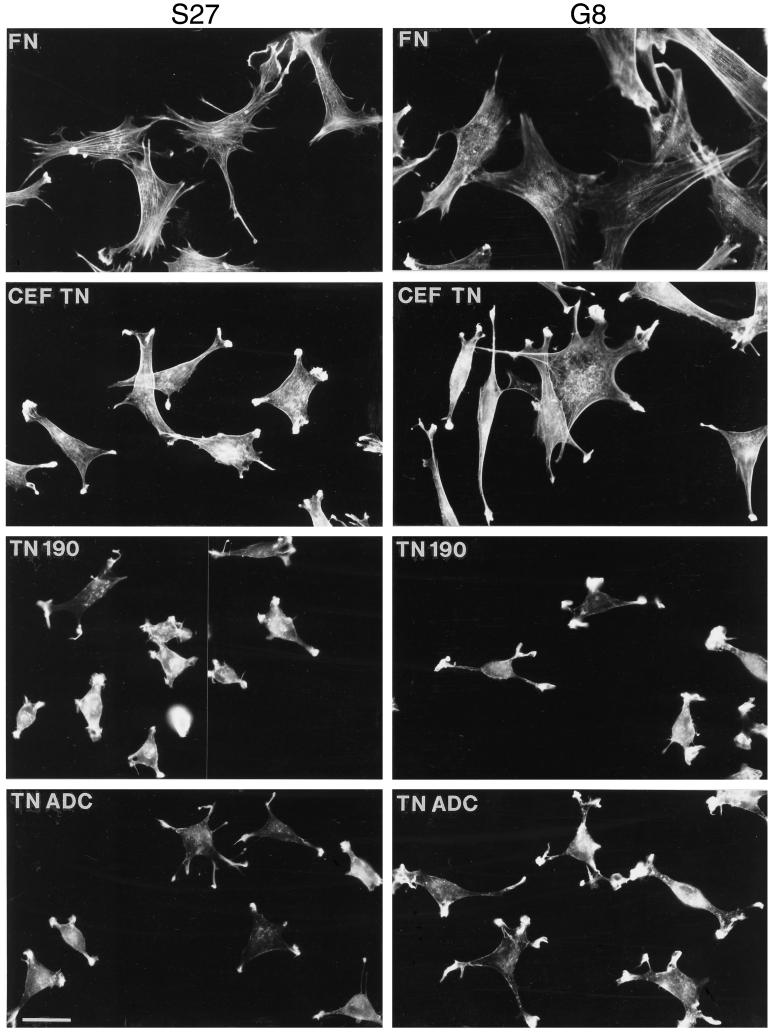
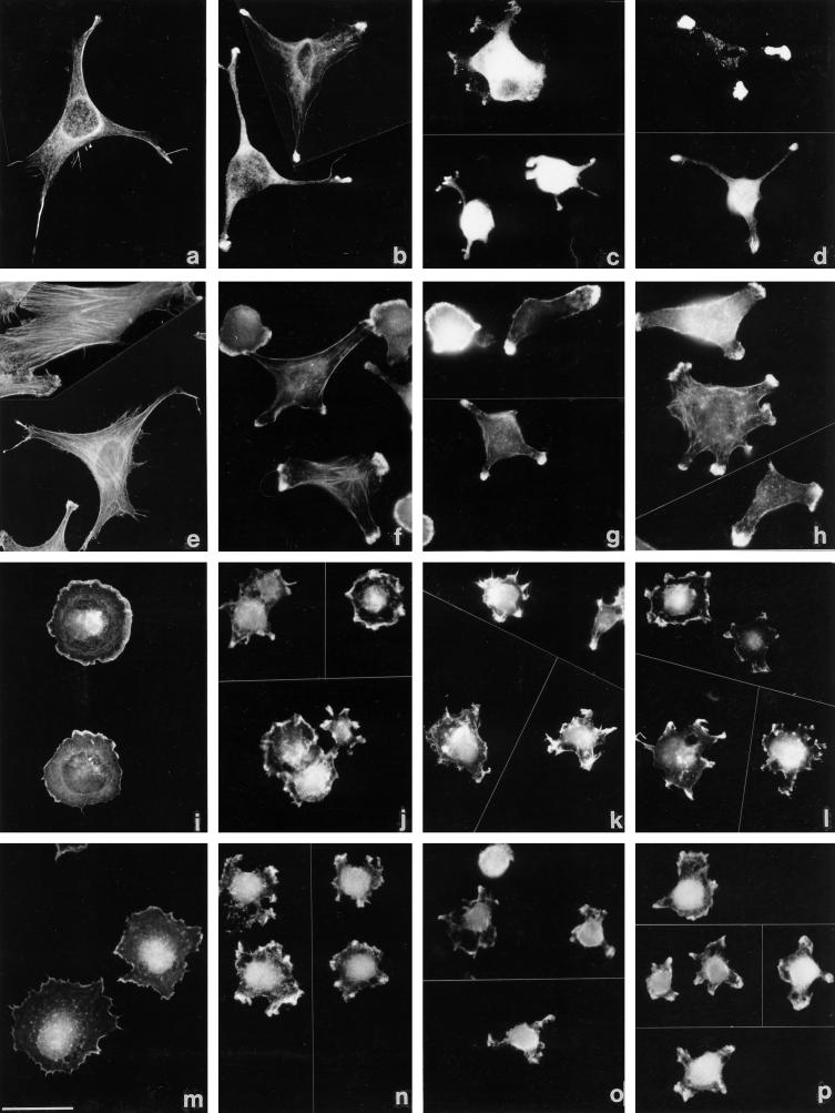
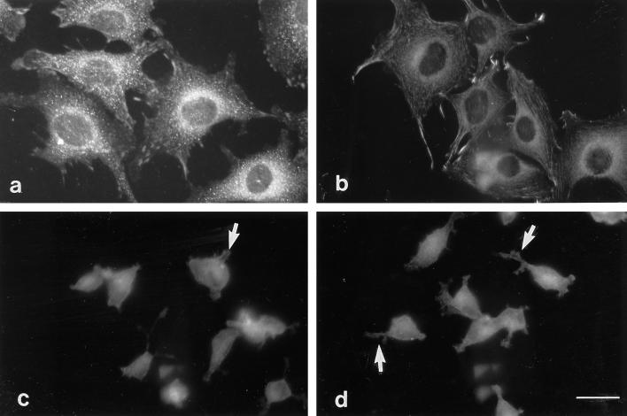
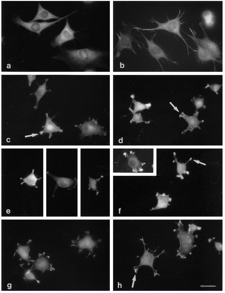
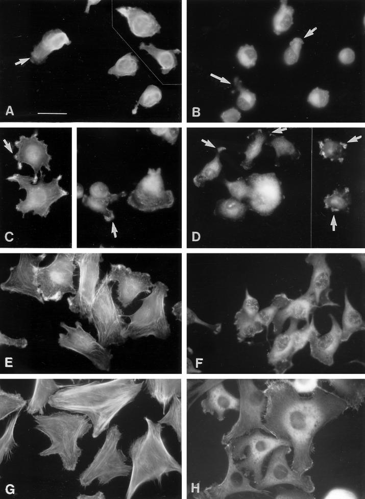
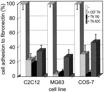
Tenascin-C is an adhesion-modulating matrix glycoprotein that has multiple effects on cell behavior. Tenascin-C transcripts are expressed in motile cells and at sites of tissue modeling during development, and alternative splicing generates variants that encode different numbers of fibronectin type III repeats. We have examined the in vivo expression and cell adhesive properties of two full-length recombinant tenascin-C proteins: TN-190, which contains the eight constant fibronectin type III repeats, and TN-ADC, which contains the additional AD2, AD1, and C repeats. In situ hybridization with probes specific for the AD2, AD1, and C repeats shows that these splice variants are expressed at sites of active tissue modeling and fibronectin expression in the developing avian feather bud and sternum. Transcripts incorporating the AD2, AD1, and C repeats are present in embryonic day 10 wing bud but not in embryonic day 10 lung. By using a panel of nine cell lines in attachment assays, we have found that C2C12, G8, and S27 myoblastic cells undergo concentration-dependent adhesion to both variants, organize actin microspikes that contain the actin-bundling protein fascin, and do not assemble focal contacts. On a molar basis, TN-ADC is more active than TN-190 in promoting cell attachment and irregular cell spreading. The addition of either TN-190 or TN-ADC in solution to C2C12, COS-7, or MG-63 cells adherent on fibronectin decreases cell attachment and results in decreased organization of actin microfilament bundles, with formation of cortical membrane ruffles and retention of residual points of substratum contact that contain filamentous actin and fascin. These data establish a biochemical similarity in the processes of cell adhesion to tenascin-C and thrombospondin-1, also an "antiadhesive" matrix component, and also demonstrate that both the adhesive and adhesion-modulating properties of tenascin-C involve similar biochemical events in the cortical cytoskeleton. In addition to these generic properties, TN-ADC is less active in adhesion modulation than TN-190. The coordinated expression of different tenascin-C transcripts during development may, therefore, provide appropriate microenvironments for regulated changes in cell shape, adhesion, and movement.


Figures

Figure 3
Figure 3
Analysis of recombinant chick tenascin-C proteins. (A) SDS-PAGE analysis. Lane 1, molecular mass standards (from the top), 200 kDa, 116 kDa, 97 kDa, and 66 kDa; lane 2, recombinant tenascin-C variant that includes the fibronectin type III repeats AD2, AD1, and C (TN-ADC); lane 3, recombinant tenascin-190 (TN-190); lane 4, tenascin-C from chicken embryo fibroblast (CEF-TN; this contains a mixture of TN-190, TN-200, and TN-230 protein variants). All samples were resolved on a 6% polyacrylamide gel under reducing conditions. (B and C) Electron microscopy of recombinant tenascin-Cs. TN -190 (B) or TN-ADC (C) were sprayed onto mica and rotary-shadowed. Bar, 50 nm. (D) Schematic diagram showing ordering of the variable fibronectin type III repeats in chicken (top) and the repeat combinations in CEF-TN, TN-190, or TN-ADC proteins.
Figure 6
Figure 6
Confocal microscopy of actin microfilament organization in C2C12 and S27 cells adherent on fibronectin or tenascin-C. Cells were plated onto coverslips coated with 50 nM fibronectin (a and b), 50 nM CEF-TN (c and d), 50 nM recombinant TN-190 (e and f), or 50 nM recombinant TN-ADC (g and h) for 90 min in serum-free medium. After incubation cells were fixed, stained with TRITC-phalloidin, and photographed on a confocal microscope. Bar, 5 μm.
Figure 1
Figure 1
Localization of tenascin-C and fibronectin in feather buds. (A) Antibody to tenascin-C stains the mesenchyme at the base of the feather buds. (B) Antibody to cellular fibronectin stains all of the dermal extracellular matrix and the underlying blood vessels. (C–F) In situ hybridization on adjacent cross-sections through the dorsal feather tract of an E10 chick. (C) Tenascin C probe specific to fibronectin type III repeat AD1. (D) Universal tenascin-C probe TN-EGF. (E) Probe to repeat AD2. (F) pUC negative control probe. All the tenascin-C probes hybridize in the mesenchyme at the base of the feather buds. Bar, 100 μm. (G) RT-PCR detection of AD2AD1C-containing tenascin-C transcript in embryonic tissues. Primers corresponding to the beginning of repeat AD2 and the end of repeat C were used to amplify products from mRNA isolated from E10 chicken lung (lane 1) or wing (lane 2). Lane 3 was loaded with PCR product corresponding to repeat C. The blot was probed with cAD2. A single band of about 550 bp (i.e., two fibronectin type III repeats) was detected in amplification products from lung. In contrast, two bands corresponding to products containing 2 and 3 fibronectin type III repeats were detected in wingbud (arrows). DNA standards from bottom to top are 100 bp, 200 bp, 300 bp, 400 bp, 600 bp, 800 bp, and 2000 bp.
Figure 2
Figure 2
Localization of tenascin-C and fibronectin in sternum and keel. Cross-sections through sternum and keel of an E10 chick were processed for immunohistochemistry (A and B) or in situ hybridization (C–G). (A) Antibody to tenascin-C stains the perichondrium surrounding the sternal anlages (s) and the keel (k) intensely. (B) Antibody to cellular fibronectin stains the fusion point between the two sternal anlage (arrowhead), matrix surrounding the pectoral muscles (p) and the keel (k). (C) Universal probe TN-EGF hybridizes in sternal perichondrium (arrowhead) and in the keel (k). (D) AD1 probe. (E) AD2 probe. (F) C probe. All three probes hybridize in the keel. (G) pUC negative control. Bar, 100 μm.
Figure 4
Figure 4
Quantitation of C2C12 cell adhesion to tenascin-Cs. (A) C2C12 cells were allowed to adhere for 1 h to the indicated concentrations of the various tenascin-C preparations or fibronectin, then fixed, stained, and counted. Each data point is the mean of triplicate experiments; bars indicate SEM. (B) Inhibition of cell adhesion to chick tenascin-C by antibodies. C2C12 cells were plated onto coverslips coated with 50 nM fibronectin (FN), CEF-TN (TN), recombinant TN-ADC, or recombinant TN-190 in the presence or absence of antiserum directed against fibronectin or tenascin-C. Solid bars, controls without antiserum; open bars, cells plated in the presence of anti-fibronectin serum; shaded bars, cells plated in the presence of antiserum to chick tenascin-C. Adherent cells were fixed and counted after a 90-min incubation at 37°C.
Figure 5
Figure 5
Organization of actin microfilaments in S27 and G8 cells adherent on fibronectin or tenascin-C. Cells were plated onto coverslips coated with 50 nM fibronectin, 50 nM CEF-TN, 50 nM recombinant TN-190, or 50 nM recombinant TN-ADC for 90 min in serum-free medium. After incubation cells were fixed, stained with TRITC-phalloidin, and examined under epifluorescence. Bar, 10 μm.
Figure 11
Figure 11
Cytoskeletal reorganization in response to soluble tenascin-Cs. C2C12 cells (a–h), COS-7 cells (i–l), or MG-63 cells (m–p) adherent on fibronectin (a, e, i, and m), on fibronectin in the presence of 35 nM CEF-TN (b, f, j, and n), in the presence of 35 nM TN-190 (c, g, k, and o), or in the presence of 35 nM TN-ADC (d, h, l, and p) were stained after 1 h with TRITC-phalloidin (e–p) or for fascin (a–d). Bar, 15 μm.
Figure 7
Figure 7
Cells adherent on tenascin-C substrata do not assemble focal contacts. C2C12 (a and c) or S27 (b and d) cells were stained for vinculin after a 90-min adhesion to fibronectin (a and b) or CEF-TN (c and d). Focal contacts are present in the cells on fibronectin, but the cytoplasmic extensions and microspikes of cells adherent on CEF-TN show no localization of vinculin (small arrows in c and d indicate positions of protrusions). Bar, 10 μm.
Figure 8
Figure 8
Presence of fascin-positive microspikes in cells adherent on tenascin-C substrata. C2C12 (a, c, e, and g) or S27 (b, d, f, and h) cells were stained for fascin after a 90-min adhesion to fibronectin (a and b), CEF-TN (c and d), TN-190 (e and f), or TN-ADC (g and h). The arrays of microspikes formed by cells adherent on the tenascin-C substrata stain positively for fascin (examples are indicated by arrows in c, d, f, and h). Bar, 10 μm.
Figure 9
Figure 9
F-actin and fascin distributions in C2C12 cells adherent on low concentrations of fibronectin. C2C12 cells were allowed to attach for 1 h to substrata coated with 1.25 μg/ml (A and B), 2.5 μg/ml (C and D), 5 μg/ml (E and F), or 20 μg/ml fibronectin (G and H) for 1 h then fixed and stained with TRITC-phalloidin (A, C, E, and G) or with antibody to fascin (B, D, F, and H). Arrows in A–D indicate examples of cells with F-actin- or fascin-containing membrane protrusions. Bar, 10 μm.
Figure 10
Figure 10
Modulation of cell attachment to fibronectin by tenascin-Cs in solution. C2C12, MG63, or COS-7 cells were allowed to attach to fibronectin substrata for 60 min in the absence or presence of 35 nM tenascin-C added in solution, as indicated, and the percentage of input cells that attached was quantitated by direct counting. Each column is the mean of three assays; bars are the SEM.

Similar articles

Cited by

References

    1. Adams JC. Formation of stable microspikes containing actin and the 55-kDa actin bundling protein, fascin, is a consequence of cell adhesion to thrombospondin-1: implications for the anti-adhesive activities of thrombospondin-1. J Cell Sci. 1995;108:1977–1990. - PubMed
    1. Adams JC, Lawler J. Diverse mechanisms for cell attachment to platelet thrombospondin. J Cell Sci. 1993;104:1061–1071. - PubMed
    1. Adams J, Lawler J. Cell-type specific adhesive interactions of skeletal myoblasts with thrombospondin-1. Mol Biol Cell. 1994;5:423–437. - PMC - PubMed
    1. Adams JC, Tucker RP, Lawler J. The Thrombospondin Gene Family. Austin, TX: R.G. Landes; 1995. and Berlin: Springer-Verlag.
    1. Asch AS, Tepler J, Silbiger S, Nachman RL. Cellular attachment to thrombospondin: cooperative interactions between receptor systems. J Biol Chem. 1991;266:1740–1745. - PubMed

Publication types

MeSH terms

LinkOut - more resources