. 1997 Nov 1;11(21):2767-79.
doi: 10.1101/gad.11.21.2767.

Regulation of the replication initiator protein p65cdc18 by CDK phosphorylation

Affiliations

Regulation of the replication initiator protein p65cdc18 by CDK phosphorylation

P V Jallepalli et al. Genes Dev. .

Abstract

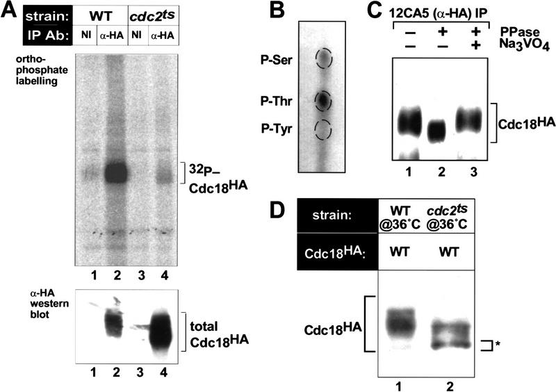
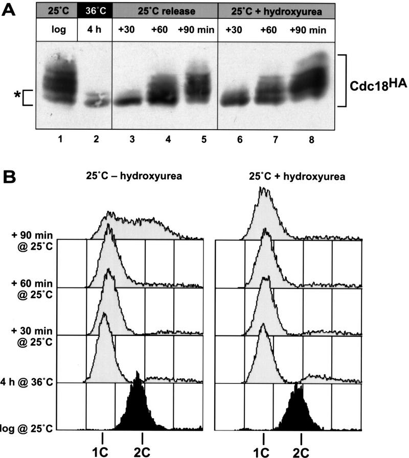
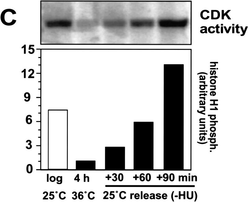
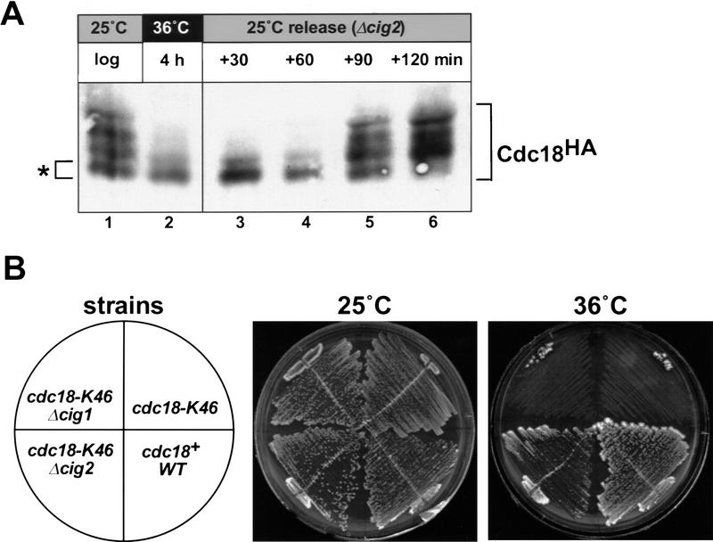
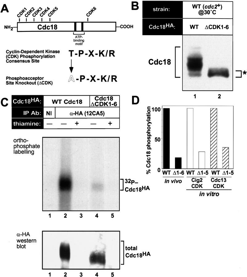
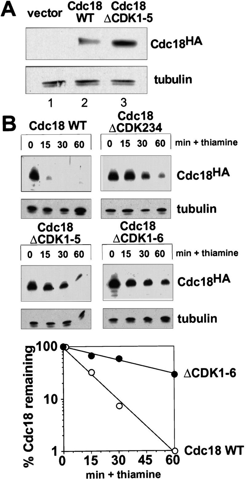
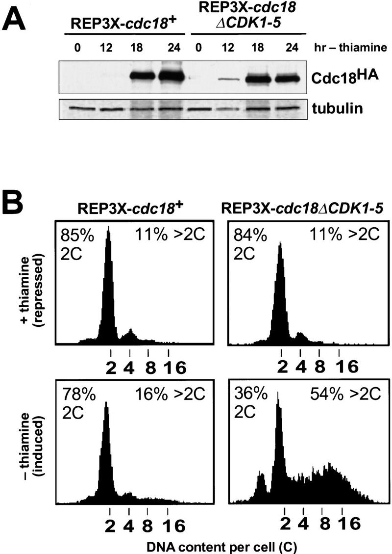
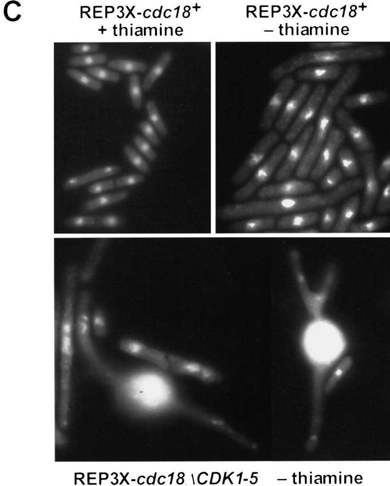
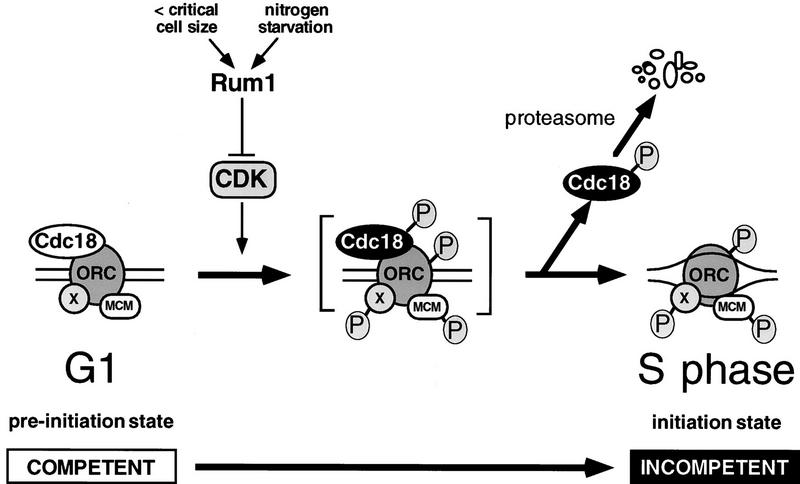
Cyclin-dependent kinases (CDKs) promote the initiation of DNA replication and prevent reinitiation before mitosis, presumably through phosphorylation of key substrates at origins of replication. In fission yeast, the p65cdc18 protein is required to initiate DNA replication and interacts with the origin recognition complex (ORC) and the p34cdc2 CDK. Here we report that p65cdc18 becomes highly phosphorylated as cells undergo the G1 --> S phase transition. This modification is dependent on p34cdc2 protein kinase activity, as well as six consensus CDK phosphorylation sites within the p65cdc18 polypeptide. Genetic interactions between cdc18+ and the S-phase cyclin cig2+ suggest that CDK-dependent phosphorylation antagonizes cdc18+ function in vivo. Using site-directed mutagenesis, we show that phosphorylation at CDK consensus sites directly targets p65cdc18 for rapid degradation and inhibits its replication activity, as strong expression of a constitutively hypophosphorylated mutant form of p65cdc18 results in large amounts of DNA over-replication in vivo. Furthermore, the over-replication phenotype produced by this mutant p65cdc18 is resistant to increased mitotic cyclin/CDK activity, a known inhibitor of over-replication. Therefore, p65cdc18 is the first example of a cellular initiation factor directly regulated in vivo by CDK-dependent phosphorylation and proteolysis. Regulation of p65cdc18 by CDK phosphorylation is likely to contribute to the CDK-driven "replication switch" that restricts initiation at eukaryotic origins to once per cell cycle.


Figures

Figure 1
Figure 1
In vivo phosphorylation of p65cdc18 depends on the p34cdc2 protein kinase. (A) Wild-type fission yeast cells (WT, lanes 1 and 2) or temperature-sensitive cdc2-33 mutant cells (cdc2ts, lanes 3 and 4) expressing hemagglutinin (HA) epitope-tagged cdc18+ from the full-strength nmt1+ promoter (REP3X) were labeled metabolically with [32P]orthophosphate at 36°C (restrictive temperature for cdc2-33). After immunoprecipitation with monoclonal anti-HA antibody 12CA5 (lanes 2,4) or non-immune (NI) antibody (lanes 1,3), samples were resolved by SDS-PAGE. (Top) 32P-labeled p65cdc18 was detected using a PhosphorImager. (Bottom) Total p65cdc18 in each immunoprecipitate was visualized by immunoblotting with HA-specific antibodies. (B) Phosphoamino acid analysis of p65cdc18 phosphorylated in vivo was performed essentially as described (Lin and Desiderio 1993). (P-Ser) Phosphoserine; (P-Thr) phosphothreonine; (P-Tyr) phosphotyrosine. (C) Extracts of cells expressing HA-tagged cdc18+ were immunoprecipitated with anti-HA antibodies. p65cdc18 immunoprecipitates were then incubated without additions (lane 1), with λ protein phosphatase (PPase) (lane 2), or with phosphatase plus the phosphatase inhibitor vanadate (lane 3). Samples were resolved by 6% SDS-PAGE and immunoblotted with antibodies to HA. (D) Dependence of p65cdc18 modification on p34cdc2 kinase activity. Wild-type cells (lane 1) and temperature-sensitive cdc2-33 mutant cells (cdc2ts, lane 2) expressing HA-tagged cdc18+ from the weak nmt1 promoter (REP42X) were shifted to 36°C for 4 hr. Total cell extracts were subjected to modified SDS-PAGE followed by immunoblotting with anti-HA antibodies. The position of the fastest migrating p65cdc18 species is marked by an asterisk.
Figure 2
Figure 2
Phosphorylation of p65cdc18 occurs at the beginning of S phase and coincides with activation of p34cdc2 kinase. HA-tagged cdc18+ was expressed from the weak nmt1 promoter (REP42X) in temperature-sensitive cdc10-V50 mutant cells. Logarithmically growing cells were synchronized in late G1 by incubation at the restrictive temperature (36°C) for 4 hr, then released by rapid downshift to the permissive temperature (25°C). (A) Extracts were prepared from cells grown asynchronously (lane 1), synchronized in G1 (lane 2), or released from G1 into S phase (lanes 3–5). Additional extracts were prepared from cells released into medium containing the DNA synthesis inhibitor hydroxyurea (lanes 6–8). p65cdc18 modification was resolved using a modified SDS-PAGE protocol; the highest mobility form of p65cdc18 is marked as a reference (*). (B) Flow cytometry analysis of DNA content. Because DNA replication coincides with cytokinesis during normal vegetative growth, asynchronously growing fission yeast cells display a single peak equal to fully replicated (2C) DNA, whereas cells arrested in G1 yield a single peak corresponding to unreplicated (1C) DNA. In untreated cells, this 1C peak was rapidly converted to 2C (left). In contrast, the 1C peak was maintained in cells arrested in early S phase with hydroxyurea (right). (C) Activation of p34cdc2 kinase at the G1 → S transition. p34cdc2 kinase complexes were isolated using p13suc1-agarose from cells synchronized as in A, and kinase reactions performed as described (see Materials and Methods). Histone H1 phosphorylation was quantitated using a PhosphorImager and is presented relative to the activity in G1-arrested cells.
Figure 2
Figure 2
Phosphorylation of p65cdc18 occurs at the beginning of S phase and coincides with activation of p34cdc2 kinase. HA-tagged cdc18+ was expressed from the weak nmt1 promoter (REP42X) in temperature-sensitive cdc10-V50 mutant cells. Logarithmically growing cells were synchronized in late G1 by incubation at the restrictive temperature (36°C) for 4 hr, then released by rapid downshift to the permissive temperature (25°C). (A) Extracts were prepared from cells grown asynchronously (lane 1), synchronized in G1 (lane 2), or released from G1 into S phase (lanes 3–5). Additional extracts were prepared from cells released into medium containing the DNA synthesis inhibitor hydroxyurea (lanes 6–8). p65cdc18 modification was resolved using a modified SDS-PAGE protocol; the highest mobility form of p65cdc18 is marked as a reference (*). (B) Flow cytometry analysis of DNA content. Because DNA replication coincides with cytokinesis during normal vegetative growth, asynchronously growing fission yeast cells display a single peak equal to fully replicated (2C) DNA, whereas cells arrested in G1 yield a single peak corresponding to unreplicated (1C) DNA. In untreated cells, this 1C peak was rapidly converted to 2C (left). In contrast, the 1C peak was maintained in cells arrested in early S phase with hydroxyurea (right). (C) Activation of p34cdc2 kinase at the G1 → S transition. p34cdc2 kinase complexes were isolated using p13suc1-agarose from cells synchronized as in A, and kinase reactions performed as described (see Materials and Methods). Histone H1 phosphorylation was quantitated using a PhosphorImager and is presented relative to the activity in G1-arrested cells.
Figure 3
Figure 3
S-phase CDK activity promotes phosphorylation of p65cdc18 and antagonizes its function in vivo. (A) Deletion of the S-phase cyclin cig2+ delays the onset of p65cdc18 modification. The G1 synchronization protocol used in Fig. 2 was performed with a similar cdc10–V50 strain also harboring a deletion of the S-phase cyclin cig2+. Note that the appearance of slower migrating forms of p65cdc18 is delayed in Δcig2 cells by ∼30 min (cf. Fig. 2A). (B) Deletion of cig2+ suppresses the cell cycle defect of a temperature-sensitive cdc18 mutation. Fission yeast strains with the indicated genotypes were constructed by standard genetic methods and tested for viability at 25°C (permissive temperature for cdc18–K46) or 36°C (restrictive temperature).
Figure 4
Figure 4
CDK consensus sites are required for phosphorylation of p65cdc18. (A) Primary structure of the p65cdc18 polypeptide. The threonine residue at each of the six CDK consensus sites was replaced with alanine. (B) Dependence of p65cdc18 modification on CDK consensus sites. Extracts from wild-type cells expressing either HA-tagged cdc18+ (WT, lane 1) or a mutant gene lacking all six consensus CDK sites (ΔCDK1–6, lane 2) from the weak nmt1 promoter (REP42X) were analyzed as in Fig. 1D. (C) In vivo phosphorylation of p65cdc18. Cells expressing HA-tagged cdc18+ (lanes 1–3) or cdc18ΔCDK1-6 (lanes 4 and 5) from the full-strength, thiamine-repressible nmt1+ promoter (REP3X) were grown in the absence (−) or presence (+) of thiamine and labeled with [32P]orthophosphate. Extracts were immunoprecipitated and analyzed as in Fig. 1A. (D) In vivo 32P incorporation is shown as a percent of that in the wild type (solid bars). Wild-type and ΔCDK1-5 mutant forms of p65cdc18 purified from yeast as GST fusion proteins (Brown et al. 1997) were subjected to in vitro phosphorylation by exogenous p34cdc2 kinases (p45cig2 CDK, open bars; p56cdc13 CDK, hatched bars). 32P incorporation was quantitated using a PhosphorImager.
Figure 5
Figure 5
Characterization of cdc18ΔCDK mutants in fission yeast. Wild-type cells (TK2) were transformed with plasmids expressing wild-type cdc18+ or cdc18ΔCDK1-5 from the cdc18+ promoter, or with vector alone (pIRTL) as a control. Following transformation, cells were plated on EMM plates and incubated at 30°C for 3 days. Examples of small, slow-growing cdc18ΔCDK1-5 transformant colonies are marked by the white arrows. Further growth was observed after several more days at 30°C, yielding colonies large enough for subsequent manipulation and analysis.
Figure 6
Figure 6
Phosphorylation down-regulates the stability of p65cdc18 in vivo. (A) Increased abundance of p65cdc18ΔCDK1-5. Fission yeast cells were transformed with plasmids expressing HA-tagged cdc18+ or cdc18ΔCDK1-5 under the control of the cdc18+ promoter, or with vector alone (pIRTL) as a control. Extracts were resolved by standard SDS-PAGE and immunoblotted with monoclonal antibodies to HA (to detect p65cdc18) and tubulin (to provide a loading control). (B) Increased stability of mutant p65cdc18 proteins. Wild-type cdc18+ or various cdc18ΔCDK mutants, each under the control of the weak nmt1 promoter (REP42X) and containing the HA epitope tag, were expressed in wild-type cells in the absence of thiamine. At time 0, cdc18 expression was repressed by addition of thiamine (Muzi-Falconi et al. 1996a). Samples harvested at the indicated times were analyzed by SDS-PAGE and immunoblotted with monoclonal antibodies to HA or tubulin. (Bottom) The abundance of wild-type p65cdc18 (○) and p65cdc18ΔCDK1-6 (•) was quantitated using 35S-labeled secondary antibodies and a PhosphorImager. These data were used to calculate the apparent half-life (t1/2) of each protein.
Figure 7
Figure 7
Increased replication activity associated with p65cdc18ΔCDK1-5. (A) Expression of HA-tagged cdc18+ (lanes 1–4) or cdc18ΔCDK1-5 (lanes 5–8) from the full-strength nmt1+ promoter (REP3X) was induced by growth in thiamine-free medium for the indicated times. Cell extracts were separated by SDS-PAGE and immunoblotted with monoclonal antibodies to HA and tubulin. Quantitation of p65cdc18 abundance as in Fig. 5B demonstrated that the wild-type protein accumulated to the same or slightly higher level as the ΔCDK mutant polypeptide by 18 hr. (B) The same strains were grown in the presence (upper panels) or absence of thiamine (lower panels) for 30 hr. DNA content per cell was measured by flow cytometry and is presented on a logarithmic scale. The percent of cells with normal (2C) and over-replicated (>2C) DNA content is indicated. (C) Cells in B were examined by DAPI staining of nuclei and fluorescence microscopy. The fields shown illustrate qualitatively the range of phenotypes observed in several independent experiments. When grown in the presence of thiamine, cells containing REP3X–cdc18ΔCDK1-5 were indistinguishable from those containing REP3X–cdc18+ (data not shown). (D) Wild-type cells transformed with the indicated plasmids were streaked onto EMM plates either with (+) or without (−) thiamine and incubated for 4 days at 30°C.
Figure 7
Figure 7
Increased replication activity associated with p65cdc18ΔCDK1-5. (A) Expression of HA-tagged cdc18+ (lanes 1–4) or cdc18ΔCDK1-5 (lanes 5–8) from the full-strength nmt1+ promoter (REP3X) was induced by growth in thiamine-free medium for the indicated times. Cell extracts were separated by SDS-PAGE and immunoblotted with monoclonal antibodies to HA and tubulin. Quantitation of p65cdc18 abundance as in Fig. 5B demonstrated that the wild-type protein accumulated to the same or slightly higher level as the ΔCDK mutant polypeptide by 18 hr. (B) The same strains were grown in the presence (upper panels) or absence of thiamine (lower panels) for 30 hr. DNA content per cell was measured by flow cytometry and is presented on a logarithmic scale. The percent of cells with normal (2C) and over-replicated (>2C) DNA content is indicated. (C) Cells in B were examined by DAPI staining of nuclei and fluorescence microscopy. The fields shown illustrate qualitatively the range of phenotypes observed in several independent experiments. When grown in the presence of thiamine, cells containing REP3X–cdc18ΔCDK1-5 were indistinguishable from those containing REP3X–cdc18+ (data not shown). (D) Wild-type cells transformed with the indicated plasmids were streaked onto EMM plates either with (+) or without (−) thiamine and incubated for 4 days at 30°C.
Figure 8
Figure 8
Over-replication induced by p65cdc18ΔCDK1-5 does not involve inhibition of mitotic CDKs and is resistant to increased mitotic CDK activity. (A) Strains expressing HA-tagged cdc18ΔCDK1-5 as in Fig. 7 were transformed with a plasmid expressing the mitotic cyclin cdc13+ (also HA-tagged) from the full-strength, thiamine-repressible nmt1+ promoter (REP4X–cdc13HA), or with empty vector (REP4X) as a control. Strains were subsequently grown in the presence (+) or absence (−) of thiamine for 24 hr. (Upper panels) Cell extracts were resolved by SDS-PAGE and immunoblotted with either monoclonal antibodies to HA (12CA5) or polyclonal antiserum to p56cdc13 (min56). Immunoblots were subsequently probed with monoclonal antibodies to p34cdc2 (Y100) as a loading control. (Bottom panel) Cell extracts were immunoprecipitated with antiserum specific for p56cdc13, and immobilized mitotic cyclin/CDK complexes were incubated with histone H1 and [γ-32P]ATP as described (Jallepalli and Kelly 1996). Histone H1 phosphorylation was quantitated using a PhosphorImager and is presented relative to the activity in normal diploid cells (lane 1). (B) The DNA content of cells in A was measured by flow cytometry. The percent of cells with normal (2C) and over-replicated (>2C) DNA content is indicated.
Figure 8
Figure 8
Over-replication induced by p65cdc18ΔCDK1-5 does not involve inhibition of mitotic CDKs and is resistant to increased mitotic CDK activity. (A) Strains expressing HA-tagged cdc18ΔCDK1-5 as in Fig. 7 were transformed with a plasmid expressing the mitotic cyclin cdc13+ (also HA-tagged) from the full-strength, thiamine-repressible nmt1+ promoter (REP4X–cdc13HA), or with empty vector (REP4X) as a control. Strains were subsequently grown in the presence (+) or absence (−) of thiamine for 24 hr. (Upper panels) Cell extracts were resolved by SDS-PAGE and immunoblotted with either monoclonal antibodies to HA (12CA5) or polyclonal antiserum to p56cdc13 (min56). Immunoblots were subsequently probed with monoclonal antibodies to p34cdc2 (Y100) as a loading control. (Bottom panel) Cell extracts were immunoprecipitated with antiserum specific for p56cdc13, and immobilized mitotic cyclin/CDK complexes were incubated with histone H1 and [γ-32P]ATP as described (Jallepalli and Kelly 1996). Histone H1 phosphorylation was quantitated using a PhosphorImager and is presented relative to the activity in normal diploid cells (lane 1). (B) The DNA content of cells in A was measured by flow cytometry. The percent of cells with normal (2C) and over-replicated (>2C) DNA content is indicated.
Figure 9
Figure 9
Regulation of p65cdc18 by CDK phosphorylation as an element of the CDK-driven replication switch (Jallepalli and Kelly 1997). Under this model, CDK phosphorylation of origin-bound replication proteins alters their activities in two important ways. First, CDK phosphorylation converts one or more of these factors from a latent pre-initiation state to an active initiation state, which promotes origin unwinding and subsequent events required for DNA replication. Second, CDK phosphorylation of several key replication proteins (including p65cdc18) prevents them from regenerating the pre-initiation state by inhibiting their enzymatic activity and/or promoting their degradation. Because establishment of the pre-initiation state precedes and is required for initiation, origins fired previously are incompetent for re-initiation during the same cell cycle. However, in cells in which hypophosphorylated forms of p65cdc18 are in abundance, fired origins fail to remain incompetent, and high levels of over-replication are observed (Fig. 7). Under normal circumstances, CDK-dependent inhibition of origin competence persists until anaphase, when mitotic cyclins are degraded and CDK activity falls to very low levels. This event allows unphosphorylated replication factors (including p65cdc18) to accumulate during the subsequent G1 interval and re-establish the pre-initiation state at each chromosomal origin, which therefore becomes competent for a new round of DNA replication. This CDK-driven replication switch can explain why DNA replication occurs once and only once per cell cycle.

References

    1. Ayscough K, Warren G. Inhibition of protein synthesis disrupts the Golgi apparatus in the fission yeast, Schizosaccharomyces pombe. Yeast. 1994;10:1–11. - PubMed
    1. Basi G, Schmid E, Maundrell K. TATA box mutations in the Schizosaccharomyces pombe nmt1 promoter affect transcription efficiency but not the transcription start point or thiamine repressibility. Gene. 1993;123:131–136. - PubMed
    1. Bell SP, Mitchell J, Leber J, Kobayashi R, Stillman B. The multidomain structure of Orc1p reveals similarity to regulators of DNA replication and transcriptional silencing. Cell. 1995;83:563–568. - PubMed
    1. Boyle WJ, van der Geer P, Hunter T. Phosphopeptide mapping and phosphoamino acid analysis by two-dimensional separation on thin-layer cellulose plates. Methods Enzymol. 1991;201:110–148. - PubMed
    1. Broek D, Bartlett R, Crawford K, Nurse P. Involvement of p34cdc2 in establishing the dependency of S phase on mitosis. Nature. 1991;349:388–393. - PubMed

Publication types

MeSH terms