. 1997 Nov 1;11(21):2883-96.
doi: 10.1101/gad.11.21.2883.

end-1 encodes an apparent GATA factor that specifies the endoderm precursor in Caenorhabditis elegans embryos

Affiliations

end-1 encodes an apparent GATA factor that specifies the endoderm precursor in Caenorhabditis elegans embryos

J Zhu et al. Genes Dev. .

Abstract

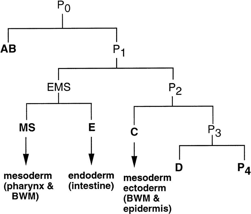
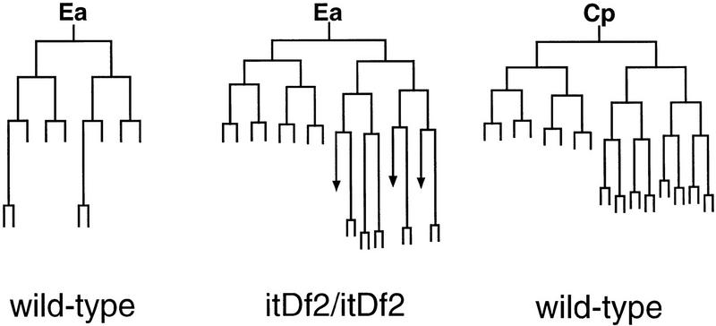
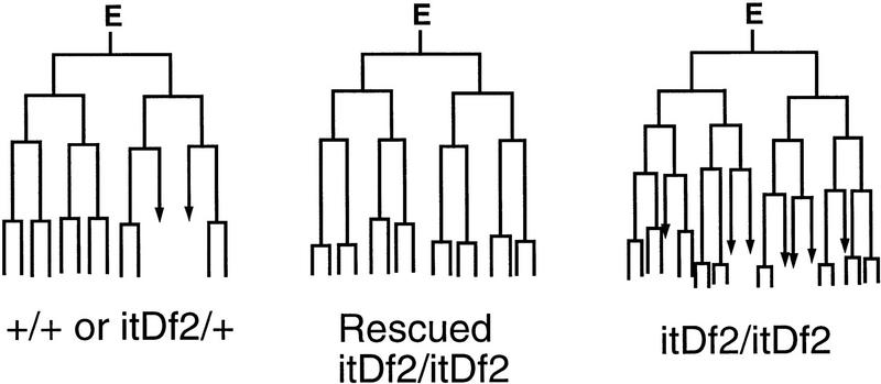
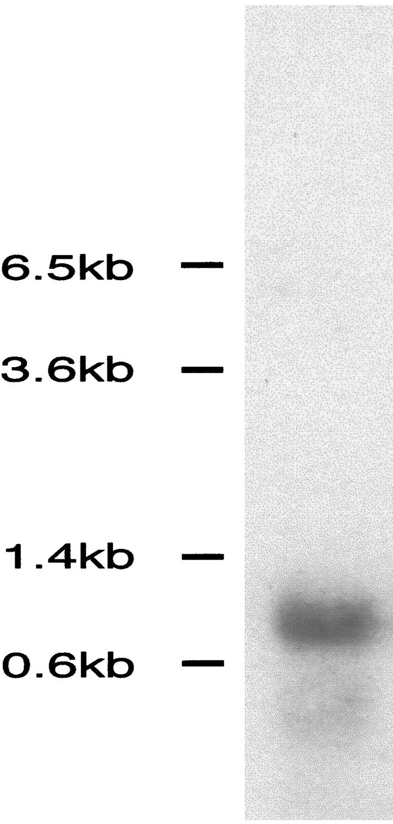
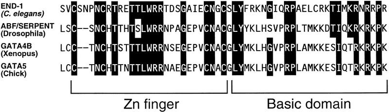
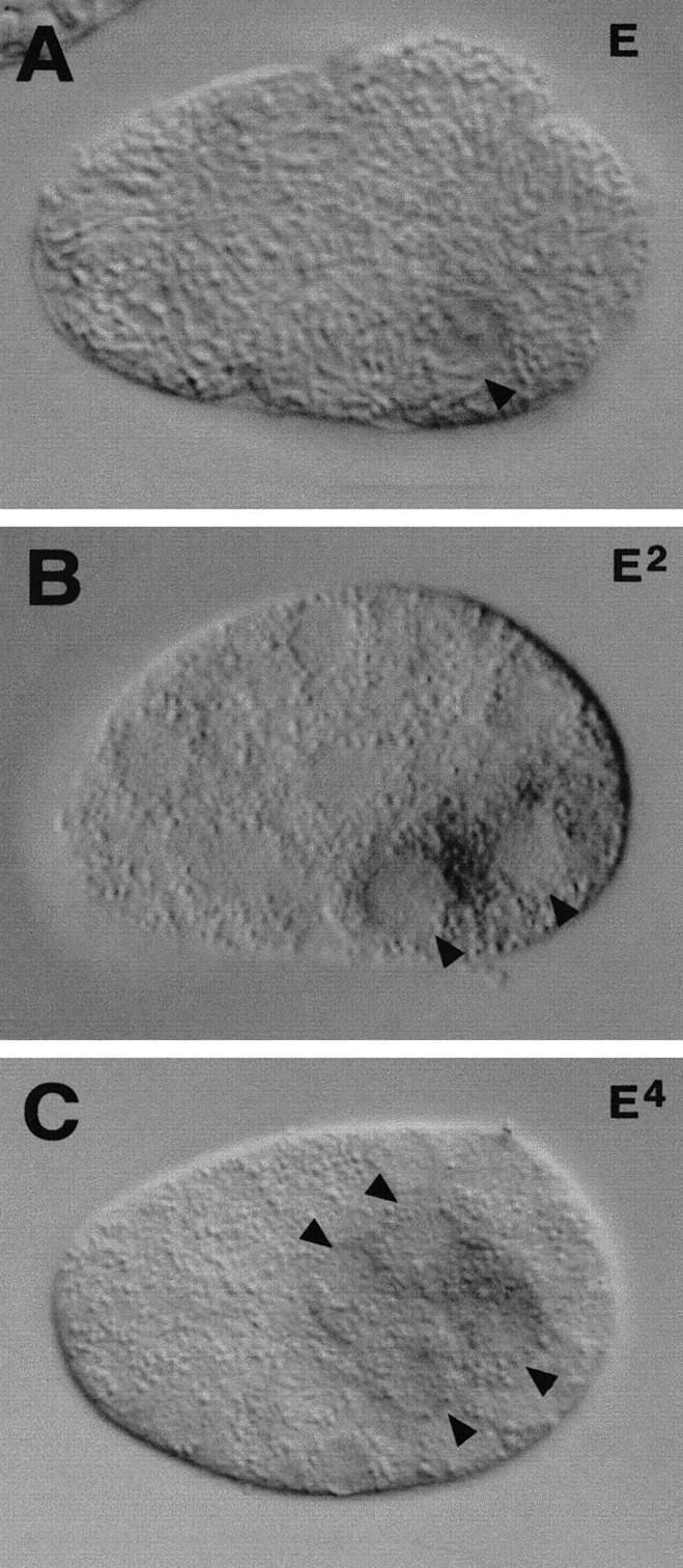
The endoderm in the nematode Caenorhabditis elegans is clonally derived from the E founder cell. We identified a single genomic region (the endoderm-determining region, or EDR) that is required for the production of the entire C. elegans endoderm. In embryos lacking the EDR, the E cell gives rise to ectoderm and mesoderm instead of endoderm and appears to adopt the fate of its cousin, the C founder cell. end-1, a gene from the EDR, restores endoderm production in EDR deficiency homozygotes. end-1 transcripts are first detectable specifically in the E cell, consistent with a direct role for end-1 in endoderm development. The END-1 protein is an apparent zinc finger-containing GATA transcription factor. As GATA factors have been implicated in endoderm development in other animals, our findings suggest that endoderm may be specified by molecularly conserved mechanisms in triploblastic animals. We propose that end-1, the first zygotic gene known to be involved in the specification of germ layer and founder cell identity in C. elegans, may link maternal genes that regulate the establishment of the endoderm to downstream genes responsible for endoderm differentiation.


Figures

Figure 1
Figure 1
Origin of the E blastomere and the intestine. The diagram shows the early lineage and the six founder cells (labeled in boldface type) of the C. elegans embryo. The germ layers and major cell types produced by E, MS, and C during wild-type development are indicated by arrows. (BWM) Body-wall muscle. For a complete description of the embryonic lineages and cell fates, see Sulston et al. (1983).
Figure 6
Figure 6
Genetic and molecular identification of the end-1 region and the end-1 gene. (A) Genetic map of a portion of the right arm of linkage group (chromosome) V and several deficiencies in the region. The region required for endoderm specification, henceforth called the “EDR,” was delimited by mapping the endpoints of the overlapping deficiencies on the physical map and comparing their intestine differentiation phenotypes. arDf1, yDf8, and yDf9 homozygotes make intestine, whereas nDf42, zuDf2, itDf2, wDf3, and wDf4 homozygotes (labeled in boldface) do not. Based on PCR analysis, itDf2 does not delete sequences including and to the left of the JZ 9,10 primers, which are derived from cm01h10, a cDNA sequence available from ACeDB. wDf4 complements him-5, which is contained in cosmid K02A12, but deletes the sequence amplified by the JZ 17,18 primers, which are derived from the org-1 sequence on the cosmid T08G5. The genomic region required for endoderm specification, which is shown by broken lines, is therefore defined by the left endpoint of itDf2 and the right endpoint of wDf4. We estimate that the interval of the end-1 region is <200 kb. (B) Cosmid clones in the end-1 region. A subset of cosmids in the area was used in the transformation rescue experiments, and three overlapping cosmids (labeled in boldface), K10F6, R7, and T26F2, were found to carry rescuing activity. (C) The location of end-1 was further refined by testing whether subclones derived from K10F6 contained rescuing activity. The ∼4-kb KpnI–SacI genomic fragment indicated by a thicker line was found to be the minimum fragment containing rescuing activity. (□) Exons as deduced by comparing the sequence of the 4-kb rescuing fragment and the nearly full-length 0.85-kb cDNA. (D) The end-1 minigene insert. The insert contains an ∼1.8-kb end-1 upstream genomic sequence and the end-1 cDNA and end-1 3′-untranslated region (UTR) sequences.
Figure 2
Figure 2
Absence of a differentiated intestine in a terminally arrested zuDf2 homozygous embryo. Shown are micrographs of live embryos viewed under Nomarski optics (A,B) or dark field with polarized light (C,D). A wild-type embryo at an early stage in morphogenesis (1.5-fold) is shown in A and C. A terminally arrested homozygous zuDf2 embryo is shown in B and D. When viewed by Nomarski microscopy, intestinal cells containing large nuclei with a prominent nucleolus (arrowhead) are evident in the wild-type embryo (A) but not in the mutant (B). The pharynx primordium (arrow) is visible in both wild-type and homozygous zuDf2 embryos. Although birefringent granules characteristic of a differentiated intestine are obvious under polarized light in wild type (C), they are absent in the homozygous zuDf2 embryo (D). Similar observations were made in itDf2 homozygous embryos. Other intestinal differentiation markers, including a gut esterase (Edgar and McGhee 1986), were also found to be absent in itDf2 homozygous embryos (not shown). Anterior is to the left and dorsal is to the top in each panel. Bar, ∼10 μm.
Figure 3
Figure 3
Tissue differentiation in wild-type and zuDf2 mutant embryos. Immunofluorescence analysis of tissue differentiation in partially elongated (1.5-fold) wild-type (A,C,G), elongated (pretzel stage) wild-type (E), and terminal homozygous zuDf2 (B,D,F,H) embryos. (A,B) Embryos stained with mAb 1CB4, which reacts with the 20 embryonic intestinal cells (Okamoto and Thomson 1985) and the intestinal–rectal valve cells. The intestinal cells are absent in homozygous zuDf2 embryos although the two intestinal–rectal valve cells, which arise from a nonendodermal precursor (the AB founder cell) are present (arrows). (C,D) Embryos stained with 3NB12, a monoclonal antibody that recognizes a subset of pharynx muscles (Priess and Thomson 1987). (E,F) Embryos stained with NE8/4C6, a monoclonal antibody that recognizes body-wall muscle cells (Goh and Bogaert 1991). (G,H) Surface view of embryos stained with MH27, a monoclonal antibody that recognizes epithelial adherens junctions (Priess and Hirsh 1986); only the epidermal cells are visible in the focal plane shown. Although the nonendodermal tissues are disorganized in terminal zuDf2 embryos, all appear to be made in approximately normal amounts. Large numbers of cells with the characteristic appearance of neurons were also apparent by Nomarski microscopy in zuDf2 and itDf2 embryos (data not shown). Bar, ∼10 μm.
Figure 4
Figure 4
The intestinal precursor E produces epidermis and body-wall muscle in zuDf2 mutants. Each row shows a single embryo stained with rabbit anti-LIN-26 (left), which recognizes the nuclei of epidermal cells, and with mAb 5.6 (right), which recognizes the cytoplasm of body-wall muscle. (A,B) An intact wild-type embryo at the two-fold stage produces epidermis (A) and body-wall muscle (B). (C–J) Partial embryos were produced by ablating all founder cells except the C blastomere (“C” isolation) (C,D) or the E blastomere (E–J) in either wild-type (C–F) or F1 embryos from a strain heterozygous for zuDf2 (G–J). An isolated wild-type C blastomere produces both epidermis (C) and body-wall muscle (D), whereas an isolated wild-type E blastomere produces neither epidermis (E) nor body-wall muscle (F). E isolations from zuDf2 heterozygotes produce two classes. Approximately one in four do not produce gut but instead produce epidermis (G) and body-wall muscle (H); these are putative zuDf2 homozygotes. Approximately three in four produce intestine but fail to produce epidermis (I) and body-wall muscle (J). Embryos shown in C–J are representative individuals used to generate data in Table 1. Bar, ∼10 μm.
Figure 5
Figure 5
The E lineage is altered in homozygous itDf2 embryos. The Ea branch of the E lineage in wild-type (left) and itDf2 embryos (middle) is shown. The Ea lineage of the mutant is dramatically different from the wild-type Ea lineage (left) and most closely resembles the wild-type lineage of Ca and Cp (Although the wild-type Ca and Cp lineages are similar, the Cp lineage, which most closely resembles the mutant lineage, is shown here at right). Termination of a vertical line indicates that the cell did not divide for at least 200 min after its birth. The arrows in the mutant lineage indicate cells that were lost during the lineage tracing. The cells derived from E in the itDf2 mutant divide at a faster rate than normal and give rise to extra divisions (see text). Similar observations were made in one other homozygous itDf2 embryo (not shown). Portions of the MS and C lineages were also followed in the itDf2 mutant embryos and appeared normal. Wild-type lineages are taken from Sulston et al. (1983). The timings of cell division in the mutant embryo were normalized to that of wild type.
Figure 7
Figure 7
Intestinal differentiation in rescued homozygous itDf2 embryos carrying the end-1 gene. Three two- or four-cell embryos were collected from heterozygous itDf2 hermaphrodites carrying the end-1 gene on extrachromosomal arrays, mounted on an agar pad, and allowed to develop at 20 ± 2°C. Nomarski (A) and dark-field with polarized light (B) micrographs of these embryos were taken 10 hr after they were mounted. The genotypes of these embryos were inferred 15 hr later based on their terminal morphology and the presence of gut granules seen under polarized light: One hundred percent of itDf2 mutant embryos fail to complete elongation and produced neither intestine nor programmed cell deaths. Thus, embryos with incomplete elongation and no programmed cell deaths but with an intestine are inferred to be itDf2 homozygotes rescued by K10F6, which contains end-1. (B) (1) Rescued homozygous itDf2 mutant; (2) nonrescued homozygous itDf2 mutant; (3) wild-type or heterozygous itDf2 embryo. The intestinal cells have lined up in the middle posterior region of the embryo (a) and show the characteristic appearance of intestinal cells (A, arrow). The normal internal location of the intestinal cells suggests that gastrulation within the E-cell lineage was normal. (C) A terminally arrested homozygous itDf2 embryo bearing extrachromosomal K10F6 and containing gut granules was immunostained with mAb 1CB4, showing that differentiated intestinal cells were made in this embryo. Bar, ∼10 μm. Similar results (not shown) were observed with a subclone containing only the end-1 gene (see description of end-1 minigene construct).
Figure 8
Figure 8
E-cell lineage in rescued homozygous itDf2 embryos carrying end-1. The first four divisions in the E lineage of a rescued homozygous itDf2 embryo are shown (middle). The E lineages in both a wild-type embryo (left; the genotype was not determined but is either +/+ or Df/+) and an unrescued itDf2 homozygous embryo (right) were also determined for comparison. The development of all three embryos was recorded simultaneously on the same slide. Arrows indicate cells that were lost during the lineage analysis. The E-lineage pattern in the rescued homozygous itDf2 mutant looks similar to a wild-type E lineage. For example, the cell cycle time of E descendants is prolonged such that the E lineage undergoes four rounds of division in the same time that a itDf2 mutant E lineage completes five rounds of division. Similar lineage observations were made with two other rescued itDf2 homozygotes (not shown).
Figure 9
Figure 9
Sequence of the end-1 gene. Genomic sequence of the region surrounding the end-1 gene is shown, with the predicted protein sequence indicated below. The first nucleotide of the 0.85-kb cDNA clone is indicated by an arrow. The proposed translational start site determined by 5′-RACE is boxed. Another possible in-frame ATG located downstream is also boxed. Splice sites are indicated by arrowheads. The zinc finger region is indicated by boldface letters. The termination codon is indicated by three asterisks (***). Two different polyadenylation sites are indicated by unfilled arrows. The (A/T)GATA(A/G) consensus binding sites (i.e., GATA sites) are enclosed by ovals. The putative SKN-1 binding sites are underlined. The putative HMG protein binding site is indicated with double underlines.
Figure 10
Figure 10
Northern analysis of end-1 transcript. A single transcript of ∼0.85 kb is detected in poly(A)+-enriched mRNA from mixed stage wild-type embryos using the end-1 cDNA insert from pJZ10 as a probe.
Figure 11
Figure 11
Comparison of the zinc finger region of the predicted END-1 protein with the single zinc finger region of SERPENT/dABF in Drosophila and the second zinc finger of GATA-4B in Xenopus and GATA-5 in chick. Residues that END-1 shares with at least one of these proteins are shown as white on black letters.
Figure 12
Figure 12
end-1 transcript expression in wild-type embryos. In situ hybridization shows that end-1 mRNA is expressed only in the E lineage. end-1 mRNAs are first detected in the E cell of an eight-cell embryo (A). Expression is also seen in Ea and Ep of a 15-cell embryo (B) and in the four granddaughters of E in an ∼50-cell embryo (C). The arrowheads point to E and its descendants. Anterior is at the left, and dorsal is at the top.

Similar articles

Cited by

References

    1. Aamodt EJ, Chung MA, McGhee JD. Spatial control of gut-specific gene expression during Caenorhabditis elegans development. Science. 1991;252:579–582. - PubMed
    1. Albertson DG, Sulston JE, White JG. Cell cycling and DNA replication in a mutant blocked in cell division in the nematode Caenorhabditis elegans. Dev Biol. 1978;63:165–178. - PubMed
    1. Altschul SF, Gish W, Miller W, Myers W, Lipman DJ. Basic local alignment search tool. J Mol Biol. 1990;215:403–410. - PubMed
    1. Arceci R, King AAJ, Simon MC, Orkin SH, Wilson DB. Mouse GATA-4: A retinoic acid-inducible GATA-binding transcription factor expressed in endodermally derived tissues and heart. Mol Cell Biol. 1993;13:2235–2246. - PMC - PubMed
    1. Austin J, Kimble J. Transcript analysis of glp-1 and lin-12, homologous genes required for cell interactions during development of C. elegans. Cell. 1989;58:565–571. - PubMed

Publication types

MeSH terms

Associated data