. 1997 Nov 11;94(23):12297-302.
doi: 10.1073/pnas.94.23.12297.

RNA-peptide fusions for the in vitro selection of peptides and proteins

Affiliations

RNA-peptide fusions for the in vitro selection of peptides and proteins

R W Roberts et al. Proc Natl Acad Sci U S A. .

Abstract

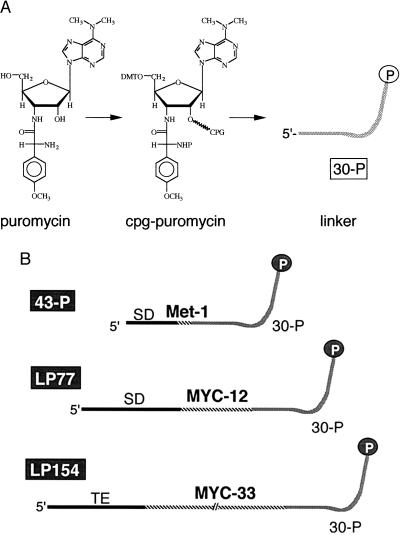
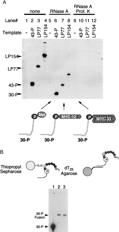
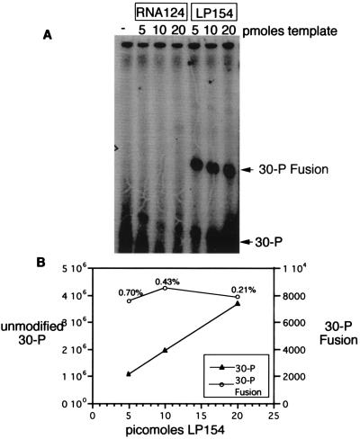
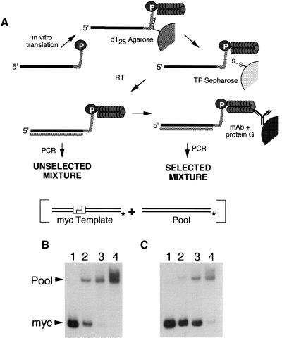
Covalent fusions between an mRNA and the peptide or protein that it encodes can be generated by in vitro translation of synthetic mRNAs that carry puromycin, a peptidyl acceptor antibiotic, at their 3' end. The stable linkage between the informational (nucleic acid) and functional (peptide) domains of the resulting joint molecules allows a specific mRNA to be enriched from a complex mixture of mRNAs based on the properties of its encoded peptide. Fusions between a synthetic mRNA and its encoded myc epitope peptide have been enriched from a pool of random sequence mRNA-peptide fusions by immunoprecipitation. Covalent RNA-peptide fusions should provide an additional route to the in vitro selection and directed evolution of proteins.


Figures

Figure 1
Figure 1
Proposed mechanism for RNA-peptide fusion formation on the ribosome. (A) The ribosome initiates protein synthesis on the mRNA and translocates toward the end of the template. (B) When the ribosome reaches the end of the RNA ORF, translation stalls at the RNA/DNA junction. The puromycin then enters the A site of the ribosome and accepts the nascent peptide. (C) Once formed the mRNA-peptide fusion is purified from the translation reaction by affinity chromatography.
Figure 2
Figure 2
Synthesis of translation templates containing 3′ puromycin. (A) Puromycin⋅(HCl)2 was protected and attached to a synthesis support to give CPG-puromycin, and the linker 30-P (dA27dCdCP) was made by automated synthesis (see Materials and Methods). (B) Translation templates used were: 43-P (Met template), LP77 (short myc template), and LP154 (long myc template) containing ORFs of 1, 12, and 33 codons, respectively, and no stop codon. The 5′ untranslated regions (UTRs) of 43-P and LP77 contain a Shine–Dalgarno sequence complementary to five bases of 16S rRNA (33) and spaced similarly to ribosomal protein sequences (34). The 5′ UTR of LP154 contains a 22-nt sequence derived from the tobacco mosaic virus 5′ UTR encompassing two ACAAAUUAC direct repeats (26). LP154 codes for the peptide used to raise the mAb 9E10 and LP77 codes for the recognition epitope EQKLISEEDL (24).
Figure 3
Figure 3
Incorporation of [35S]Met into the Met (43-P), short myc (LP77), and long myc (LP154) translation templates. (A) Denaturing PAGE analysis of templates isolated from translation reactions. The gel shows templates isolated by dT25 affinity chromatography with no treatment (lanes 1–4), treatment with RNase A (lanes 5–8), and treatment with both RNase A and Proteinase K (lanes 9–12). The positions of unmodified 30-P, 43-P, LP77, and LP154 are indicated by arrows at the side of the gel. The schematic below indicates the proposed composition of the fragments in the RNase-treated lanes. (B) Sequential isolation of long myc linker fusion (30-P fusion) fragments by thiopropyl (TP) Sepharose and dT25 affinity chromatography. Lane 2 is fusion product purified on TP Sepharose followed by dT25, whereas lane 3 is material purified by dT25 only.
Figure 4
Figure 4
Analysis and quantitation of fusion formation by immunoprecipitation. (A) Denaturing urea PAGE analysis of the 32P-labeled linker-peptide fusions isolated by immunoprecipitation-RNase A-kinase treatment of the translation reactions. Reactions containing no template or only the RNA portion of the long myc template (RNA124) show no fusion formation. Reactions containing the long myc template contain bands corresponding to long myc linker fusions (30-P fusion) indicated by the arrow. The amount of input template is indicated at the top. (B) Recovered unmodified linker 30-P (left y axis) is linearly proportional to input template (x axis), whereas linker-peptide fusion (right y axis) is constant, as determined by PhosphorImager counting.
Figure 5
Figure 5
Enrichment of myc dsDNA vs. pool dsDNA by in vitro selection. (A) Schematic of the selection protocol. Four mixtures of the myc and pool templates were translated in vitro and isolated on dT25 agarose followed by thiopropyl (TP) Sepharose to purify the template fusions from unmodified templates. The mRNA-peptide fusions then were reverse-transcribed to suppress any secondary or tertiary structure present in the templates. Aliquots of each mixture were removed before (B) and after (C) affinity selection, amplified by PCR in the presence of a labeled primer, and digested with a restriction enzyme that cleaves only the myc DNA. The input mixtures of templates were: lane 1, pure myc; lanes 2–4, 1:20, 1:200, and 1:2,000 myc/pool. The unselected material deviates from the input ratios because of preferential translation and reverse transcription of the myc template. The enrichment of the myc template during the selective step was calculated from the change in the pool/myc ratio before and after selection.

Comment in

References

    1. Breaker R R. Chem Rev (Washington, DC) 1997;97:371–390. - PubMed
    1. Osborne S E, Ellington A D. Chem Rev (Washington, DC) 1997;97:349–370. - PubMed
    1. Williams K P, Bartel D P. Nucleic Acids Mol Biol. 1996;10:367–381.
    1. Lorsch J R, Szostak J W. Acc Chem Res. 1996;29:103–110. - PubMed
    1. Gold L, Polisky B, Uhlenbeck O, Yarus M. Annu Rev Biochem. 1995;64:763–797. - PubMed

Publication types

LinkOut - more resources