. 1997 Nov 11;94(23):12378-83.
doi: 10.1073/pnas.94.23.12378.

A method that allows the assembly of kinetochore components onto chromosomes condensed in clarified Xenopus egg extracts

Affiliations

A method that allows the assembly of kinetochore components onto chromosomes condensed in clarified Xenopus egg extracts

A Desai et al. Proc Natl Acad Sci U S A. .

Abstract

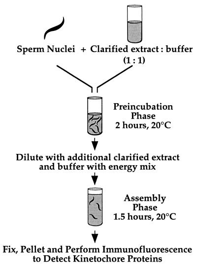
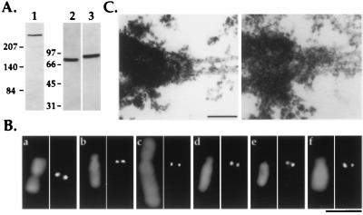
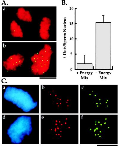
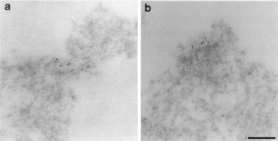
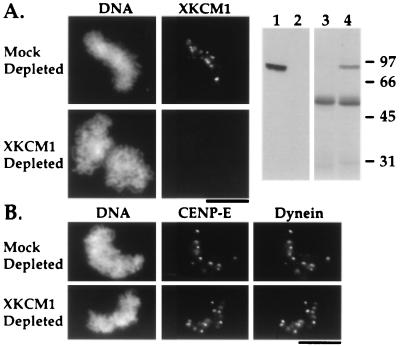
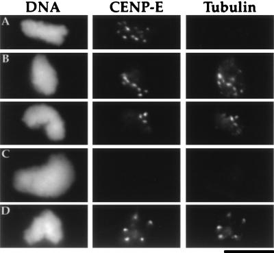
Kinetochores are complex macromolecular structures that link mitotic chromosomes to spindle microtubules. Although a small number of kinetochore components have been identified, including the kinesins CENP-E and XKCM1 as well as cytoplasmic dynein, neither how these and other proteins are organized to produce a kinetochore nor their exact functions within this structure are understood. For this reason, we have developed an assay that allows kinetochore components to assemble onto discrete foci on in vitro-condensed chromosomes. The source of the kinetochore components is a clarified cell extract from Xenopus eggs that can be fractionated or immunodepleted of individual proteins. Kinetochore assembly in these clarified extracts requires preincubating the substrate sperm nuclei in an extract under low ATP conditions. Immunodepletion of XKCM1 from the extracts prevents the localization of kinetochore-associated XKCM1 without affecting the targeting of CENP-E and cytoplasmic dynein or the binding of monomeric tubulin to the kinetochore. Extension of this assay for the analysis of other components should help to dissect the protein-protein interactions involved in kinetochore assembly and function.


Figures

Figure 2
Figure 2
Schematic of the two-step kinetochore assembly reaction in clarified Xenopus egg extracts (see text for details).
Figure 1
Figure 1
Reagents for following kinetochore assembly in Xenopus egg extracts. (A) Immunoblots of Xenopus extracts using 1 μg/ml affinity-purified anti-human CENP-E (lane 1), anti-chicken cytoplasmic dynein intermediate chain mAb 70.1 ascites fluid (lane 2), and 0.1 μg/ml affinity-purified anti-XKCM1 (lane 3) antibodies. Extracts were fractionated by 6% SDS/PAGE for CENP-E immunoblots and by 10% SDS/PAGE for dynein and XKCM1 immunoblots. Molecular mass markers (in kDa) are indicated to the left of each set of panels. (B) Comparative immunofluorescence of CHO (a, c, and e) and A6 (b, d, and f) cell chromosomes using antibodies to CENP-E (a and b), cytoplasmic dynein intermediate chain (c and d), and XKCM1 (e and f). Antibody localizations are shown in the right panel of each pair and DNA in the left panel. (Bar = 5 μm.) (C) Analysis of mitotic XL177 cells by electron microscopy demonstrating the presence of a trilaminar plate kinetochore structure at the point of interaction of kinetochore microtubules with the chromosome. Two separate examples are shown. (Bar = 0.2 μm.)
Figure 3
Figure 3
Characterization of in vitro kinetochore assembly. (A) Effect of presence (a) or absence (b) of energy mix in preincubation buffer on kinetochore assembly. DNA is in red and CENP-E is in green. The same result is obtained with anti-dynein and anti-XKCM1 antibodies. There is no striking difference in the morphology of the condensed DNA at the end of the assembly reaction between the two conditions. (B) Quantitation of kinetochore assembly and effect of energy mix in preincubation buffer. The bars represent the mean number of dots of kinetochore marker proteins per sperm nucleus ± 1 SD. For the left column (+ Energy Mix in preincubation buffer) n = 103 sperm nuclei from two experiments on two different extract preparations. For the right column (− Energy Mix in preincubation buffer; standard kinetochore assembly reaction) n = 303 sperm nuclei from four experiments on three different extract preparations. The absence of energy mix in the preincubation buffer results in >90% of the sperm nuclei demonstrating colocalization of all three markers at the reported density. The residual sperm nuclei show no localization of any of the three markers. (Bars = 10 μm.) (C) Double label immunofluorescence of in vitro assembled kinetochores demonstrating colocalization of all three mitotic markers to discrete foci on the DNA. Panels show 4′,6′diamidino-2-phenylindole (DAPI)-labeled sperm nucleus DNA (a and d) and corresponding double label localization of dynein and CENP-E (b and c) or dynein and XKCM1 (e and f). Dots associated with the chromosome clusters are distributed over several focal planes; the images shown are photomicrographs of single focal planes and do not account for all dots present on these chromosomal clusters.
Figure 4
Figure 4
Morphological analysis of in vitro assembled kinetochores using immunoelectron microscopy. Anti-CENP-E antibody was used for the immunoelectron microscopy. Two examples of gold clusters (a and b) show the specificity of labeling, the absence of a striking lamellar morphology and differentiation of the chromatin at the region of labeling. The cluster in a appears to show a slight degree of constriction; the cluster in b shows clear organization of the underlying chromatin fibers. (Bar = 0.2 μm.)
Figure 5
Figure 5
XKCM1 depletion does not affect targeting of dynein and CENP-E to kinetochores. (A) XKCM1 localization to condensed chromosomes is abolished by XKCM1 depletion. Right two panels show an immunoblot (lanes 1 and 2) of equal amounts of mock-depleted (lane 1) and XKCM1-depleted (lane 2) extracts probed with anti-XKCM1 antibody, and a Coomassie-stained gel (lanes 3 and 4) of the corresponding immunoprecipitates: control IgG (lane 3) and anti-XKCM1 (lane 4). (B) XKCM1 depletion does not affect CENP-E and dynein localization. CENP-E and dynein targeting to kinetochores was compared in XKCM1-depleted vs. mock-depleted extracts. Quantitation of number of dots of CENP-E and dynein per sperm nucleus show no difference between mock- and XKCM1-depleted extracts (n = 100 sperm nuclei from two experiments on two different extract preparations). (Bars = 10 μm.)
Figure 6
Figure 6
Tubulin binding by in vitro-assembled kinetochores. Kinetochores were identified using the anti-CENP-E antibody. (A) Purified condensed chromosome clusters lack associated monomeric tubulin. (B) In vitro-assembled kinetochores bind exogenously added monomeric tubulin. Purified condensed chromosome clusters were incubated with 10 μM tubulin and then assayed for binding of monomeric tubulin. As shown here for two separate sperm nuclei, every CENP-E dot was associated with monomeric tubulin (100 sperm nuclei, two experiments). The tubulin also shows faint punctate background staining of the DNA and, occasionally, a few larger dots which do not correspond to CENP-E dots. (C) In vitro-assembled kinetochores fail to bind monomeric tubulin after salt extraction. Assembled kinetochores were extracted using 100 mM extra NaCl before being purified and assayed for binding of monomeric tubulin. This extraction removes kinetochore-associated CENP-E, XKCM1, and dynein but does not remove chromosomal histones (data not shown). (D) XKCM1 depletion does not affect tubulin binding by in vitro-assembled kinetochores. XKCM1 depletion was assayed in parallel by immunoblots and immunofluorescence as described in Fig. 5. (Bar = 10 μm.)

References

    1. Brinkley B R, Ouspenski I, Zinkowski R P. Trends Cell Biol. 1992;2:15–21. - PubMed
    1. Earnshaw W C, Tomkiel J E. Curr Opin Cell Biol. 1992;4:86–93. - PubMed
    1. Rieder C L. Int Rev Cytol. 1982;79:1–58. - PubMed
    1. Wordeman L, Steuer E R, Sheetz M P, Mitchison T. J Cell Biol. 1991;114:285–294. - PMC - PubMed
    1. Palmer D K, O’Day K, Wener M H, Andrews B S, Margolis R L. J Cell Biol. 1987;104:805–815. - PMC - PubMed

Publication types

Substances

LinkOut - more resources