. 1997 Nov 11;94(23):12407-12.
doi: 10.1073/pnas.94.23.12407.

Nonmuscle myosin II-B is required for normal development of the mouse heart

Affiliations

Nonmuscle myosin II-B is required for normal development of the mouse heart

A N Tullio et al. Proc Natl Acad Sci U S A. .

Abstract

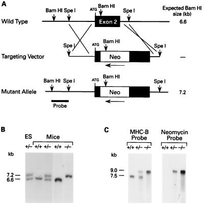
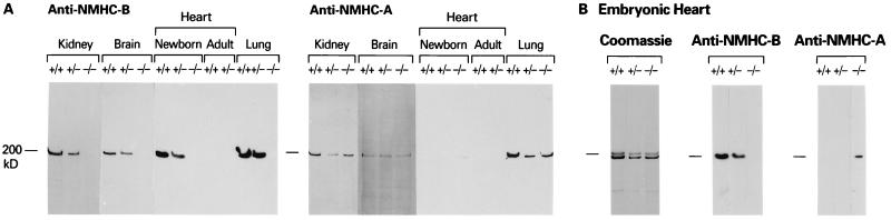
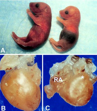
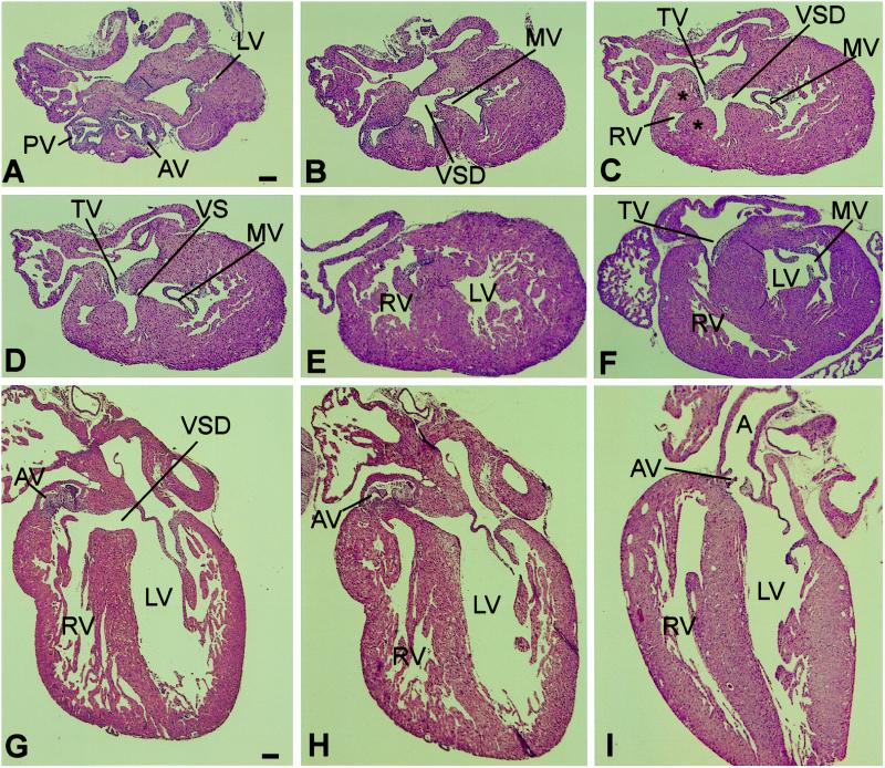
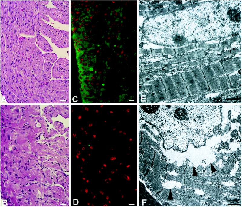
We used targeted gene disruption in mice to ablate nonmuscle myosin heavy chain B (NMHC-B), one of the two isoforms of nonmuscle myosin II present in all vertebrate cells. Approximately 65% of the NMHC-B-/- embryos died prior to birth, and those that were born suffered from congestive heart failure and died during the first day. No abnormalities were detected in NMHC-B+/- mice. The absence of NMHC-B resulted in a significant increase in the transverse diameters of the cardiac myocytes from 7.8 +/- 1.8 micron (right ventricle) and 7.8 +/- 1.3 micron (left ventricle) in NMHC-B+/+ and B+/- mice to 14.7 +/- 1.1 micron and 13.8 +/- 2.3 micron, respectively, in NMHC-B-/- mice (in both cases, P < 0.001). The increase in size of the cardiac myocytes was seen as early as embryonic day 12.5 (4.5 +/- 0.2 micron for NMHC-B+/+ and B+/- vs. 7. 2 +/- 0.6 micron for NMHC-B-/- mice (P < 0.01)). Six of seven NMHC-B-/- newborn mice analyzed by serial sectioning also showed structural cardiac defects, including a ventricular septal defect, an aortic root that either straddled the defect or originated from the right ventricle, and muscular obstruction to right ventricular outflow. Some of the hearts of NMHC-B-/- mice showed evidence for up-regulation of NMHC-A protein. These studies suggest that nonmuscle myosin II-B is required for normal cardiac myocyte development and that its absence results in structural defects resembling, in part, two common human congenital heart diseases, tetralogy of Fallot and double outlet right ventricle.


Figures

Figure 1
Figure 1
Targeted disruption of the mouse NMHC-B locus. (A) Genomic map of the mouse NMHC-B locus surrounding exon 2. The locations of the crossover sites are indicated at either end of the targeting vector. Exon 2 contains the initiating ATG, after which a PGK-Neo cassette was inserted in the 3′–5′ direction. The probe used for analysis is indicated on the mutant allele. The fragment size after digestion of genomic DNA with BamHI is indicated on the right. The map is not drawn to scale. (B) Southern analysis of an ES clone (lane 1) and mouse genomic DNA (lanes 2–5). DNA derived from targeted ES cells and mice was digested with BamHI and hybridized with the probe indicated in A. A 6.6-kb fragment indicates a + (wild-type) allele, whereas a 7.2-kb fragment indicates a − (recombinant) allele. (C) Analysis of mouse brain mRNA. A 1.6-kb mouse cDNA probe specific for NMHC-B detected both a 7.5-kb message (+ allele) and a 9.0 kb message (− allele), whereas a 1.0-kb cDNA probe specific for the neomycin cassette detected a 9.0-kb message (− allele) only.
Figure 2
Figure 2
Immunoblot analyses of tissues from +/+, +/−, and −/− mice (A) and evidence for up-regulation of NMHC-A (B). (A) The blot on the left was analyzed with antibodies specific for the carboxyl-terminal sequence of NMHC-B and the one on the right with antibodies specific for the carboxyl-terminal sequence of NMHC-A (13). All samples, except those marked adult, are from newborn mice. (B) These samples are from an E18 mouse heart. The Coomassie stain shows that +/+, +/−, and −/− mice contain α and β cardiac myosin and the immunoblot demonstrates up-regulation of NMHC-A in the −/− heart.
Figure 3
Figure 3
Gross features of newborn mice. (A) Newborn +/+ (left) and −/− (right) mice from the same litter. Note the darkened abdominal region, indicative of a congested liver, and the dome-shaped head, due to hydrocephalus involving the lateral ventricles, in the −/− mouse. The size of the newborn −/− mice varied, and all of them failed to nurse. (B and C) Unopened +/+ (B) and −/− (C) hearts. The −/− heart is more rounded in shape and shows right atrial (RA) dilatation and bulging of the right ventricular outflow tract (∗).
Figure 4
Figure 4
Histologic sections of newborn −/− and +/+ hearts. (A–E) Selected serial transverse sections, from cephalad to caudad, of a −/− heart. (F) Transverse section of a +/+ heart at approximately the same level as C. (G and H) Sagittal sections of another −/− heart. (I) Sagittal section of a +/+ heart. (Hematoxylin and eosin stain; scale bar = 100 μm.) (A) Cross-sectional view of the aorta and the pulmonary artery at the levels of the semilunar valves, showing the positions of the two vessels, and the uppermost portion of the lateral wall of the left ventricle (LV). AV, aortic valve; PV, pulmonary valve. (B) This section shows the uppermost portion of the ventricular septal defect (VSD). The mitral valve (MV) is also shown. Comparison with A shows that the VSD is located beneath the aortic valve. (C) Shown are the VSD, the MV, the tricuspid valve (TV), and the right ventricular outflow tract (RV), which is narrowed by hypertrophic muscle (∗). (D) The edge of the membranous portion of the ventricular septum (VS), the MV, and the TV are demonstrated. (E) The left and right ventricular walls and cavities are evident, as is the muscular portion of the VS. (F) Transverse section of +/+ heart. (G) Sagittal sections of a −/− heart showing the VSD and the rightward malposition of the AV. (H) View showing the relationships of the AV to the left ventricular outflow tract in the area adjacent to the VS. (I) Sagittal section of +/+ heart showing the normal relationships of the VS to the AV and aorta (A).
Figure 5
Figure 5
Histology (A and B), immunocytochemistry (C and D), and ultrastructure (E and F) of +/+ (A, C, and E) and −/− (B, D, and F) hearts. (A and B) Myocytes from the right ventricular wall of newborn hearts. Note the disarray and larger size of the myocytes in the −/− heart (B). (Hematoxylin and eosin stain; bar = 20 μm.) (C and D) Fluorescence photomicrographs of heart sections from E18.5 embryos. The green color indicates the presence of NMHC-B in the +/+ heart (C) and the lack of green, its absence in the −/− heart (D). Nuclei are stained red with propidium iodide. (Bar = 10 μm.) (E and F) Electron micrographs demonstrating myofibrillar disarray in the −/− heart (F) compared with the normal heart (E). Although there is myofibrillar disarray, the myofibrillar components appear normal in the −/− heart. Arrowheads indicate the perinuclear and interfibrillar areas where glycogen particles are present. (Bar = 1 μm.)

Similar articles

Cited by

References

    1. Sellers J R, Goodson H V. Protein Profile. 1995;2:1323–1423. - PubMed
    1. Goodson H V, Spudich J A. Proc Natl Acad Sci USA. 1993;90:659–663. - PMC - PubMed
    1. Saez C G, Myers J C, Shows T B, Leinwand L A. Proc Natl Acad Sci USA. 1990;87:1164–1168. - PMC - PubMed
    1. Simons M, Wang M, McBride O W, Kawamoto S, Yamakawa K, Gdula D, Adelstein R S, Weir L. Circ Res. 1991;69:530–539. - PubMed
    1. Katsuragawa Y, Yanagisawa M, Inoue A, Masaki T. Eur J Biochem. 1989;184:611–616. - PubMed

Substances