. 1997 Nov 11;94(23):12413-8.
doi: 10.1073/pnas.94.23.12413.

Dystrobrevin and dystrophin: an interaction through coiled-coil motifs

Affiliations

Dystrobrevin and dystrophin: an interaction through coiled-coil motifs

H M Sadoulet-Puccio et al. Proc Natl Acad Sci U S A. .

Abstract

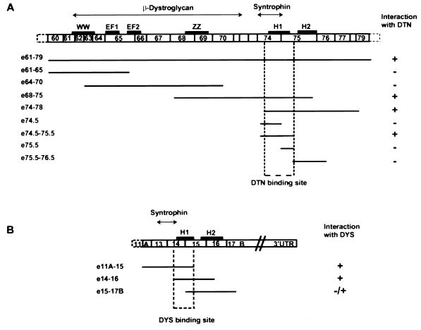
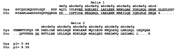
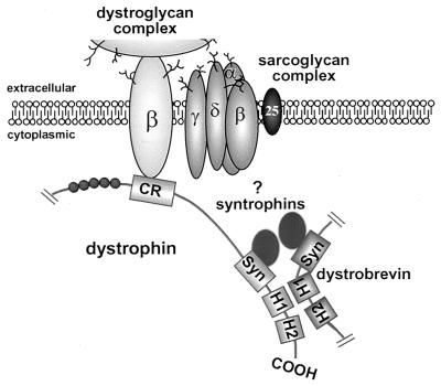
Dystrobrevin, a dystrophin-related and -associated protein, has been proposed to be important in the formation and maintenance of the neuromuscular junction. Dystrobrevin coprecipitates with both the acetylcholine receptor complex as well as the dystrophin glycoprotein complex. Although the nature of dystrobrevin's association with the dystrophin glycoprotein complex remains unclear, it is known that dystrobrevin binds directly to the syntrophins, a heterologous group of dystrophin-associated proteins. Using the yeast two-hybrid system to identify protein-protein interactions, we present evidence for the heterodimerization of dystrobrevin directly with dystrophin. The C terminus of dystrobrevin binds specifically to the C terminus of dystrophin. We further refined this site of interaction to these proteins' homologous coiled-coil motifs that flank their respective syntrophin-binding sites. We also show that the interaction between the dystrobrevin and dystrophin coiled-coil domains is specific and is not due to a nonspecific coiled-coil domain interaction. From the accumulated evidence of protein-protein interactions presented here and elsewhere, we propose a partially revised model of the organization of the dystrophin-associated glycoprotein complex.


Figures

Figure 1
Figure 1
Dystrobrevin–syntrophin and dystrobrevin–dystrophin interactions by coimmunoprecipitation. In vitro translated proteins were incubated together and precipitated with a specific antibody in the presence of G-Sepharose. The pellet was washed three times and resuspended in sample buffer for analysis on SDS/PAGE. The radiolabeled translated products were detected by autoradiography (see Experimental Procedures). (A) Lanes: 1, [14C]methylated molecular weight standards; 2, translated radiolabeled β1-syntrophin incubated with dystrobrevin-2 alone; 3, translated radiolabeled β1-syntrophin incubated with an antidystrobrevin antibody (AB433) alone; 4, coprecipitation of dystrobrevin-2 (DTN-2) with translated radiolabeled β1-syntrophin (Tβ1-S), with an antidystrobrevin antibody (AB433); 5, translated radiolabeled dystrobrevin-2 incubated with an antisyntrophin antibody (SYN1351) alone; 6, coprecipitation of β1-syntrophin (Tβ1-S) with translated radiolabeled dystrobrevin-2 (DTN-2), with an antisyntrophin antibody (SYN1351); 7, translated radiolabeled dystrobrevin-2 incubated with β1-syntrophin alone. A background of nonspecific aggregation of β1-syntrophin, dystrobrevin, or dystrophin was seen regardless of whether a specific antibody was used as has been previously reported. This background is variable from different experiments but is never higher than the specific coprecipitation reactions (37). The dystrobrevin–dystrophin coprecipitation was accomplished a minimum of 20 times, and no significant differences were observed. (B) Lanes: 1, translated radiolabeled dystrobrevin-2 incubated with C-terminal dystrophin polypeptide (C2979) alone; 2, [14C]methylated molecular weight standards; 3, translated C-terminal dystrophin polypeptide (C2979) and radiolabeled dystrobrevin-2 incubated with a nonspecific antibody to the N terminus of dystrophin (Dys1); 4, coprecipitation of C-terminal dystrophin polypeptide (C2979) with translated radiolabeled dystrobrevin-2 (DTN-2), by using an antibody against dystrophin C-terminus (Dys2); 5, translated radiolabeled dystrobrevin-2 incubated with Dys2 antibody alone; 6, translated radiolabeled dystrophin polypeptide (C2979) incubated with dystrobrevin-2 alone; 7, coprecipitation of dystrobrevin-2 (DTN-2) with translated radiolabeled dystrophin polypeptide(C2979), by using an antidystrobrevin antibody (AB433); 8, translated radiolabeled dystrophin polypeptide (C2979) incubated with AB433 alone.
Figure 2
Figure 2
Schematic diagram of dystrophin and dystrobrevin overlapping peptide fusion constructs used in the two hybrid assay to map the narrowest region of interaction. (A) The open boxes represent a schematic of dystrophin C-terminal exons showing the domain organization [WW domain, calcium-binding EF hand (EF1 and EF2), ZZ domain, and the coiled-coil motif (H1 and H2)]. The regions for β-dystroglycan and syntrophin binding are indicated above (double ended arrow horizontal lines). Each fusion construct tested in the two-hybrid assay for interaction with dystrobrevin (DTN) is schematically represented below. The status of interaction with dystrobrevin (+ or −) as determined by the transcriptional activity of both Gal4-inducible histidine and LacZ is indicated (see Experimental Procedures). The dystrobrevin-binding site on dystrophin was delineated to the first helix of the coiled-coil motif (H1). (B) The open boxes represent a schematic of dystrobrevin-2 C-terminal exons showing the domain organization and the coiled-coil motif (H1 and H2). The syntrophin-binding site is indicated above (double-ended arrow horizontal line). Each fusion construct tested in the two-hybrid assay for interaction with dystrophin is schematically represented below. The status of interaction with dystrophin (+ or −) as determined by the transcriptional activity of both Gal4-inducible histidine and LacZ is indicated. The dystrophin-binding site on dystrobrevin was delineated to the first helix of the coiled-coil motif (H1).
Figure 3
Figure 3
Sequence alignment of part of the C terminus of the dystrophin (amino acids 3,474–3,603) and dystrobrevin (amino acids 436–551) proteins. The letters a–g designate the positions of residues within the heptad of the coiled-coil domain. The dystrobrevin-binding site on dystrophin is represented by a single underline, whereas the dystrophin-binding site on dystrobrevin is represented by a double underline. The respective pIs of the coiled-coil domains are given below.
Figure 4
Figure 4
Partial schematic model of the dystrophin glycoprotein complex. Schematic diagram of some of the proposed intermolecular interactions of dystrophin. Dystrophin as a monomer associates at its N terminus with actin (data not shown), and the dystrophin-associated glycoprotein complex near the C terminus. Dystrophin interacts with the dystroglycan complex via its CR, binds a syntrophin molecule through exon 74, and finally heterodimerizes with dystrobrevin via the first helix of its coiled-coil motif. Together, dystrobrevin and dystrophin are proposed to recruit two syntrophins per complex. The pair of syntrophins (α-, β1-, or β2-syntrophin) present in the complex is unclear (as indicated by ?) as no intrinsic binding specificity can be allocated to either dystrophin or dystrobrevin (see Discussion).

Similar articles

Cited by

References

    1. Wagner K R, Cohen J B, Huganir R L. Neuron. 1993;10:511–522. - PubMed
    1. Carr C, Fischbach G D, Cohen J B. J Cell Biol. 1989;109:1753–1764. - PMC - PubMed
    1. Butler M H, Douville K, Murnane A A, Kramarcy N R, Cohen J B, Sealock R, Froehner S C. J Biol Chem. 1992;267:6213–6218. - PubMed
    1. Ervasti J M, Campbell K P. Cell. 1991;66:1121–1131. - PubMed
    1. Ibraghimov-Beskrovnaya O, Ervasti J M, Leveille C J, Slaughter C A, Sernett S W, Campbell K P. Nature (London) 1992;355:696–702. - PubMed

Publication types

LinkOut - more resources