. 1997 Nov 11;94(23):12425-30.
doi: 10.1073/pnas.94.23.12425.

Prominin, a novel microvilli-specific polytopic membrane protein of the apical surface of epithelial cells, is targeted to plasmalemmal protrusions of non-epithelial cells

Affiliations

Prominin, a novel microvilli-specific polytopic membrane protein of the apical surface of epithelial cells, is targeted to plasmalemmal protrusions of non-epithelial cells

A Weigmann et al. Proc Natl Acad Sci U S A. .

Abstract

Using a new mAb raised against the mouse neuroepithelium, we have identified and cDNA-cloned prominin, an 858-amino acid-containing, 115-kDa glycoprotein. Prominin is a novel plasma membrane protein with an N-terminal extracellular domain, five transmembrane segments flanking two short cytoplasmic loops and two large glycosylated extracellular domains, and a cytoplasmic C-terminal domain. DNA sequences from Caenorhabditis elegans predict the existence of a protein with the same features, suggesting that prominin is conserved between vertebrates and invertebrates. Prominin is found not only in the neuroepithelium but also in various other epithelia of the mouse embryo. In the adult mouse, prominin has been detected in the brain ependymal layer, and in kidney tubules. In these epithelia, prominin is specific to the apical surface, where it is selectively associated with microvilli and microvilli-related structures. Remarkably, upon expression in CHO cells, prominin is preferentially localized to plasma membrane protrusions such as filopodia, lamellipodia, and microspikes. These observations imply that prominin contains information to be targeted to, and/or retained in, plasma membrane protrusions rather than the planar cell surface. Moreover, our results show that the mechanisms underlying targeting of membrane proteins to microvilli of epithelial cells and to plasma membrane protrusions of non-epithelial cells are highly related.


Figures
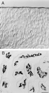
Figure 1
Figure 1
Immunoperoxidase localization of the 13A4 antigen (prominin) in the neuroepithelium and kidney tubules. Triton X-100-permeabilized cryosections (8 μm) of paraformaldehyde-fixed telencephalic neuroepithelium of a 12-day-old mouse embryo (A) and adult mouse kidney cortex (B) were stained with mAb 13A4 (1 μg/100 μl) followed by peroxidase-coupled goat anti-rat IgG/IgM and observed using Nomarski optics. In A, the apical and the basal side of the neuroepithelium are at the top and bottom, respectively. Immunoreactivity is confined to the apical, ventricular side. In B, most tubules are cross-sectioned. Immunoreactivity is restricted to their apical, lumenal side. (Bars = 20 μm.)
Figure 2
Figure 2
Immunoelectron microscopy (26) of the 13A4 antigen (prominin) on the apical surface of neuroepithelial cells and the brush border of kidney tubules. Ultrathin cryosections (26) of paraformaldehyde-fixed tissue were stained with mAb 13A4 (0.5–1 mg/ml) followed by rabbit anti-rat IgG/IgM and 9-nm protein A-gold. Neuroepithelial cells of 9-day-old (A and C) and 10-day-old (B and D) mouse embryos and proximal tubule cells of adult mouse kidney (E and F) are shown. In neuroepithelial cells (AD), immunoreactivity is confined to protrusions of the apical plasma membrane including microvilli-like structures. Junctional complexes are indicated by white arrowheads in C and D. In the kidney proximal tubule (E and F), immunoreactivity is restricted to the brush border where it appears to be concentrated at the tips of the microvilli. A tight junction is indicated by white arrowheads in E. (Bars = 0.5 μm.)
Figure 3
Figure 3
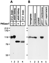
Glycosylation and membrane association of prominin. (A) Membranes (50 μg protein) prepared from either the brains of 12-day-old mouse embryos (E12) or adult mouse kidneys (40,000 × g, 30 min) were dissolved in SDS, incubated in the absence (−) or presence (+) of 0.8 units of PNGase F (Boehringer Mannheim) and analyzed by immunoblotting using mAb 13A4 (1 μg/ml) followed by the ECL system (Amersham). (B) A homogenate of adult mouse kidney was fractionated into a total membrane and cytosol fraction and the total membranes were treated with carbonate at pH 11 (27), yielding pH11 membranes and the pH11 supernatant. Corresponding aliquots of these fractions were analyzed by immunoblotting using mAb 13A4.
Figure 4
Figure 4
Amino acid sequence of mouse prominin deduced from the corresponding cDNA. Doubly underlined italic letters, putative signal peptide; doubly underlined plain letters, predicted membrane-spanning segments; bold letters, cysteine cluster and N-glycosylation sites.
Figure 5
Figure 5
Expression of prominin in CHO cells. CHO cells were stably transfected with either the expression vector containing the mouse prominin cDNA (CHO-PRO) or, as a control, vector DNA alone (lanes C). (A) Lysates from the CHO cells and, for comparison, from adult mouse kidney membranes were incubated in the absence (−) or presence of 10 milliunits endo H (H) or 1 unit PNGase F (F) (Boehringer Mannheim), and analyzed by immunoblotting with mAb 13A4. Arrowheads, endo H-resistant, PNGase F-sensitive form; arrow, endo H- and PNGase F-sensitive form; asterisks, product after N-deglycosylation. (B) The CHO cells were labeled for 2 h with [35S]sulfate, followed by immunoprecipitation of prominin with the antiserum against the C-terminal domain and incubation of the solubilized immunoprecipitate without (−) or with (F) 1 unit PNGase F. (C) The CHO cells were incubated for 1 h at 4°C without (−) or with (lanes B) sulfo-NHS-LC-biotin (27), solubilized, and biotinylated proteins adsorbed to streptavidin-Agarose followed by immunoblot analysis of the adsorbed material using the antiserum against the C-terminal domain (1:5000).
Figure 6
Figure 6
Intracellular localization of the C-terminal domain of prominin. CHO cells were stably transfected with the expression vector containing the mouse prominin cDNA. Intact cells at 4°C (27) (A and B) or paraformaldehyde-fixed, Triton X-100-permeabilized cells (C and D) were incubated with the mAb 13A4 (10 μg/ml) and the antiserum against the C-terminal domain (1:3000), followed by appropriate fluorescein- and rhodamine-conjugated secondary antibodies and double immunofluorescence analysis using confocal microscopy. Single optical sections at the level of the coverslip are shown. (Bars = 10 μm.)
Figure 7
Figure 7
Immunoelectron microscopy of prominin on the plasma membrane of transfected, paraformaldehyde-fixed CHO cells. Ultrathin cryosections were stained with mAb 13A4 followed by rabbit anti-rat IgG/IgM and 9-nm protein A-gold. The white asterisk in B indicates a filopodium extending from the cell shown in this panel. All panels are the same magnification. (Bar in F = 0.2 μm.)

Similar articles

Cited by

References

    1. Nelson W J. Science. 1992;258:948–955. - PubMed
    1. Rodriguez-Boulan E, Powell S K. Annu Rev Cell Biol. 1992;8:395–427. - PubMed
    1. Simons K, Dupree P, Fiedler K, Huber L A, Kobayashi T, Kurzchalia T, Olkkonen V, Pimplikar S, Parton R, Dotti C. Cold Spring Harbor Symp Quant Biol. 1992;57:611–619. - PubMed
    1. Eaton S, Simons K. Cell. 1995;82:5–8. - PubMed
    1. Mays R W, Nelson W J, Marrs J A. Cold Spring Harbor Symp Quant Biol. 1995;60:763–773. - PubMed

Publication types

MeSH terms

Associated data