

Biological characterization of Drosophila Rapgap1, a GTPase activating protein for Rap1
- PMID: 9356476
- PMCID: PMC25011
- DOI: 10.1073/pnas.94.23.12485
Biological characterization of Drosophila Rapgap1, a GTPase activating protein for Rap1
Abstract
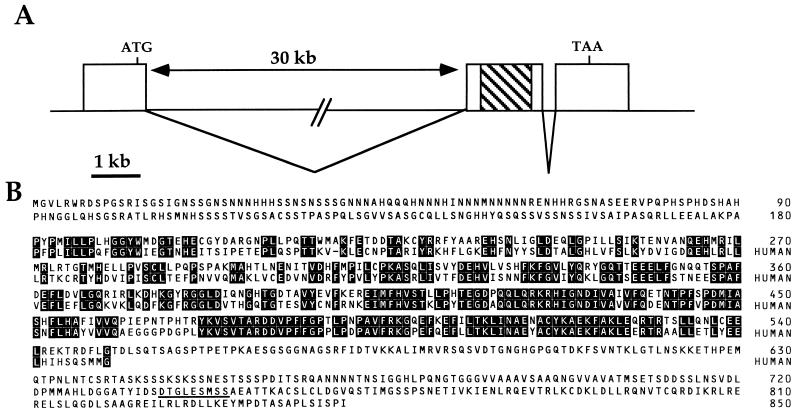
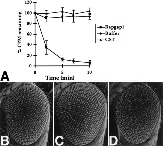
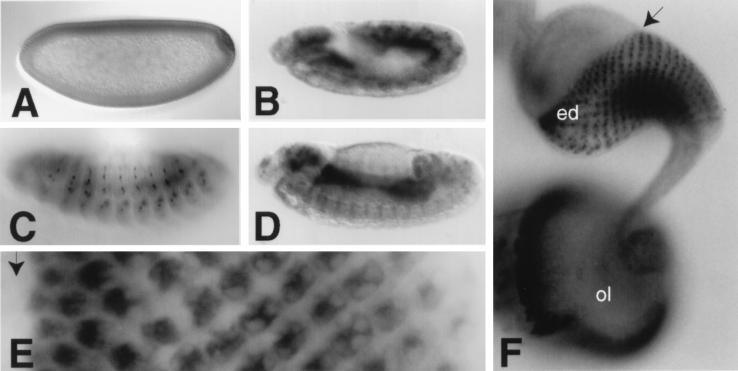
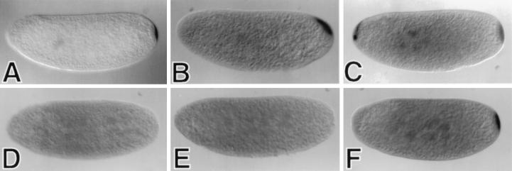
The activity of Ras family proteins is modulated in vivo by the function of GTPase activating proteins, which increase their intrinsic rate of GTP hydrolysis. We have isolated cDNAs encoding a GAP for the Drosophila Rap1 GTPase. Drosophila Rapgap1 encodes an 850-amino acid protein with a central region that displays substantial sequence similarity to human RapGAP. This domain, when expressed in Escherichia coli, potently stimulates Rap1 GTPase activity in vitro. Unlike Rap1, which is ubiquitously expressed, Rapgap1 expression is highly restricted. Rapgap1 is expressed at high levels in the developing photoreceptor cells and in the optic lobe. Rapgap1 mRNA is also localized in the pole plasm in an oskar-dependent manner. Although mutations that completely abolish Rapgap1 function display no obvious phenotypic abnormalities, overexpression of Rapgap1 induces a rough eye phenotype that is exacerbated by reducing Rap1 gene dosage. Thus, Rapgap1 can function as a negative regulator of Rap1-mediated signaling in vivo.
Figures
References
-
- Bourne H R, Sanders D A, McCormick F. Nature (London) 1990;348:125–132. - PubMed
-
- Trahey M, McCormick F. Science. 1987;238:542–545. - PubMed
-
- Vogel U S, Dixon R A F, Schaber M D, Diehl R E, Marshall M S, Scolnick E M, Sigal I S, Gibbs J B. Nature (London) 1988;335:90–93. - PubMed
-
- Yatani A, Okabe K, Polakis P, Halenbeck R, McCormick F, Brown A M. Cell. 1990;61:769–776. - PubMed
-
- Kitayama H, Sugimoto Y, Matsuzaki T, Ikawa Y, Noda M. Cell. 1989;56:77–84. - PubMed
Publication types
MeSH terms
Substances
Associated data
- Actions
LinkOut - more resources
Full Text Sources
Molecular Biology Databases
Miscellaneous
