. 1997 Nov 11;94(23):12497-502.
doi: 10.1073/pnas.94.23.12497.

The absence of effect of gid or mioC transcription on the initiation of chromosomal replication in Escherichia coli

Affiliations

The absence of effect of gid or mioC transcription on the initiation of chromosomal replication in Escherichia coli

D B Bates et al. Proc Natl Acad Sci U S A. .

Abstract

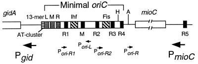
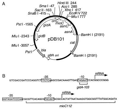
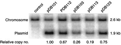
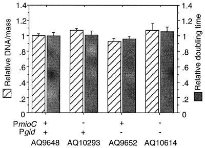
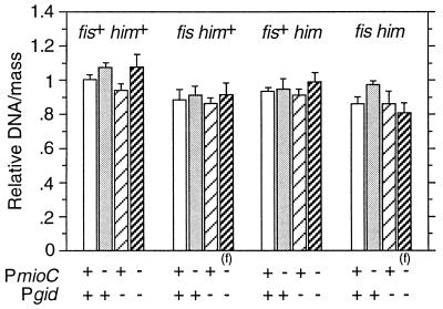
Despite the widely accepted view that transcription of gid and mioC is required for efficient initiation of cloned oriC, we show that these transcriptions have very little effect on initiation of chromosome replication at wild-type chromosomal oriC. Furthermore, neither gid nor mioC transcription is required in cells deficient in the histone-like proteins Fis or IHF. However, oriC that is sufficiently impaired for initiation by deletion of DnaA box R4 requires transcription of at least one of these genes. We conclude that transcription of mioC and especially gid is needed to activate oriC only under suboptimal conditions. We suggest that either the rifampicin-sensitive step of initiation is some other transcription occurring from promoter(s) within oriC, or the original inference of transcriptional activation derived from the rifampicin experiments is incorrect.


Figures

Figure 1
Figure 1
The minimal oriC and surrounding transcription. The position of the six DnaA boxes R1–R5 and M; 13-mer repeats L, M, and R; A+T-rich cluster; and binding sites for IHF and Fis proteins are indicated. Large arrows represent location and direction of major promoters near oriC. Small arrows represent weaker promoters within oriC. H, HindIII (+244); A, AccI (+285) (ref. and references therein).
Figure 2
Figure 2
Structure of oriC plasmid and promoter sequences. (A) The plasmid (pDB101) from which other oriC plasmids are derived is shown with relative restriction sites and genetic map (open arcs). Thick (solid) and thin arcs denote the oriC region and vector sequences, respectively. Shaded arcs denote antibiotic markers. All oriC coordinates are according to refs. and . (B) DNA sequences of the gid (7) and mioC (14) promoter regions with promoter mutations are shown. The −10 and −35 consensus homologies and transcriptional start sites are indicated.
Figure 3
Figure 3
Copy numbers of oriC plasmids carrying promoter mutations. polA1 cells (AQ2178) were transformed with the indicated modified oriC plasmids and grown in the presence of chloramphenicol (50 μg/ml). Total DNA was extracted from exponentially growing cells and digested with PstI and electrophoresed in a 0.8% agarose gel. DNA was blotted, and probed with a 714-bp MluI(−2343) to MluI(−3057) 32P-labeled oriC DNA fragment. Band intensities were quantified by using a PhosphorImager, and the plasmid-to-chromosome ratio relative to pDB101 is indicated under each lane. For relevant genotypes of plasmids, see Table 2. Control lane is untransformed AQ2178.
Figure 4
Figure 4
DNA synthesis and growth rates of promoter mutants. Cells were grown in LB plus glucose at 37°C with aeration. DNA/mass values were obtained from flow cytometric analysis of exponentially growing cells. Doubling times were determined from cell counts with a particle counter. Values are averages of three independent analyses plotted relative to AQ9648 (18.8-min doubling time). Error bars indicate the standard deviation.
Figure 5
Figure 5
DNA synthesis of fis::km himA::tet cells carrying promoter mutations. Cells were grown and DNA/mass values were obtained as in Fig. 4. An (f) indicates that these cells exhibit extreme filamentation during exponential growth. Values shown are averages of three independent analyses. Error bars indicate the standard deviation.
Figure 6
Figure 6
Lethal effect of expression of an rnhA+ gene on rnhA224 PmioC112 oriC207::bla Pgid-103 mutant. Cells were transformed with pRNH-Km (carries lacZ::rnhA gene fusion) or pHK (vector) and selected for kanamycin resistance. Transformants were then transformed with pLacIq, selecting for tetracycline resistance. Equal amounts of cells were spread on minimal plates with or without 5 mM IPTG as indicated. (A) rnhA224 PmioC112 Pgid-103/pHK, pLacIq. (B) rnhA224 PmioC112 Pgid-103/pRNH-Km, pLacIq. (C) rnhA224 oriC207::bla PmioC112 Pgid-103/pHK, pLacIq. (D) rnhA224 oriC207::bla PmioC112 Pgid-103/pRNH-Km, pLacIq.

References

    1. Messer W, Weigel C. In: Escherichia coli and Salmonella typhimurium: Cellular and Molecular Biology. Neidhardt F C, Curtiss R, Ingraham J L, Lin E C C, Low K B, Magasanik B, Reznikoff W S, Riley M, Schaechter M, Umbarger H E, editors. Washington, DC: Am. Soc. Microbiol.; 1996. pp. 1579–1601.
    1. Lark K G. J Mol Biol. 1972;64:47–60. - PubMed
    1. Messer W. J Bacteriol. 1972;112:7–12. - PMC - PubMed
    1. Rasmussen K V, Atlung T, Kerszman G, Hansen G E, Hansen F G. J Bacteriol. 1983;154:443–451. - PMC - PubMed
    1. Tanaka M, Ohmori H, Hiraga S. Mol Gen Genet. 1983;192:51–60. - PubMed

Publication types

LinkOut - more resources