. 1997 Nov 11;94(23):12551-6.
doi: 10.1073/pnas.94.23.12551.

Dendritic cell ontogeny: a human dendritic cell lineage of myeloid origin

Affiliations

Dendritic cell ontogeny: a human dendritic cell lineage of myeloid origin

J Olweus et al. Proc Natl Acad Sci U S A. .

Abstract

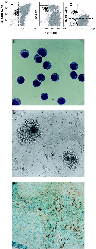
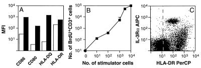
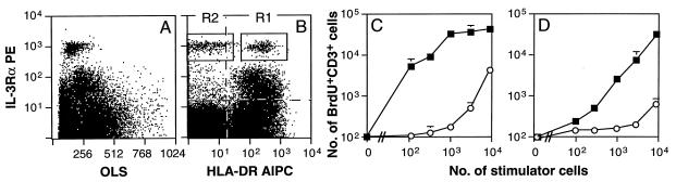
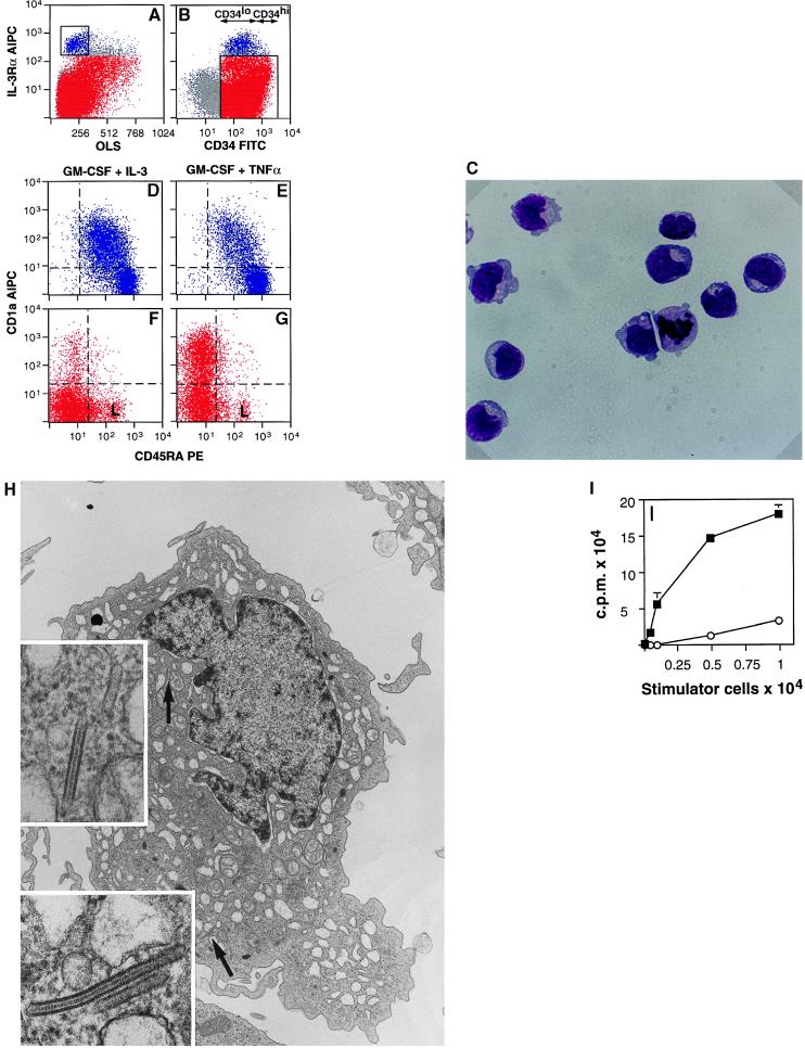
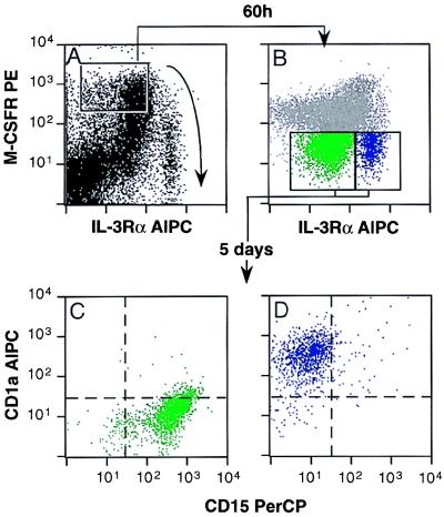
Dendritic cells (DC) have been thought to represent a family of closely related cells with similar functions and developmental pathways. The best-characterized precursors are the epidermal Langerhans cells, which migrate to lymphoid organs and become activated DC in response to inflammatory stimuli. Here, we demonstrate that a large subset of DC in the T cell-dependent areas of human lymphoid organs are nonactivated cells and belong to a separate lineage that can be identified by high levels of the interleukin 3 receptor alpha chain (IL-3Ralphahi). The CD34+IL-3Ralphahi DC progenitors are of myeloid origin and are distinct from those that give rise to Langerhans cells in vitro. The IL-3Ralphahi DC furthermore appear to migrate to lymphoid organs independently of inflammatory stimuli or foreign antigens. Thus, DC are heterogeneous with regard to function and ontogeny.


Figures

Figure 1
Figure 1
Antibodies to IL-3Rα selectively stain DC in extrafollicular regions of human tonsils. (AC) Tonsillar mononuclear cells were stained with anti-HLA-DR peridinin-chlorophyll protein (PerCP), CD4 PE, anti-IL-3Rα biotin + Streptavidin allo-phycocyanin (ALPC), and a mixture of FITC-conjugated lineage markers (“lin”) for lymphocytes and monocytes (CD3, CD14, CD16, CD19, CD20, CD56, and goat-anti-human IgM). The cells were analyzed by four-color flow cytometry. Dendritic cells were identified as HLA-DR+CD4+lin—i.e., cells that simultaneously satisfy the criteria of the box regions in A and B, and are represented by large black dots. Dashed lines represent isotype control levels. (D) Wright–Giemsa staining of a cytocentrifuge slide of freshly FACS-sorted tonsillar HLA-DR+linIL-3Rαhi cells. (×600.) (E) FACS-sorted tonsillar HLA-DR+linIL-3Rαhi cells were cultured for 24 h with GM-CSF and IL-3 and photographed in situ. (×400.) (F) A frozen section of tonsillar tissue was stained with anti-IL-3Rα, biotinylated anti-mouse IgG and streptavidin peroxidase. (×100). Staining was visualized by diaminobenzidine and hydrogen-peroxide, and the section was counterstained with methylene blue.
Figure 2
Figure 2
IL-3Rαhi DC are immature and appear in lymphoid organs independently of stimuli that cause up-regulation of major histocompatibility complex class II and costimulatory molecules. (A) Mononuclear cells from tonsil were stained with anti-HLA-DR, anti-IL-3Rα and lineage markers, and either CD86, CD80, HLA-DQ, or isotype control mAbs before (open bars) and after (filled bars) a 16-h incubation at 37°C in Yssel’s medium (30). The HLA-DR+linIL-3Rαhi population was analyzed for mean fluorescence intensity (MFI) staining with the markers indicated on the figure, and the bars represent MFI after isotype control levels were subtracted. (B) T cells (105) were cocultured with indicated numbers of allogeneic IL-3RαhilinHLA-DR+ cells from tonsil (stimulator cells). T cell proliferation was measured as the total number of CD3+ BrdU+ cells per well at day 6 of coculture. (C) Fetal lymph node cells were stained with anti-HLA-DR, anti-IL-3Rα, and lineage markers (data not shown) and analyzed by flow cytometry as described in Fig. 1 AC. Data are representative of three experiments.
Figure 3
Figure 3
HLA-DR+linIL-3Rαhi DC are present in peripheral blood. (A and B) PBMC were stained with anti-HLA-DR, anti-IL-3Rα, and lineage markers (not shown) and analyzed by flow cytometry as described in Fig. 1 AC. R1 and R2 in B represent regions used to sort IL-3Rαhi blood cells that were positive or negative for HLA-DR, respectively. (C and D) HLA-DR+linIL-3Rαhi cells sorted from blood according to R1 in B were first cultured separately with IL-3 and GM-CSF for 36 h and then incubated with allogeneic (C) or autologous (D) CD4+ T cells. T cell proliferation was measured as total number of CD3+BrdU+ cells per well at day 6 of coculture with indicated numbers of IL-3RαhilinHLA-DR+ cells (▪) or CD14hi monocytes (○) from the same donor (stimulator cells). Individual displays show data that are representative of three experiments. OLS, ortogonal light scatter.
Figure 4
Figure 4
Proliferating progenitors for IL-3Rαhi DC are found as a discrete CD34+IL-3Rαhi population that is distinct from the cells that give rise to Langerhans cells in response to GM-CSF and TNF-α. (A and B) Isolated CD34+ fetal bone marrow cells were stained with CD34 and anti-IL-3Rα. The cells were analyzed by flow cytometry as described in Fig. 1 AC. The CD34+IL-3Rαhi population was defined according to the region in A (blue dots). CD34+IL-3Rαlo cells were defined according to the region in B (red dots). OLS, ortogonal light scatter. (C) Freshly sorted Wright–Giemsa-stained CD34+IL-3Rαhi cells display mitotic figures. (×600.) (DG) CD34+IL-3Rαhi cells (blue) and CD34+IL-3Rαlo cells (red) were sorted according to the regions in A and B, respectively, and cultured in the presence of indicated cytokines, stained with CD1a and CD45RA after 5 days of culture, and analyzed by FACS. L = B lymphoid cells staining brightly with CD19 (data not shown). None of the cultured CD34+IL-3Rαhi cells (D and E) stained positively with CD19 (data not shown). Dashed lines indicate isotype control levels. (H) Transmission electron microscograph (×5,000) of a CD1a+ cell sorted from CD34+IL-3Rαlo cells cultured with GM-CSF and TNF-α, as described in G. The arrows point to areas containing Birbeck granules, (shown in Insets, ×45,000). (I) CD4+ T cells (105) were cocultured with indicated numbers of allogeneic DC (▪) or macrophages (○) from the same donor. The DC were generated by culturing sorted CD34+IL-3Rαhi cells for 5 days with GM-CSF and IL-3 (▪). The macrophages were generated by culture of CD34+M-CSFR+ cells for 5 days with M-CSF and purified by FACS sorting of CD14+ cells. T cell proliferation was measured as incorporation of [3H]thymidine at day 6 of coculture. c.p.m; counts per minute. Data are representative of three experiments.
Figure 5
Figure 5
IL-3Rαhi DC follow the myeloid differentiation pathway to the branching point of the granulocytic and monocytic lineages. (A) CD34+ cells were incubated for 12 h in serum-free medium (25) to allow up-regulation of the M-CSFR, and stained with CD34, anti-M-CSFR, and anti-IL-3Rα. The arrow shows the suggested differentiation pathway of CD34+IL-3Rαhi cells. The region shows sorting criteria for M-CSFRhi/IL-3Rαlo cells. An additional gate was set to include only CD34hi cells, shown in Fig. 4B, to restrict the sort to immature progenitors (29). (B) After a 60-h culture of CD34hiM-CSFR+ cells in serum-free medium (25) containing stem cell factor, G-CSF, GM-CSF, IL-3, and IL-6, the cells were stained with anti-M-CSFR and anti-IL-3Rα. The regions indicate criteria for sorting of the two populations that had downmodulated the M-CSFR during the culture period. (C and D) After a 5-day secondary culture of IL-3RαloM-CSFRlo cells (green) and IL-3RαhiM-CSFRlo cells (blue) with IL-3 and GM-CSF, the cells were stained with CD1a and CD15 and analyzed by FACS. The small subset of CD15lo cells in C represent basophilic granulocytes (27, 29).

References

    1. Steinman R M. Annu Rev Immunol. 1991;9:271–296. - PubMed
    1. Schuler G, Steinman R. J Exp Med. 1985;161:526–546. - PMC - PubMed
    1. Silberberg-Sinakin I, Thorbecke J, Baer R, Rosenthal S, Berezowsky V. Cell Immunol. 1976;25:137–151. - PubMed
    1. Macatonia S, Knight S, Edwards A, Griffiths S, Fryer P. J Exp Med. 1987;166:1654–1667. - PMC - PubMed
    1. Kripke M, Munn C, Jeevan A, Tang J, Bucana C. J Immunol. 1990;145:2833–2838. - PubMed

Publication types

LinkOut - more resources