. 1997 Nov 11;94(23):12580-5.
doi: 10.1073/pnas.94.23.12580.

The affected gene underlying the class K glycosylphosphatidylinositol (GPI) surface protein defect codes for the GPI transamidase

Affiliations

The affected gene underlying the class K glycosylphosphatidylinositol (GPI) surface protein defect codes for the GPI transamidase

J Yu et al. Proc Natl Acad Sci U S A. .

Abstract

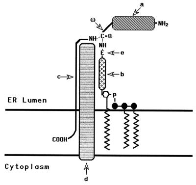
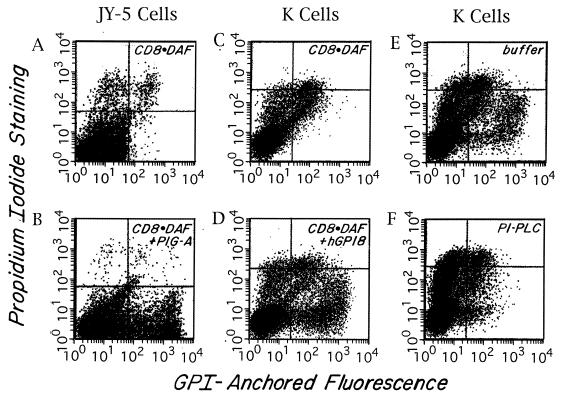
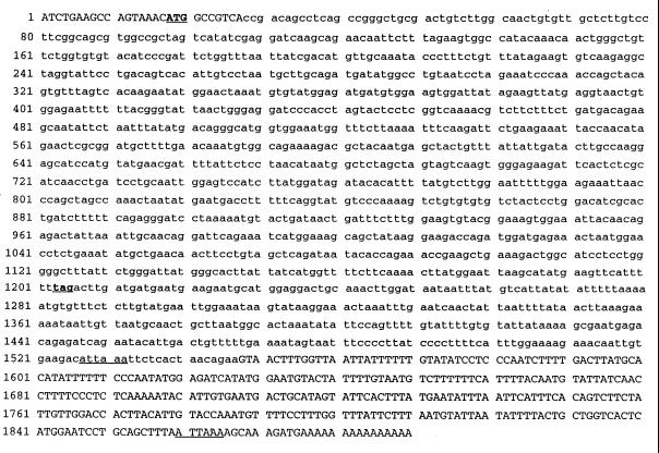
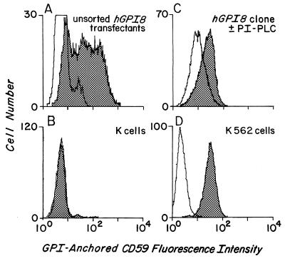
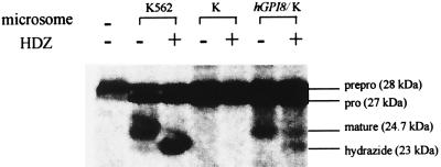
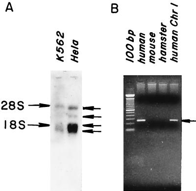
The final step in glycosylphosphatidylinositol (GPI) anchoring of cell surface proteins consists of a transamidation reaction in which preassembled GPI donors are substituted for C-terminal signal sequences in nascent polypeptides. In previous studies we described a human K562 cell mutant, termed class K, that accumulates fully assembled GPI units but is unable to transfer them to N-terminally processed proproteins. In further work we showed that, unlike wild-type microsomes, microsomes from these cells are unable to support C-terminal interaction of proproteins with the small nucleophiles hydrazine or hydroxylamine, and that the cells thus are defective in transamidation. In this study, using a modified recombinant vaccinia transient transfection system in conjunction with a composite cDNA prepared by 5' extension of an existing GenBank sequence, we found that the genetic element affected in these cells corresponds to the human homolog of yGPI8, a gene affected in a yeast mutant strain exhibiting similar accumulation of GPI donors without transfer. hGPI8 gives rise to mRNAs of 1.6 and 1.9 kb, both encoding a protein of 395 amino acids that varies in cells with their ability to couple GPIs to proteins. The gene spans approximately 25 kb of DNA on chromosome 1. Reconstitution of class K cells with hGPI8 abolishes their accumulation of GPI precursors and restores C-terminal processing of GPI-anchored proteins. Also, hGPI8 restores the ability of microsomes from the mutant cells to yield an active carbonyl in the presence of a proprotein which is considered to be an intermediate in catalysis by a transamidase.


Figures

Figure 1
Figure 1
Schematic representation of the GPI transfer reaction. The GPI transamidase catalyzes substitution of the GPI donor for the C-terminal peptide at the ω site in the N-terminally processed proprotein. a, Proprotein; ω, site of cleavage and GPI addition to the proprotein; b, carbohydrate portion of the GPI donor; c, C-terminal signal peptide; d, transamidase; e, terminal ethanolamine group of the GPI.
Figure 2
Figure 2
Rescue of GPI anchoring as assessed in the Vaccinia transient transfection system. Two-color FACS (Becton Dickinson) analyses of anti-CD8 and propidium iodide-stained cells. FL1 (x axis) = CD8 fluorescence intensity; FL2 (y axis) = propidium iodide fluorescence. Cells in the upper quadrants are propidium iodide-positive dead cells. Cells in the lower right quadrant are CD8+ and propidium iodide-negative rescued cells. The small number of cells in the lower right quadrant seen with CD8⋅DAF alone also stained with nonrelevant antibody. (A and B) JY-5 cells transfected with CD8⋅DAF alone or CD8⋅DAF and PIG-A cDNA. (C and D) K cells transfected with CD8⋅DAF alone or CD8⋅DAF and hGPI8. (E and F) CD8⋅DAF and hGPI8 transfected transients treated with buffer or PI-PLC. About 70% of the CD8+ cells were cleaved.
Figure 3
Figure 3
Cloning of hGPI8 sequence. The accession uppercase sequence is the newly cloned and corrected sequence and the lowercase sequence was PCR cloned from a GenBank sequence (accession no. Y07596). The bold, underlined, capitalized ATG gives the start codon and the bold, underlined, lowercase tag gives the stop codon. The underlines show the alternative polyadenylation signals.
Figure 4
Figure 4
Stable hGPI8/K cell transfectants. The x axis shows CD59 fluorescence, and the y axis shows cell number. (A) Unsorted Neo-resistant stable transfectants. (B) Nontransfected mutant K cells. (C) Cloned transfectant treated with buffer (shaded) or PI-PLC (unshaded) prior to staining. (D) Wild-type K562 cells. Shaded areas represent staining with anti-CD59 mAb and, except for C, unshaded areas represent staining with nonrelevant RPC5 control mAb.
Figure 5
Figure 5
Effect of hGPI8 on C-terminal processing of miniPLAP. Cotranslational processing of miniPLAP mRNA by microsomes of parental K562, untransfected K, and hGPI8/K transfectants in the absence and presence of HDZ. The microsome preparations were made at different times and fewer microsomes (35 vs. 50 OD) were used in the case of hGPI8/K transfectants. The positions of 28-kDa preprominiPLAP, 27-kDa prominiPLAP, 24.7-kDa GPI-anchored miniPLAP, and the 23.0-kDa miniPLAP hydrazide are shown.
Figure 6
Figure 6
Northern blot analyses and chromosomal localization of hGPI8. (A) Poly(A)+ mRNA probed with 1.2-kb coding segment of hGPI8. (B) DNA from chromosome-specific mouse– or hamster–human hybrids probed with a 257-bp 3′ untranslated region hGPI8 segment.

Similar articles

Cited by

References

    1. Englund P T. Annu Rev Biochem. 1993;62:121–138. - PubMed
    1. Stevens V L. Biochem J. 1995;310:361–370. - PMC - PubMed
    1. Gerber L D, Kodukula K, Udenfriend S. J Biol Chem. 1992;267:12168–12173. - PubMed
    1. Moran P, Raab H, Kohr W J, Caras I W. J Biol Chem. 1991;266:1250–1257. - PubMed
    1. Maxwell S E, Ramalingam S, Gerber L D, Udenfriend S. Proc Natl Acad Sci USA. 1995;92:1550–1554. - PMC - PubMed

Publication types

Substances

Associated data

LinkOut - more resources