. 1997 Nov 11;94(23):12661-6.
doi: 10.1073/pnas.94.23.12661.

Localization of neuropeptide Y Y1 receptors in cerebral blood vessels

Affiliations

Localization of neuropeptide Y Y1 receptors in cerebral blood vessels

L Bao et al. Proc Natl Acad Sci U S A. .

Abstract

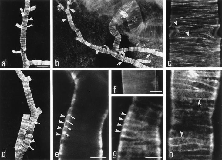
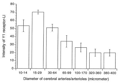
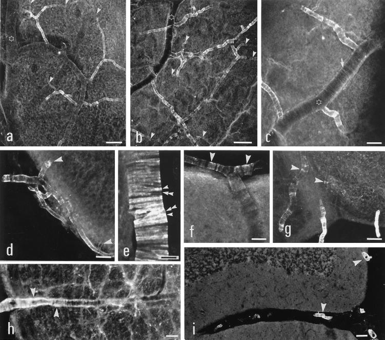
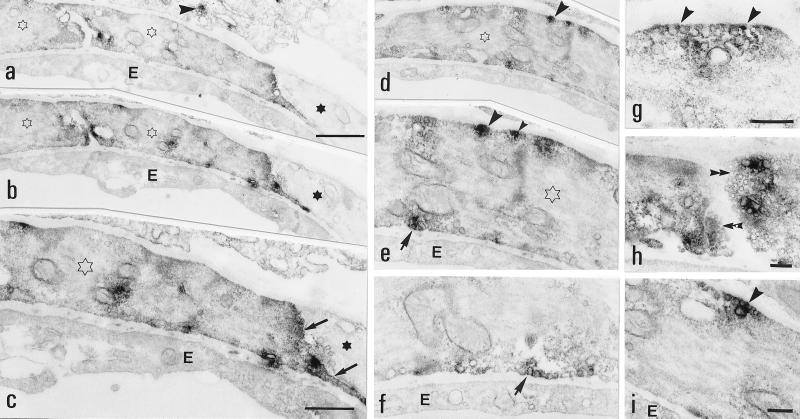
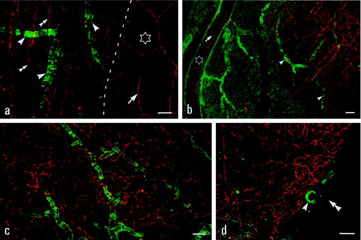
The localization of neuropeptide Y (NPY) Y1 receptor (R) -like immunoreactivity (LI) has been studied in cerebral arteries and arterioles of the rat by immunohistochemistry using fluorescence, confocal, and electron microscopy. High levels of Y1-R-LI were observed in smooth muscle cells (SMCs) in the small arterioles of the pial arterial network, especially on the basal surface of the brain, and low levels in the major basal cerebral arteries. The levels of Y1-R-LI varied strongly between adjacent SMCs. Y1-R-LI was associated with small endocytosis vesicles, mainly on the outer surface of the SMCs, but also on their endothelial side and often laterally at the interface between two SMCs. NPY-immunoreactive (Ir) nerve fibers could not be detected in association with the Y1-R-rich small arterioles but only around arteries with low Y1-R levels. A dense network of central NPY-Ir nerve fibers in the superficial layers of the brain was lying close to the strongly Y1-R-Ir small arterioles. The results indicate that NPY has a profound effect on small arterioles of the brain acting on Y1-Rs, both on the peripheral and luminal side of the SMCs. However, the source of the endogenous ligand, NPY, remains unclear. NPY released from central neurons may play a role, in addition to blood-borne NPY.


Figures

Figure 1
Figure 1
Immunofluorescence (a–d and f) and confocal (e, g, and h) micrographs of arterioles and arteries isolated from the basal surface of rat brain. (a, b, and d) Many strong Y1-R+, circularly oriented SMCs (arrowheads) are seen along the small arteries with intermingled Y1-R-negative cells (double arrowheads). The number of Y1-R-Ir SMCs is high and the intensity of labeling is relatively stronger along the small branches of the first branch of the middle cerebral artery (star). (c) Weak Y1-R-LI is observed in some circularly oriented SMCs (arrowheads) of the basilar artery. (f) Incubation with pre-absorbed antibody completely abolishes immunostaining. (e, g, and h) Confocal micrographs of Y1-R-LI in SMCs in isolated arteriole scanned at different levels. Y1-R-LI is seen on the perivascular side (arrowheads) with the highest intensity along the lateral portion (arrows) of SMCs (e). The pattern of labeling extends along the surface of SMCs (arrowheads) (g). Y1-R-LI is seen on the plasmalemma of entire SMCs (arrowheads) (h). [Bars = 100 μm (a–d, f) and 5 μm (e, g, and h).]
Figure 2
Figure 2
Histogram plotting the intensity of Y1-R-LI in basal arteries and arterioles against vessel diameter.
Figure 3
Figure 3
Confocal (a–g) and immunofluorescence (h and i) micrographs of basal (a–e, g and h), dorsal (f), and cerebellar (i) vessels. (a and b) Strong Y1-R-LI is located on the small arterioles given off by second- (stars) or third-order branches of the middle cerebral artery. Y1-R-LI decreases in the smallest arterioles (arrowheads). (c) Weak Y1-R-LI in SMCs (arrow) on a second branch artery (stars) of the middle cerebral artery. (d) Y1-R-LI disappears when the arteriole has entered hypothalamus (arrowheads). (e) Strongly Y1-R-Ir SMCs (arrowheads) with interspersed negative cells (double arrowheads) in an arteriole. (f) Weak Y1-R+ SMCs (arrowheads) in an arteriole on the dorsal cerebral cortex. (g) Y1-R-LI is seen within the brain along arterioles (arrowheads) around the interpeduncular nucleus. (h) Y1-R-LI (arrowheads) is present on an arteriole at some distance from the medullary surface. (i) Small arterioles (arrowheads) on the cerebellar surface are Y1-R+. [Bars = 100 μm (a, and b), 50 μm (d, g, and i) and 25 μm (c, e, f, and h).]
Figure 4
Figure 4
Electron micrographs of arterioles. (a–c) A Y1-R+ (open stars) and a Y1-R-negative (stars) SMC in two adjacent sections. The Y1-R-LI is distributed in the cytoplasm and more intensely at lateral portions (arrows) of the SMC. The scattered labeling (arrowhead) in the cell peripheral to the SMC is unspecific. (c) Higher magnification from b. (d–i) Several intensely Y1-R-Ir sites are seen along the plasmalemma facing the perivascular space (arrowheads), the endothelium (arrows), or laterally toward another SMC (double arrowheads). Note close association with vesicles in many cases. E, endothelium. [Bars = 1 μm (a, b, and d), 500 nm (c and e), 250 nm (f and i; g and h).]
Figure 5
Figure 5
Confocal micrographs showing Y1-R (green) and NPY (red) -LIs. (a) NPY+ nerve fibers (arrows) but no Y1-R-LI are seen around the inferior anterior cerebral artery (stars). Strong Y1-R-LI around some small arterioles directly derived from the basilar artery, but no NPY+ nerve fibers are seen along these arterioles. Double arrowheads pointing to central NPY+ nerve fibers. (b) Strongly Y1-R-Ir arterioles lacking NPY+ nerve fibers (arrowheads). NPY+ fibers are seen around a first branch (stars) of the middle cerebral artery. (c) A dense network of central NPY+ nerve fibers intermingles with strong Y1-R+ arterioles (basal cortex). (d) A Y1-R+ superficial arteriole (arrowheads) adjacent to NPY+ nerve fibers in the basal hypothalamus. The pia mater (double arrowheads) is not labeled. [Bars = 25 μm (a, b), 50 μm (c), and 100 μm (d).]

Similar articles

Cited by

References

    1. Nielsen K C, Owman C. Brain Res. 1967;6:773–776. - PubMed
    1. Edvinsson L, MacKenzie E T, McCulloch J. Cerebral Blood Flow and Metabolism. New York: Raven; 1993.
    1. Tatemoto K, Carlquist M, Mutt V. Nature (London) 1982;296:659–660. - PubMed
    1. Lundberg J M, Terenius L, Hökfelt T, Goldstein M. Neurosci Lett. 1983;42:167–172. - PubMed
    1. Edvinsson L, Emson P, McCulloch J, Tatemoto K, Uddman R. Neurosci Lett. 1983;43:79–84. - PubMed

Publication types