. 1997 Nov;8(11):2241-51.
doi: 10.1091/mbc.8.11.2241.

Galectin-4 and small intestinal brush border enzymes form clusters

Affiliations
Free PMC article

Galectin-4 and small intestinal brush border enzymes form clusters

E M Danielsen et al. Mol Biol Cell. 1997 Nov.
Free PMC article

Abstract

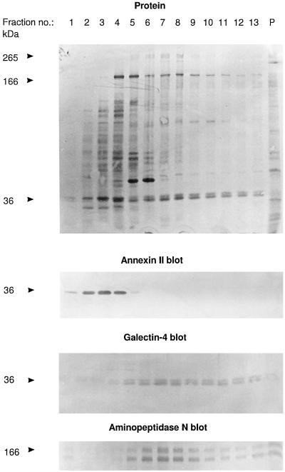
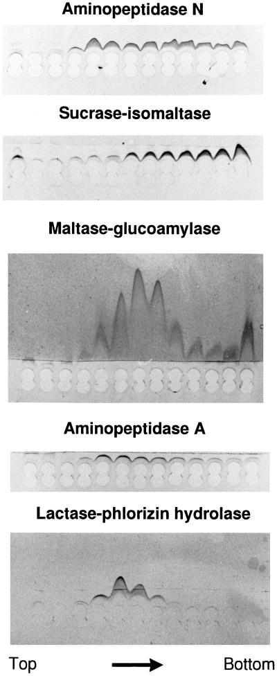
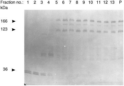
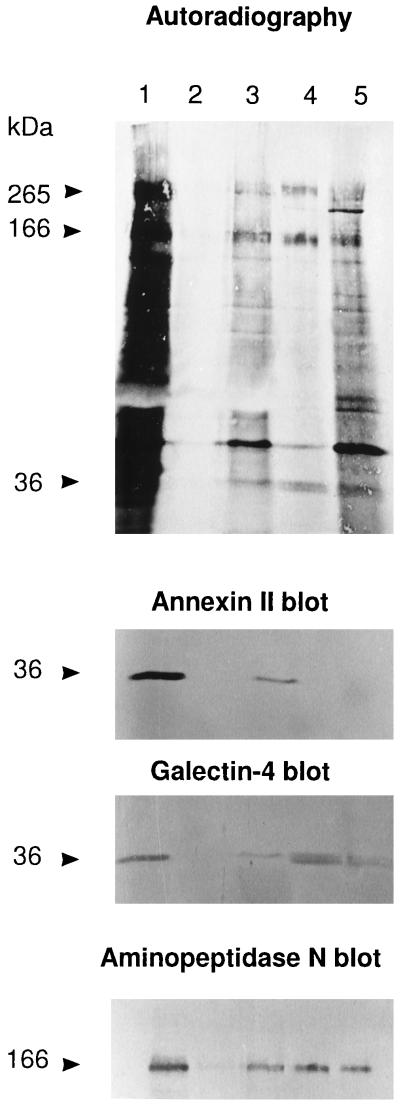
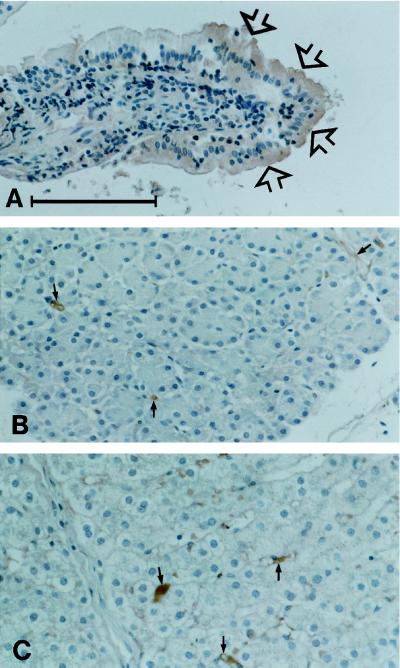
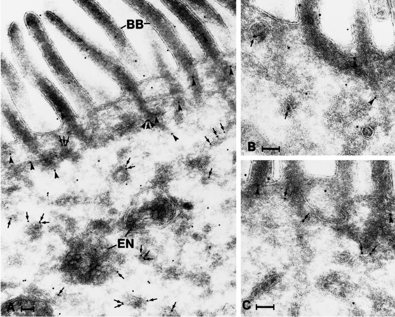
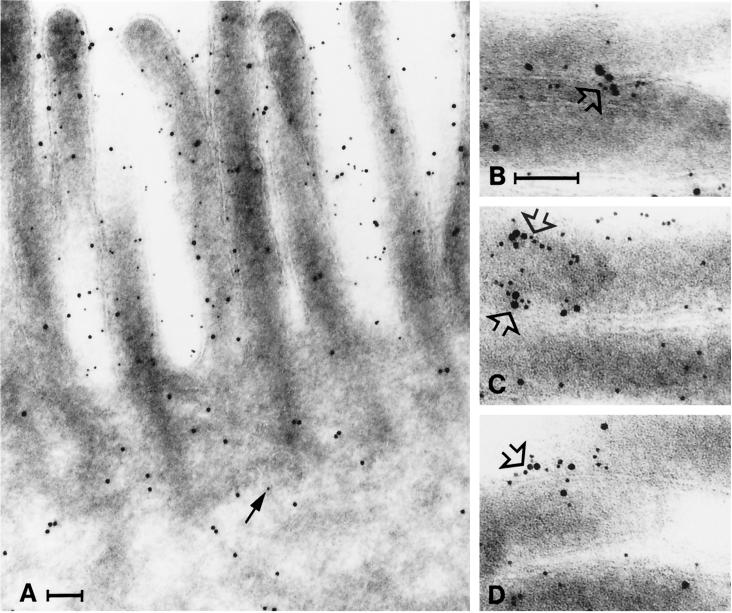
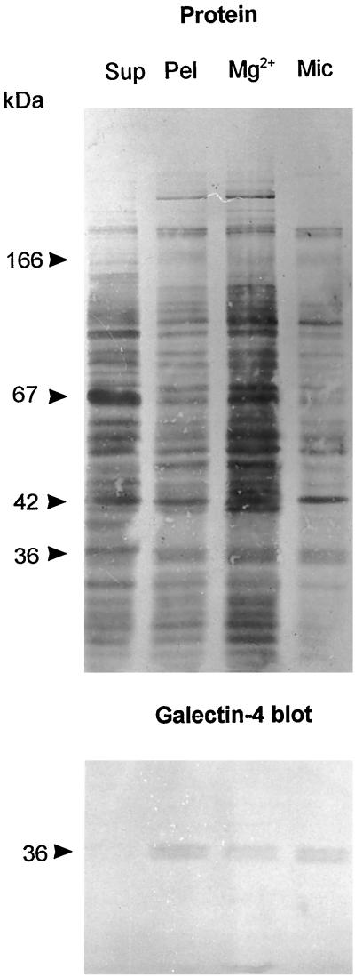
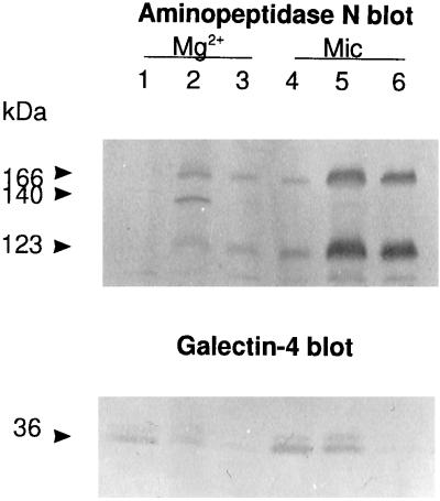
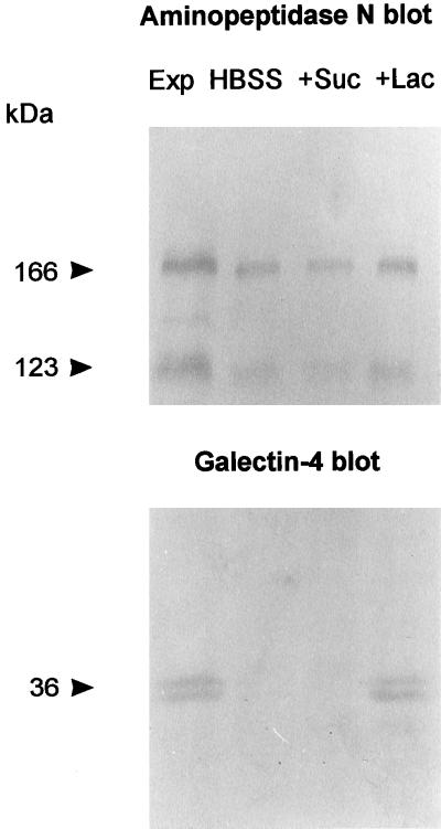
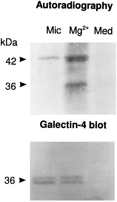
Detergent-insoluble complexes prepared from pig small intestine are highly enriched in several transmembrane brush border enzymes including aminopeptidase N and sucrase-isomaltase, indicating that they reside in a glycolipid-rich environment in vivo. In the present work galectin-4, an animal lectin lacking a N-terminal signal peptide for membrane translocation, was discovered in these complexes as well, and in gradient centrifugation brush border enzymes and galectin-4 formed distinct soluble high molecular weight clusters. Immunoperoxidase cytochemistry and immunogold electron microscopy showed that galectin-4 is indeed an intestinal brush border protein; we also localized galectin-4 throughout the cell, mainly associated with membraneous structures, including small vesicles, and to the rootlets of microvillar actin filaments. This was confirmed by subcellular fractionation, showing about half the amount of galectin-4 to be in the microvillar fraction, the rest being associated with insoluble intracellular structures. A direct association between the lectin and aminopeptidase N was evidenced by a colocalization along microvilli in double immunogold labeling and by the ability of an antibody to galectin-4 to coimmunoprecipitate aminopeptidase N and sucrase-isomaltase. Furthermore, galectin-4 was released from microvillar, right-side-out vesicles as well as from mucosal explants by a brief wash with 100 mM lactose, confirming its extracellular localization. Galectin-4 is therefore secreted by a nonclassical pathway, and the brush border enzymes represent a novel class of natural ligands for a member of the galectin family. Newly synthesized galectin-4 is rapidly "trapped" by association with intracellular structures prior to its apical secretion, but once externalized, association with brush border enzymes prevents it from being released from the enterocyte into the intestinal lumen.


Figures

Figure 1
Figure 1
Identification of galectin-4 as a 36-kDa protein present in high molecular weight clusters. Intestinal mucosa was homogenized in a Potter-Elvehjem homogenizer in ice-cold 25 mM HEPES and 150 mM NaCl, pH 7.0, containing 10 μg/ml aprotinin and 10 μg/ml leupeptin, and centrifuged at 500 × g for 5 min. The supernatant was centrifuged at 48,000 × g for 30 min. The resulting pellet of total membranes was resuspended in the above buffer and solubilized by extraction at 37°C for 10 min with 20 mM CHAPS and 1 mM EDTA. One milliliter of the extract was analyzed by velocity sedimentation in a sucrose gradient as described in MATERIALS AND METHODS. After centrifugation, 0.25 ml of each fraction was mixed with an equal volume of acetone, and after 15 min on ice, protein was pelleted by centrifugation at 20,000 × g for 10 min and analyzed by SDS-PAGE and Western blotting, using the primary antibodies indicated. Lanes 1 and 13 represent the top and bottom fractions of the gradient, respectively, and lane P shows the proteins recovered from the pellet of the centrifugation. Total protein was stained with Coomassie brilliant blue. Both the 166-kDa band of aminopeptidase N and its B-subunit, which is formed by proteolytic cleavage in vivo (Sjöström et al., 1978) are shown at the bottom. Molecular mass values are indicated.
Figure 2
Figure 2
Brush border enzymes in high molecular weight clusters. Rocket immunoelectrophoresis against antibodies to aminopeptidase N, sucrase-isomaltase, maltase-glucoamylase, aminopeptidase A, and lactase-phlorizin hydrolase of gradient fractions from top to bottom of the experiment shown in Figure 1. Twenty microliters of each fraction was applied to the wells and after electrophoresis, the immunoprecipitates were visualized by staining with Coomassie brilliant blue.
Figure 3
Figure 3
Lactose releases galectin-4 from high molecular weight clusters. An experiment similar to that shown in Figure 1 except that the membranes were detergent extracted in the presence of 100 mM lactose and that lactose (10 mM) was present in the sucrose gradient. After SDS-PAGE and electrotransfer onto Immobilon, the bands of galectin-4 and aminopeptidase N were visualized by Western blotting. Molecular mass values are indicated.
Figure 4
Figure 4
Galectin-4 is a major component of glycolipid microdomains. SDS-PAGE of Triton X-100-insoluble complexes prepared from a microvillar fraction. After electrophoresis and electrotransfer onto an Immobilon membrane, the gel tracks were either stained for protein with Coomassie brilliant blue (lane 1) or Western blotted, using a monoclonal antibody to annexin II (lane 2) or a polyclonal antibody to galectin-4 prepared as described in MATERIALS AND METHODS (lane 3). Notice that the galectin-4 antibody only reacted with the 36-kDa band and not the 265- and 166-kDa bands of sucrase-isomaltase and aminopeptidase N, respectively. Molecular mass values are indicated.
Figure 5
Figure 5
Copurification of galectin-4 and brush border enzymes. A mucosal explant, labeled for 2 h with 0.5 mCi/ml [35S]methionine, was homogenized in a Potter-Elvehjem homogenizer in 1 ml of 25 mM HEPES and 150 mM NaCl, pH 7.0, containing 10 μg/ml aprotinin and 10 μg/ml leupeptin, and centrifuged at 500 × g for 5 min. The supernatant was centrifuged for 48,000 × g for 30 min. The resulting pellet of total membranes was resuspended in 0.25 ml of HEPES buffer and solubilized for 10 min on ice by the addition of CHAPS (20 mM). The extract was centrifuged at 48,000 × g for 30 min to obtain a supernant of low temperature detergent-soluble protein and a pellet. The pellet was resuspended in 0.25 ml HEPES buffer and solubilized for 10 min at 37°C by 20 mM CHAPS and 1 mM EDTA. The extract was centrifuged at 48,000 × g for 30 min to obtain a supernatant of (solubilized) low-temperature detergent-insoluble protein and a pellet. Galectin-4 antibody (0.2 ml) was added to both the extracts of low-temperature detergent-soluble and low-temperature detergent-insoluble proteins, and after incubation at 4°C overnight, the extracts were centrifuged at 5000 × g for 5 min to pellet the immunoprecipitates. The immunoprecipitates of both the low-temperature detergent-soluble (lane 2) and low-temperature detergent-insoluble (lane 4) fractions were washed in buffer once and analyzed by SDS-PAGE together with 100 μl of the corresponding extract supernatants left after immunoprecipitation (lanes 1 and 3, respectively) and the pellet of detergent-insoluble material (lane 5). After electrophoresis and electrotransfer onto an Immobilon membrane, the gel tracks were visualized by autoradiography, and then Western blotted in three successive steps using the indicated primary antibodies. (The single sharp 36-kDa band seen in lanes 1 and 3 of the galectin-4 blot is residual staining of annexin II from the previous round of blotting because they were not observed in single blot experiments using the galectin-4 antibody alone). Molecular mass values are indicated.
Figure 6
Figure 6
Distribution of galectin-4 in a pig small intestinal villus (A), pancreas (B), and liver (C). Note that the enterocytes, in particular those at the tip of the villus (arrows in A) are clearly labeled. In the two other tissues the epithelial paranchyma is unstained although some immunoperoxidase staining may be seen in relation to stromal elements (small arrows). Bar, 100 μm.
Figure 7
Figure 7
Distribution of galectin-4 in the apical portion of enterocytes as revealed by immunogold labeling. Gold particles are seen throughout the cytoplasm, in association with tubulo-vesicular structures (arrows) and the actin rootlets of microvilli (arrowheads). Note that many of the gold particles associated with tubulo-vesicular structures are found on their cytosolic surface. BB, brush border; En, endosomes. Bar, 100 nm.
Figure 8
Figure 8
Clustering of galectin-4 and aminopeptidase N. Immunogold double labeling for aminopeptidase N (5-nm gold) and galectin-4 (10-nm gold). (A) Aminopeptidase N and galectin-4 are present in the brush border, but little aminopeptidase N is seen in the cytoplasm (arrow). (B–D) Galectin-4 and aminopeptidase N often form clusters in the microvillar membrane (open arrows). Bars, 100 nm.
Figure 9
Figure 9
Subcellular distribution of galectin-4. About 1 g of frozen intestinal mucosa was thawed and homogenized in a Potter-Elvehjem homogenizer in 10 ml of 25 mM HEPES and 150 mM NaCl, pH 7.0. The homogenate was cleared by centrifugation at 500 × g for 5 min and then centrifuged at 48,000 × g for 30 min, to obtain a pellet (Pel) and a supernatant (Sup). The pellet was resuspended in 10 ml of the above buffer and 50 μl of this fraction and of the supernatant was analyzed by SDS-PAGE. Likewise, from 1 g of intestinal mucosa, Mg2+-precipitated (Mg2+) and microvillar (Mic) fractions were prepared and resuspended in equal volumes of 25 mM HEPES and 150 mM NaCl, pH 7.0, and samples of 50 μl were examined by SDS-PAGE. After electrophoresis, the gel tracks were Western blotted with the galectin-4 antibody and afterward stained for protein with Coomassie brilliant blue.
Figure 10
Figure 10
Galectin-4 is present on the extracellular side of mivrovillar vesicles. Mg2+-precipitated (Mg2+) and microvillar (Mic) membranes were resuspended in 25 mM HEPES and 150 mM NaCl, pH 7.0, containing 100 mM lactose and incubated for 10 min on ice. After centrifugation at 48,000 × g for 30 min, the pellets were collected, resuspended in the same buffer containing 100 mM lactose and 20 mM CHAPS, and incubated for 10 min at 37°C before centrifugation again as described above. The lactose-extracted fractions (lanes 1 and 4), lactose + CHAPS-extracted fractions (lanes 2 and 5), and the insoluble fractions (lanes 3 and 6) were analyzed by SDS-PAGE, followed by Western blotting using antibodies to aminopeptidase N (top) or galectin-4 (bottom). (The 140-kDa band is the transient high-mannose glycosylated form of aminopeptidase N, which is only present in the Mg2-precipitated fraction.) Molecular mass values are indicated.
Figure 11
Figure 11
Release of galectin-4 from mucosal explants. Mucosal explants of about 0.1 g (wet weight) were excised and placed in culture dishes and immersed in 1 ml of ice-cold Hanks’ buffered salt solution (HBSS). After 15 min, the HBSS was collected and replaced by 1 ml of ice-cold HBSS containing 100 mM sucrose (+Suc). After 15 min, this solution was replaced by 1 ml of ice-cold HBSS containing 100 mM lactose (+Lac) and incubated 15 min. Protein released by the three wash solutions was precipitated by addition of an equal volume of acetone and pelleted by centrifugation at 20,000 × g for 10 min, and equal amounts of sample from each wash were analyzed by SDS-PAGE with a sample of the mucosal explant (Exp). After electrophoresis and transfer to Immobilon, galectin-4 and aminopeptidase N were visualized by Western blotting. Molecular mass values are indicated.
Figure 12
Figure 12
Newly synthesized galectin-4 resides intracellularly. Mg2+-precipitated (Mg2+) and microvillar (Mic) fractions were prepared from mucosal explants labeled for 1 h, extracted with CHAPS at 37°C for 10 min, and centrifuged through a sucrose gradient as described in MATERIALS AND METHODS. After centrifugation, the five bottom fractions of each gradient were pooled, acetone-precipitated, and analyzed by SDS-PAGE with the culture medium (Med). After electrophoresis and electrotransfer onto Immobilon, galectin-4 was visualized by autoradiography (A) and by Western blotting (B). Molecular mass values are indicated. (Notice that only the upper band of the 36-kDa doublet is radiolabeled, indicating that the lower band is generated by proteolysis after biosynthesis).

References

    1. Alpers DH. In: Physiology of the Gastrointestinal Tract. L.R. Johnson, New York: Raven Press; 1987. pp. 1469–1487.
    1. Barondes SH, Cooper DNW, Gitt MA, Leffler H. Galectins. Structure and function of a large family of animal lectins. J Biol Chem. 1994;269:20807–20810. - PubMed
    1. Bjerrum OJ, Larsen KP, Wilkien M. In: Modern Methods in Protein Chemistry. Tschesche H, editor. Berlin: Walter de Gruyter; 1983. pp. 79–124.
    1. Booth AG, Kenny AJ. A rapid method for the preparation of microvilli from rabbit kidney. Biochem J. 1974;142:575–581. - PMC - PubMed
    1. Booth AG, Kenny AJ. A morphometric and biochemical investigation of the vesiculation of kidney microvilli. J Cell Sci. 1976;21:449–463. - PubMed

Publication types

LinkOut - more resources