. 1997 Nov;8(11):2253-65.
doi: 10.1091/mbc.8.11.2253.

Integrin alpha 6A beta 1 induces CD81-dependent cell motility without engaging the extracellular matrix migration substrate

Affiliations
Free PMC article

Integrin alpha 6A beta 1 induces CD81-dependent cell motility without engaging the extracellular matrix migration substrate

S Z Domanico et al. Mol Biol Cell. 1997 Nov.
Free PMC article

Abstract

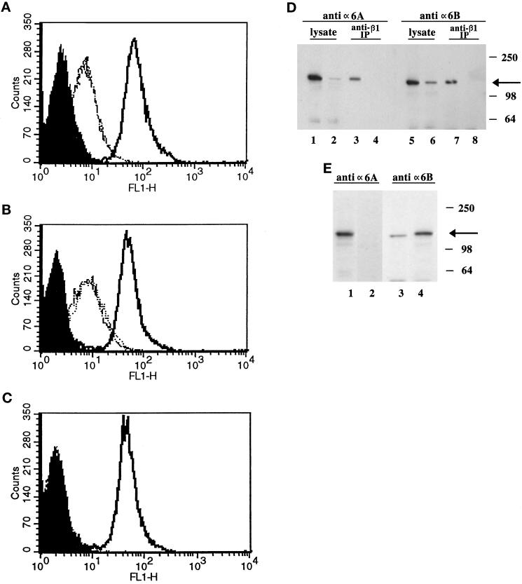
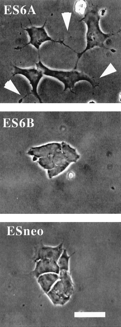
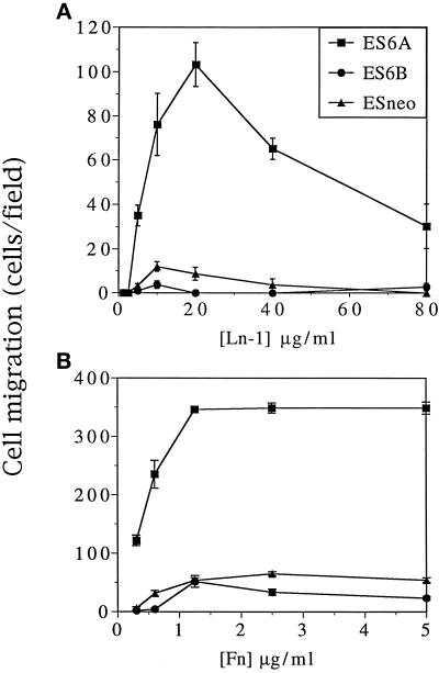
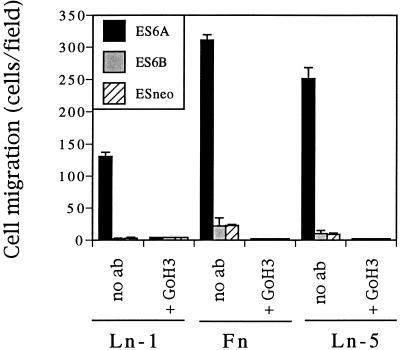
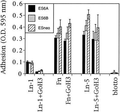
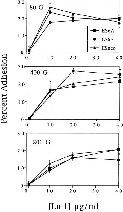
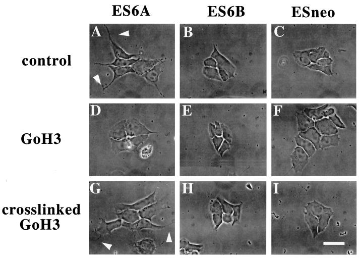
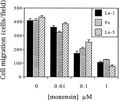
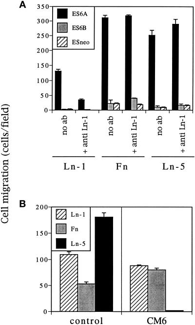
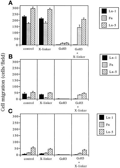
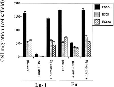
It is well established that integrins and extracellular matrix (ECM) play key roles in cell migration, but the underlying mechanisms are poorly defined. We describe a novel mechanism whereby the integrin alpha 6 beta 1, a laminin receptor, can affect cell motility and induce migration onto ECM substrates with which it is not engaged. By using DNA-mediated gene transfer, we expressed the human integrin subunit alpha 6A in murine embryonic stem (ES) cells. ES cells expressing alpha 6A (ES6A) at the surface dimerized with endogenous beta 1, extended numerous filopodia and lamellipodia, and were intensely migratory in haptotactic assays on laminin (LN)-1. Transfected alpha 6A was responsible for these effects, because cells transfected with control vector or alpha 6B, a cytoplasmic domain alpha 6 isoform, displayed compact morphology and no migration, like wild-type ES cells. The ES6A migratory phenotype persisted on fibronectin (Fn) and Ln-5. Adhesion inhibition assays indicated that alpha 6 beta 1 did not contribute detectably to adhesion to these substrates in ES cells. However, anti-alpha 6 antibodies completely blocked migration of ES6A cells on Fn or Ln-5. Control experiments with monensin and anti-ECM antibodies indicated that this inhibition could not be explained by deposition of an alpha 6 beta 1 ligand (e.g., Ln-1) by ES cells. Cross-linking with secondary antibody overcame the inhibitory effect of anti-alpha 6 antibodies, restoring migration or filopodia extension on Fn and Ln-5. Thus, to induce migration in ES cells, alpha 6A beta 1 did not have to engage with an ECM ligand but likely participated in molecular interactions sensitive to anti-alpha 6 beta 1 antibody and mimicked by cross-linking. Antibodies to the tetraspanin CD81 inhibited alpha 6A beta 1-induced migration but had no effect on ES cell adhesion. It is known that CD81 is physically associated with alpha 6 beta 1, therefore our results suggest a mechanism by which interactions between alpha 6A beta 1 and CD81 may up-regulate cell motility, affecting migration mediated by other integrins.


Figures

Figure 1
Figure 1
Alternate cytoplasmic tails of the α6 integrin subunit. Shown is the single letter amino acid sequence of the α6A and α6B cytoplasmic tails beginning at amino acid 988. The extracellular domains are identical from amino acid 1 through 994 after which the sequences are completely divergent. An arrowhead marks the point where the sequences begin to differ.
Figure 2
Figure 2
ES6A transfectant cells express human α6A on the cell surface, complexed with endogenous mouse β1. Expression of full-length human α6A or α6B cDNA was analyzed by FACS of ES6A (A), ES6B (B), and ESneo cells (C) with FITC-secondary antibody alone (solid peaks), mAb BQ16 and 2B7 (anti-human α6; dashed/dotted lines), and EA1 (anti-mouse and human α6; solid lines). Cells were analyzed on a FACScan machine (Becton Dickinson). (D) Cell lysates were subjected to immunoprecipitation using Sepharose beads coupled to the anti-β1 mAb 9EG7. Beads were eluted in nonreducing (odd-numbered lanes) or reducing (even-numbered lanes) sample buffer, separated by SDS-PAGE, and transferred to polyvinylidene difluoride membranes. Whole lysate (lanes 1, 2, 5, and 6) was electrophoresed along with the anti-β1 immunoprecipitations. Lanes 1–4, probed with anti-α6A antibody; lanes 5–8, probed with anti-α6B antibody. (E) As control to show specificity of the anti-peptide antibodies, whole lysates from ES6A (lanes 1 and 3) and ES6B (lanes 2 and 4) were separated under nonreducing conditions. Lanes 1 and 2, probed with anti-α6A antibody; lanes 3 and 4, probed with anti-α6B antibody. In both cases (D and E), the arrow indicates a band corresponding to nonreduced α6 polypeptide detected at approximately 140 kDa.
Figure 3
Figure 3
Periphery of ES6A cells, but not ES6B or ESneo cells, exhibits lamellipodia and filopodia. Cells were grown on Ln-1-coated glass coverslips overnight, washed with PBS, fixed in 2.5% paraformaldehyde in PBS, and mounted in Immunofluore (ICN). Cells were visualized in phase contrast by using a Zeiss Axiovert microscope and photographed by using TMAX 400 ASA film. Top, ES6A (arrowheads indicate lamellipodia and filopodia); middle ES6B; bottom, ESneo. Bar, 20 μm.
Figure 4
Figure 4
ES6A cells migrate on Ln-1 and Fn. Transwell filters were coated for 4 h at 37°C with various concentrations of Ln-1 or Fn. ES6A, ES6B, or ESneo cells (1 × 10⁴ cells/filter) were plated on the uncoated side. Cells were maintained at 37°C for 18 h, and then filters were fixed and stained. To quantify migration, stained cells were counted in four fields (by using a 40× objective) from each of two filters for each condition. Results of representative experiments are expressed as the number of cells counted in each field (mean ± SD).
Figure 5
Figure 5
ES6A cell migration on Ln-1, Fn, and Ln-5 is blocked by anti-α6 mAb GoH3. Transwell filters were coated for 4 h at 37°C with 20 μg/ml Ln-1, 20 μg/ml Fn, or 10 μg/ml Ln-5. ES6A, ES6B, or ESneo cells (1 × 10⁴ cells/filter) were plated on the uncoated side. For blocking experiments, cells were incubated at room temperature with 10 μg/ml GoH3 antibody in migration medium for 30 min before plating on coated filters. Cells were maintained at 37°C for 18 h, and then filters were fixed and stained. To quantify migration, stained cells were counted in four fields (by using a 40× objective) from each of two filters for each condition. Results of representative experiments are expressed as the number of cells counted in each field (mean ± SD).
Figure 6
Figure 6
ES6A, ES6B, and ESneo cells adhere to Ln-1 in an α6-dependent manner and to Fn and Ln-5 in an α6-independent manner. Ln-1 (20 μg/ml), Fn (20 μg/ml), and Ln-5 (1 μg/ml) were coated in 0.2 M carbonate buffer onto 96-well plates at 37°C for 4 h. Plates were washed and blocked for 1 h. For blocking with GoH3, cells were pre incubated with 10 μg/ml GoH3 and then plated in the presence of antibody. Cells adhered for 30 min at 37°C, washed, fixed, and stained. Stained cells were solubilized with 1% SDS and absorbance was quantified at 595 nm.
Figure 7
Figure 7
ES6A cells do not differ in strength of adhesion to Ln-1. The wells of the assay plate were coated with a titration of Ln-1 or heat-denatured BSA diluted in HBSS, incubated at 4°C for 4 h, then washed, and blocked with 2% heat-denatured BSA at 4°C overnight. [35S]Methionine-labeled ES6A, ES6B, or ESneo cells were plated (10,000 cells/well). The plates were immediately centrifuged at 80 × g to synchronize cell contact with the substratum. The cells were allowed to bind for 30 min at 37°C, and then the plates were flooded with warm PBS, sealed, inverted, and centrifuged for 8 min at 80 × g (top), 400 × g (middle), or 800 × g (bottom). The entire plate, still inverted, was submerged in ice-cold PBS and then in fixative (3.7% formaldehyde, 5% sucrose, 0.1% Triton X-100, PBS). After air drying, the bound radioactivity, representing cell adhesion, was quantified on a Molecular Dynamics PhosphorImager.
Figure 8
Figure 8
ES6A filopodia and lamellipodia are blocked by anti-α6 antibody GoH3 and are restored by cross-linking GoH3 with secondary antibody. Glass coverslips were coated for 30 min at 37°C with 40 μg/ml Fn. ES6A, ES6B, or ESneo cells (1 × 10⁴ cells/coverslip) were plated. For antibody treatments, cells were incubated at room temperature with 10 μg/ml GoH3 or 10 μg/ml goat anti-rat affinity-pure antibody in migration medium for 15 min. For cross-linking, 10 μg/ml goat anti-rat affinity-pure antibody was added for an additional 15 min to cells that had bound GoH3. No antibodies were removed before plating on the coverslips. Cells were maintained at 37°C for 18 h, then washed with PBS, fixed in 2.5% paraformaldehyde in PBS, and mounted in Immunofluore. Cells were visualized in phase contrast by using a Zeiss Axiovert microscope and photographed using TMAX 400 ASA film. (A, D, and G) ES6A cells: control, GoH3, and cross-linked GoH3, respectively. (B, E, and H) ES6B cells: control, GoH3, and cross-linked GoH3, respectively. (C, F, and I) ESneo cells: control, GoH3, and cross-linked GoH3, respectively. Bar, 20 μm.
Figure 9
Figure 9
Monensin does not abolish α6A-induced migration on Fn. Transwell filters were coated for 4 h at 37°C with 40 μg/ml Ln-1, 40 μg/ml Fn, or 10 μg/ml Ln-5. ES6A, ES6B, or ESneo cells (1 × 10⁴ cells/filter) were plated on the uncoated side of filters in migration medium, or migration medium including 0.01, 0.1, or 1 μM monensin. In all cases, cells were maintained at 37°C for 18 h, and then filters were fixed, stained, and analyzed as previously described.
Figure 10
Figure 10
ES6A migration is blocked by anti-Ln-1 on anti-Ln-5 antibodies only on the corresponding ECM substrates. Transwell filters were coated for 4 h at 37°C with 40 μg/ml Ln-1, 40 μg/ml Fn, or 10 μg/ml Ln-5. ES6A, ES6B, or ESneo cells (1 × 10⁴ cells/filter) were plated on the uncoated side. For blocking experiments, cells were incubated at room temperature with a 1:100 dilution of anti-Ln-1 antibody in migration medium for 30 min before plating on coated filters (A). For anti-Ln-5 blocking experiments, ES6A cells were incubated at room temperature with 25 μg/ml CM6 or, as control, 25 μg/ml MOPC in migration medium for 30 min before plating on coated filters (B). In all cases, cells were maintained at 37°C for 18 h, and then filters were fixed, stained, and analyzed as previously described.
Figure 11
Figure 11
Anti-α6 antibody GoH3 blocks migration on Fn and Ln-5, but cross-linking GoH3 with secondary antibody restores it. Transwell filters were coated for 4 h at 37°C with 40 μg/ml Ln-1, 40 μg/ml Fn, or 10 μg/ml Ln-5. ES6A, ES6B, or ESneo cells (1 × 10⁴ cells/filter) were plated on the uncoated side. For controls, cells were incubated at room temperature with 10 μg/ml GoH3 or 10 μg/ml purified goat anti-rat antibody in migration medium for 15 min. For cross-linking, 10 μg/ml purified goat anti-rat antibody was added for an additional 15 min to cells that had bound GoH3. No antibodies were removed before plating on the filters. In all cases, cells were maintained at 37°C for 18 h, and then filters were fixed, stained, and analyzed as previously described. (A) ES6A. (B) ES6B. (C) ESneo.
Figure 12
Figure 12
Anti-CD81 antibody blocks migration on Ln-1 and Fn. Transwell filters were coated for 4 h at 37°C with 20 μg/ml Ln-1 or 20 μg/ml Fn. ES6A, ES6B, or ESneo cells (1 × 10⁴ cells/filter) were plated on the uncoated side. Cells were incubated with 75 μg/ml hamster anti-CD81 (2F7) or, for controls, cells were incubated with 2% glycerol in PBS (since 2F7 is supplied in 20% glycerol) or with 75 μg/ml hamster anti-TNP antibody in migration medium for 30 min. No antibodies were removed before plating on the filters. In all cases, cells were maintained at 37°C for 18 h, and then filters were fixed, stained, and analyzed as previously described.

References

    1. Abe, T., Ohno, M., Sato, T., Murakami, M., Kajiki, M., and Kodaira, R. (1991). “Differentiation induction” culture of human leukemic myeloid cells stimulates high production of macrophage differentiation inducing factor. [Review]. Cytotechnology 5(suppl 2), S75–S93. - PubMed
    1. Berditchevski F, Zutter MM, Hemler ME. Characterization of novel complexes on the cell surface between integrins and proteins with 4 transmembrane domains (TM4 proteins) Mol Biol Cell. 1996;7:193–207. - PMC - PubMed
    1. Boismenu R, Rhein M, Fischer WH, Havran WL. A role for CD81 in early T cell development. Science. 1996;271:198–200. - PubMed
    1. Calof AL, Lander AD. Relationship between neuronal migration and cell-substratum adhesion: laminin and merosin promote olfactory neuronal migration but are anti-adhesive. J Cell Biol. 1991;115:779–794. - PMC - PubMed
    1. Chan BM, Kassner PD, Schiro JA, Byers HR, Kupper TS, Hemler ME. Distinct cellular functions mediated by different VLA integrin alpha subunit cytoplasmic domains. Cell. 1992;68:1051–1060. - PubMed

Publication types

MeSH terms

LinkOut - more resources