. 1997 Nov;8(11):2291-306.
doi: 10.1091/mbc.8.11.2291.

End4p/Sla2p interacts with actin-associated proteins for endocytosis in Saccharomyces cerevisiae

Affiliations
Free PMC article

End4p/Sla2p interacts with actin-associated proteins for endocytosis in Saccharomyces cerevisiae

A Wesp et al. Mol Biol Cell. 1997 Nov.
Free PMC article

Abstract

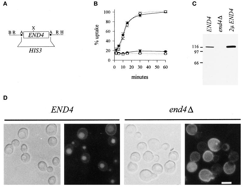
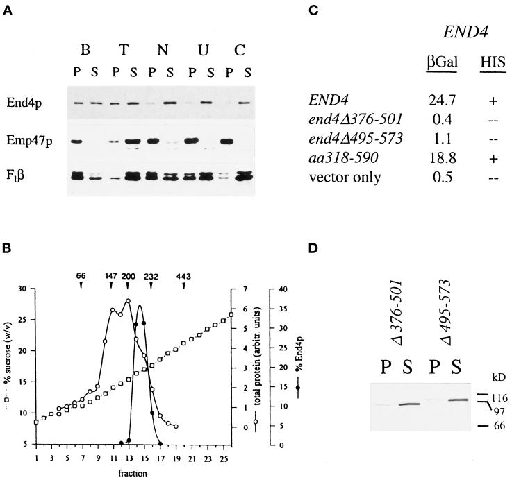
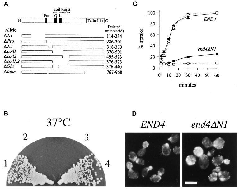
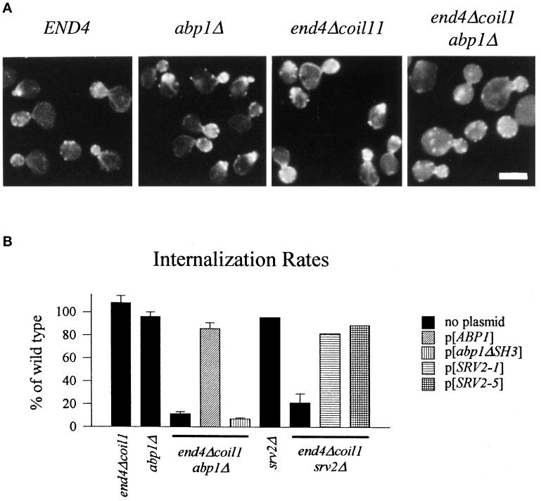
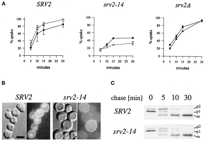
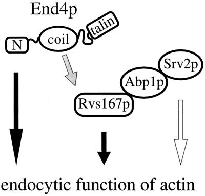
end4-1 was isolated as a temperature-sensitive endocytosis mutant. We cloned and sequenced END4 and found that it is identical to SLA2/MOP2. This gene is required for growth at high temperature, viability in the absence of Abp1p, polarization of the cortical actin cytoskeleton, and endocytosis. We used a mutational analysis of END4 to correlate in vivo functions with regions of End4p and we found that two regions of End4p participate in endocytosis but that the talin-like domain of End4p is dispensable. The N-terminal domain of End4p is required for growth at high temperature, endocytosis, and actin organization. A central coiled-coil domain of End4p is necessary for formation of a soluble sedimentable complex. Furthermore, this domain has an endocytic function that is redundant with the function(s) of ABP1 and SRV2. The endocytic function of Abp1p depends on its SH3 domain. In addition we have isolated a recessive negative allele of SRV2 that is defective for endocytosis. Combined biochemical, functional, and genetic analysis lead us to propose that End4p may mediate endocytosis through interaction with other actin-associated proteins, perhaps Rvs167p, a protein essential for endocytosis.


Figures

Figure 1
Figure 1
Deletion of END4 constitutively blocks endocytosis. (A) The complete END4 ORF on an AatII fragment was replaced with the prototrophic marker HIS3. The resulting EcoRI fragment was used to delete END4 by integration/replacement. B, BamHI; R, EcoRI; A, AatII; X, XhoI; H, HindIII. (B) Receptor-mediated internalization of α-factor is blocked at both 24°C (solid symbols) and 37°C (open symbols) in RH3391 cells cured of pGP6 (end4Δ, squares), whereas RH3393 cells (END4, circles) show no defect at either temperature. (C) End4p-specific antiserum generated against a C-terminal peptide specifically recognizes a single band with an apparent mobility of ∼116 kDa that is absent in strain RH3391 cured of pGP6 (end4Δ) extracts but more prominent in extracts prepared from strains expressing multiple copies of the gene (2μEND4). (D) In contrast to strain RH2887 (END4), strain RH3391 cured of pGP6 (end4Δ) does not accumulate LY in the vacuole after incubation with the dye at 24°C for 1 h. Bar, 5 μm.
Figure 2
Figure 2
Coiled-coil domain of End4p mediates complex formation. (A) Wild-type cell extracts were incubated on ice for 1 h in 1% Triton X-100 (lane T), 0.5 M NaCl (lane N), 2.5 M urea (lane U), or 0.1 M carbonate, pH 11.5 (lane C) or were mock treated with lysis buffer (lane B). After high-speed centrifugation, equivalent volumes of pellets (lanes P) and supernatants (lanes S) were analyzed for End4p and control proteins by immunodetection. (B) Wild-type cell lysates were fractionated on linear 9–30% sucrose gradients. Fractions were collected from the top (fraction 1) and End4p content was quantified by Western blotting. Arrowheads indicate the position of the peaks of marker proteins; numbers are given in kDa. (C) Wild-type END4 as well as mutant end4 alleles were fused in-frame to the gal4 transcription-activation domain. These fusion constructs were tested in the two-hybrid system for interaction with the wild-type END4 gene fused to the LexA DNA-binding domain. β-Galactosidase activities of extracts prepared from yeast strain L40 harboring the various combinations of plasmids were measured and expressed as units/milligram of protein. Alternatively, growth on plates lacking histidine was scored to monitor interaction of End4p. (D) Extracts of end4Δ376–501 (RH3395) and end4Δ495–573 (RH3397) mutants were prepared in lysis buffer and processed as described in A. Mutant End4p was visualized in pellets (lanes P) and supernatants (lanes S) by immunodetection.
Figure 3
Figure 3
N terminus of End4p is essential for actin organization and endocytosis. (A) Mutant end4 alleles devoid of the indicated regions (open space in the schematic representation) were generated as described in MATERIALS AND METHODS. Pro, proline-rich sequence; Q, glutamine-rich sequence; L, putative leucine-zipper; coil1 and coil2, putative coiled-coil regions 1 and 2, respectively. Proline at aa 498 divides the region from aa 376 to aa 573 into two separate coiled-coil domains (MATERIALS AND METHODS). The nomenclature of end4 alleles and the deleted amino acids are indicated. (B) END4 (section 1, RH3393), end4Δ (section 2, RH3212), end4ΔN1 (section 3, RH3769), and end4Δcoil1 (section 4, RH3395) strains were streaked onto YPUADT plates and incubated for 2 d at 37°C. Deletion of the N-terminal part of End4p but not deletion of the coiled-coil domain impairs growth at higher temperatures. (C) Receptor-mediated endocytosis of radiolabeled pheromone in END4 (RH3393) and end4ΔN1 (RH3769) strains after a 5-min preincubation at either 24°C (solid symbols) or 37°C (open symbols). (D) END4 (RH3393) and end4ΔN1 (RH3769) strains were grown at 24°C and processed for visualization of filamentous actin with rhodamine-conjugated phalloidin as described in MATERIALS AND METHODS. Bar, 5 μm.
Figure 4
Figure 4
Abp1p and Srv2p have cryptic endocytic functions. (A) Visualization of filamentous actin with rhodamine-conjugated phalloidin of END4 (RH3393), abp1Δ (RH3297), end4Δcoil1 (RH3395), and end4Δcoil1 abp1Δ (RH3407) cells grown at 24°C. (B) Internalization rates expressed as a percentage of wild-type (RH3393) in end4Δcoil1 (RH3395), abp1Δ (RH3297), and srv2Δ (RH4008) single mutants and in end4Δcoil1 abp1Δ (RH3407) or end4Δcoil1 srv2Δ (RH4009) double mutants transformed with the indicated plasmids, respectively. p[abp1ΔSH3] encodes a mutant allele of ABP1 with a deleted SH3 domain; p[SRV2–1] encodes wild-type SRV2 and p[SRV2–5] encodes a mutant allele of SRV2 that lacks both binding sites for the SH3 domain of Abp1p. All plasmids are centromere-based and described in Lila and Drubin (1997).
Figure 5
Figure 5
srv2–14, but not srv2Δ, mutations confer an endocytic defect. (A) Endocytosis of radiolabeled pheromone in SRV2 (RH978) and srv2–14 (RH3993) strains was measured at 24°C (solid symbols) or 37°C (open symbols) after a 15-min (SRV2, srv2–14) preincubation at the indicated temperature. Endocytosis in srv2Δ (RH4008, triangles) and SRV2 (RH3393, circles) was measured after a 5-min preincubation at 24°C. (B) Accumulation of LY in SRV2 (RH978) and srv2–14 (RH3993) mutants grown at 24°C and preincubated for 15 min at 37°C prior to addition of LY for 1 h. (C) Maturation of radiolabeled CPY was measured in SRV2 (RH978) and srv2–14 (RH3993) mutants grown at 24°C and preincubated for 15 min at 37°C prior to starting a pulse–chase experiment as described in MATERIALS AND METHODS. p1, endoplasmic reticulum form of CPY; p2, Golgi-modified form of CPY; m, mature vacuolar form of CPY.
Figure 6
Figure 6
Hypothetical model. Two parts of End4p participate in endocytosis: the N terminus, which is essential for endocytosis and actin organization, and the coiled-coil domain, which mediates an endocytic function redundant with Abp1p and Srv2p. We propose that this function involves binding to Rvs167p, a protein itself required for endocytosis. The shaded arrow indicates that the physical interaction of End4p and Rvs167p is based solely on two-hybrid data. The open arrow indicates that a recessive negative mutation but not a deletion of SRV2 results in a defect in endocytosis (see text for further explanations).

References

    1. Adams AE, Pringle JR. Relationship of actin and tubulin distribution to bud growth in wild type and morphogenetic mutant Saccharomyces cerevisiae. J Cell Biol. 1984;98:934–945. - PMC - PubMed
    1. Allen LH, Aderem A. A role for MARCKS, the alpha isozyme of protein kinase C and myosin I in zymosan phagocytosis by macrophages. J Exp Med. 1995;182:829–840. - PMC - PubMed
    1. Bauer F, Urdaci M, Aigle M, Crouzet M. Alteration of a yeast SH3 protein leads to conditional viability with defects in cytoskeletal and budding patterns. Mol Cell Biol. 1993;13:5070–5084. - PMC - PubMed
    1. Bénédetti H, Raths S, Crausaz F, Riezman H. The END3 gene encodes a protein that is required for the internalization step of endocytosis and for actin cytoskeleton organization in yeast. Mol Biol Cell. 1994;5:1023–1037. - PMC - PubMed
    1. Burridge K, Fath K, Kelly T, Nuckolls G, Turner C. Focal adhesions: transmembrane junctions between the extracellular matrix and the cytoskeleton. Annu Rev Cell Biol. 1988;4:487–525. - PubMed

Publication types

MeSH terms

LinkOut - more resources