. 1997 Nov;8(11):2345-63.
doi: 10.1091/mbc.8.11.2345.

Characterization of cell-matrix adhesion requirements for the formation of fascin microspikes

Affiliations
Free PMC article

Characterization of cell-matrix adhesion requirements for the formation of fascin microspikes

J C Adams. Mol Biol Cell. 1997 Nov.
Free PMC article

Abstract

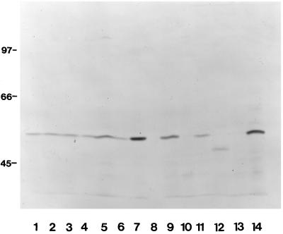
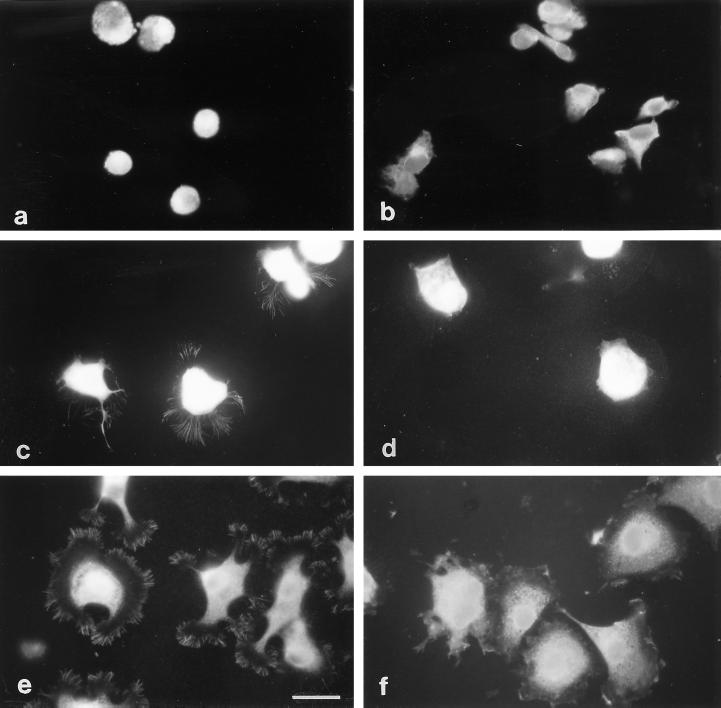
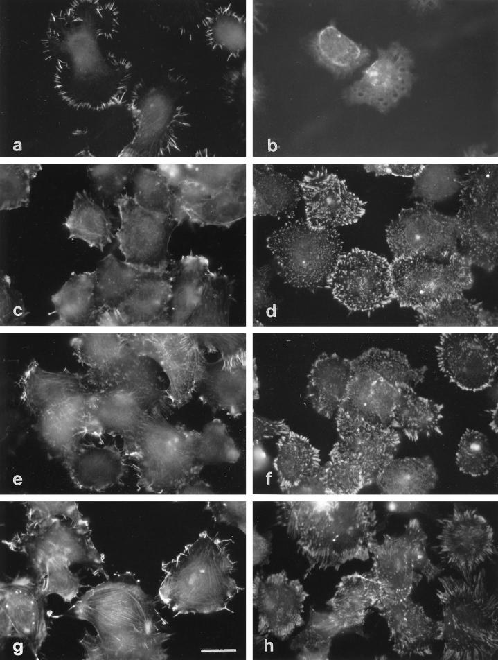
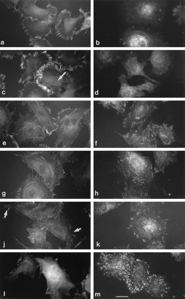
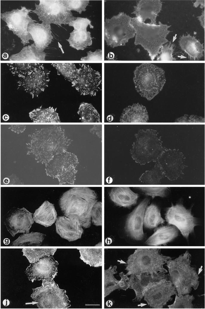
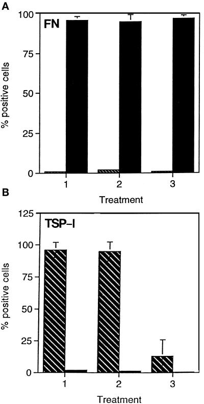
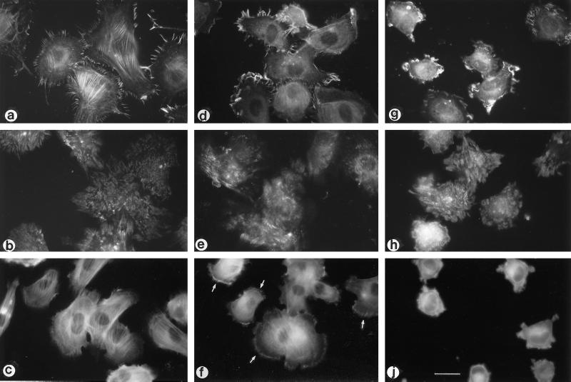
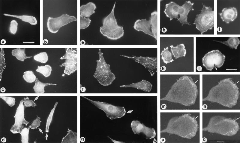
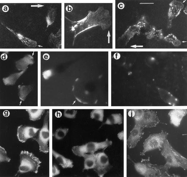
Cell adhesion to thrombospondin-1 (TSP-1) correlates with assembly of cell-substratum contact structures that contain fascin microspikes. In this analysis, cell-matrix requirements for assembly of fascin microspikes were examined in detail. In six cell lines, cell spreading on a TSP-1 substratum correlated with expression of fascin protein and formation of fascin microspikes. Microspikes were not formed by H9c2 cells adherent on fibronectin, vitronectin, collagen IV, or platelet factor 4. However, both fascin microspikes and focal contacts were assembled by cells adherent on laminin-1. Using mixed substrata containing different proportions of TSP-1, and fibronectin, fascin microspike formation by H9c2 and C2C12 cells was found to be reduced on substrata containing 25% fibronectin and abolished on substrata containing 75% fibronectin. Adhesion to intermediate mixtures of TSP-1 and fibronectin resulted in coassembly of fascin microspikes and focal contacts, colocalization of fascin with actin stress fiber bundles and altered distributions of beta 1 integrins, cortical alpha-actinin, and tropomyosin. In cells adherent on 50% TSP-1:50% fibronectin, GRGDSP peptide treatment decreased focal contact assembly and altered cytoskeletal organization but did not inhibit microspike assembly. Treatment with chondroitin sulfate A or p-nitrophenol beta-D-xylopyranoside decreased microspike formation and modified cytoskeletal organization but did not inhibit focal contact formation. In polarized migratory and postmitotic C2C12 cells, fascin microspikes and ruffles were localized at leading edges and TSP matrix deposition was also concentrated in this region. Depletion of matrix TSP by heparin treatment correlated with decreased microspike formation and cell motility. Thus, the balance of adhesive receptors ligated at the cell surface during initial cell-matrix attachment serves to regulate the type of substratum adhesion contact assembled and subsequent cytoskeletal organization. A role for fascin microspikes in cell motile behavior is indicated.


Figures

Figure 1
Figure 1
Expression of fascin by cell lines used in attachment assays. Equivalent numbers of cells were lysed in SDS-PAGE sample buffer, the extracts were resolved on a 12.5% polyacrylamide gel under reducing conditions, transferred to nitrocellulose, and probed with antibody 55K2 to fascin. Lanes: 1, A10 cells; 2, HISM cells; 3, MG-63 cells; 4, SK-N-SH cells; 5, H9c2 cells; 6, G8 cells; 7 and 14, C2C12 cells; 8, A549 cells; 9, C32 cells; 10, RT4 cells; 11, HT1080 cells; 12, G361 cells; 13, MDCK cells. Molecular mass markers are indicated in kDa.
Figure 2
Figure 2
Distribution of fascin in cells adherent on TSP-1 or fibronectin substrata. HT1080 cells (a and b), SK-N-SH cells (c and d), or A10 cells (e and f) were stained for fascin 1 h after plating on substrata coated with 50 nM TSP-1 (a, c, and e) or 50 nM fibronectin (b, d, and f) under serum-free conditions. Bar, 12 μm.
Figure 3
Figure 3
Formation of fascin microspikes and focal contacts by H9c2 cells plated on various ECM glycoproteins. H9c2 cells were stained for fascin (a, c, e, and g) or vinculin (b, d, f, and h) 1 h after plating on substrata coated with 50 nM TSP-1 (a and b), vitronectin (c and d), collagen IV (e and f), or EHS laminin (g and h). Bar, 12 μm.
Figure 4
Figure 4
Formation of fascin microspikes and focal contacts by H9c2 cells adherent on mixed TSP-1/fibronectin substrata. H9c2 cells were stained for fascin (a, c, e, g, j, and l) or vinculin (b, d, f, h, k, and m) 1 h after plating on substrata consisting of 100% TSP-1 (a and b), 90% TSP-1:10% fibronectin (c and d), 75% TSP-1:25% fibronectin (e and f), 50% TSP-1:50% fibronectin (g and h), 25% TSP-1:75% fibronectin (j and k), or 100% fibronectin (l and m). A cell displaying weak colocalization of fascin with microfilaments is shown with an arrow in c. Residual isolated microspikes are shown with arrows in j. Bar, 10 μm.
Figure 5
Figure 5
Effect of 50:50 mixed TSP-1/fibronectin substratum on cytoskeletal organization in H9c2 cells. H9c2 cells were stained for β1 integrins (a and b; apparent retraction fibers are shown with arrows), paxillin (c and d), phosphotyrosine (e and f), tropomyosin (g and h), or α-actinin (j and k) 1 h after plating on 100% fibronectin (a, c, e, g, and j) or 50% TSP-1:50% fibronectin (b, d, f, h, and k). The arrow in j indicates zone where periodic distribution of α-actinin along stress fibers is apparent; short arrows in k indicate cortical regions that show a different distribution of α-actinin on the mixed substratum. Bar, 10 μm.
Figure 6
Figure 6
Effects of xylosides on assembly of focal contacts or microspikes. Control H9c2 cells (bar 1) or cells treated for 20 h with o-nitrophenol β-d-xylopyranoside (bar 2) or p-nitrophenol β-d-xylopyranoside (bar 3) were allowed to adhere for 1 h to substrata coated with 50 nM fibronectin (A) or 50nM TSP-1 (B) and then scored for formation of microspikes (hatched columns) or focal contacts (solid columns) by staining for fascin or vinculin, respectively. Each column is the mean of duplicate experiments. Error bars, SD.
Figure 7
Figure 7
Role of integrins and proteoglycans in cytoskeletal organization of H9c2 cells adherent on 50% TSP-1:50% fibronectin substrata. H9c2 cells were allowed to adhere for 1 h to 50:50 TSP-1/fibronectin under control conditions (a–c) or in the presence of 1 mM GRGDSP peptide (d–f) or 100 μg/ml chondroitin sulfate A (g, h, j) and then stained for fascin (a, d, and g), vinculin (b, e, and h), or tropomyosin (c, f, and j). Arrows in f indicate tropomyosin localization at cell margins. Bar, 10 μm.
Figure 8
Figure 8
Presence of fascin-positive microspikes and ruffles at the protrusive margins of migratory and postmitotic C2C12 cells. Sparse cultures of C2C12 myoblasts were stained with rhodamine-phalloidin (a, b, and e), antibody to vinculin (c and f), or antibody to fascin (d, g, and h–q) and examples of small polarized cells (a, b, c, and d), larger fan-shaped cells (e, f, and g), or postmitotic pairs of cells (h–l) photographed under epifluorescence. Arrows in d and g indicate fascin microspikes and ruffles at leading edges; arrowhead in g indicates postmitotic pair of cells. (h) Asymmetric pair of cells nearing completion of cytokinesis which bear fascin microspikes and ruffles at their protrusive margins. (j and l) Examples of cells at the onset of cytokinesis: arrow in l indicates cleavage furrow. (k) Example of postmitotic pair stained 2 h after release from nocadazole block. Bars: a and l, 10um. (m–q) Series of projections from 3D reconstruction of confocal serial optical sections through one cell of a postmitotic pair stained for fascin. Projections were made at 180° (m), 210° (n), 230° (p), and 250° (q) to the original plane of section. Arrows indicate an upraised group of fascin microspikes displayed by the projections. Bar, 2.5 μm.
Figure 9
Figure 9
Deposition of extracellular TSP and development of the motile phenotype in sparse cultures of C2C12 cells. Cells from control cultures (a–e and g) or cultures treated with 500 μg/ml heparin for 6 h (f, h, and j) were stained for fibronectin (a–c), TSP (d–f), fascin (g and h), or vinculin (j). Large arrows in a–c indicate cells with no detectable fibronectin at the leading edge; small arrows in a and c indicate examples of cells with some deposition of fibronectin in the lamellar region. Arrows in d and e indicate concentrations of TSP staining at leading edges. Bar, 9 μm.

References

    1. Abercrombie M, Heaysman JEM, Pegrum SM. The locomotion of fibroblasts in culture I. Movements of the leading edge. Exp Cell Res. 1970a;59:393–398. - PubMed
    1. Abercrombie M, Heaysman JEM, Pegrum SM. The locomotion of fibroblasts in culture II. “Ruffling.”. Exp Cell Res. 1970b;60:437–444. - PubMed
    1. Abercrombie M, Heaysman JEM, Pegrum SM. The locomotion of fibroblasts in culture. IV. Electron microscopy of the leading lamella. Exp Cell Res. 1971;67:359–367. - PubMed
    1. Adams JC. Formation of stable microspikes containing actin and the 55kDa actin-bundling protein, fascin, is a consequence of cell adhesion to thrombospondin-1: implications for the anti-adhesive activities of thrombospondin-1. J Cell Sci. 1995;108:1977–1990. - PubMed
    1. Adams JC, Lawler J. Diverse mechanisms for cell attachment to platelet thrombospondin. J Cell Sci. 1993;104:1061–1071. - PubMed

Publication types

MeSH terms

LinkOut - more resources