. 1997 Oct;273(4):H1968-76.
doi: 10.1152/ajpheart.1997.273.4.H1968.

Three-dimensional residual strain in midanterior canine left ventricle

Affiliations

Three-dimensional residual strain in midanterior canine left ventricle

K D Costa et al. Am J Physiol. 1997 Oct.

Abstract

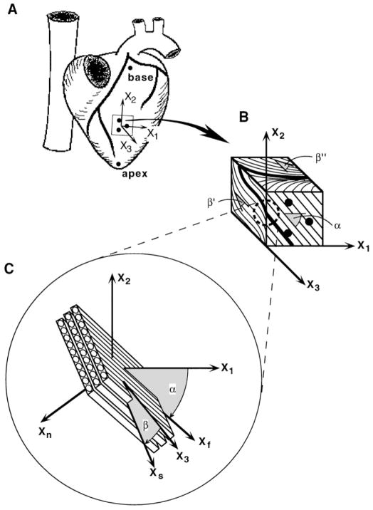
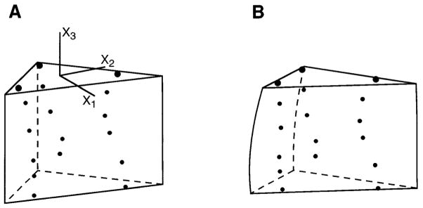
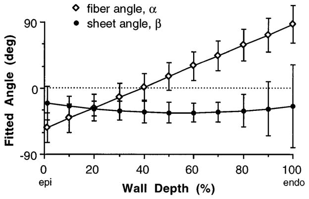
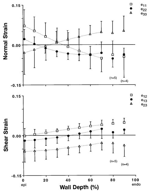
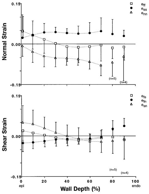
All previous studies of residual strain in the ventricular wall have been based on one- or two-dimensional measurements. Transmural distributions of three-dimensional (3-D) residual strains were measured by biplane radiography of columns of lead beads implanted in the midanterior free wall of the canine left ventricle (LV). 3-D bead coordinates were reconstructed with the isolated arrested LV in the zero-pressure state and again after local residual stress had been relieved by excising a transmural block of tissue. Nonhomogeneous 3-D residual strains were computed by finite element analysis. Mean +/- SD (n = 8) circumferential residual strain indicated that the intact unloaded myocardium was prestretched at the epicardium (0.07 +/- 0.06) and compressed in the subendocardium (-0.04 +/- 0.05). Small but significant longitudinal shortening and torsional shear residual strains were also measured. Residual fiber strain was tensile at the epicardium (0.05 +/- 0.06) and compressive in the subendocardium (-0.01 +/- 0.04), with residual extension and shortening, respectively, along structural axes parallel and perpendicular to the laminar myocardial sheets. Relatively small residual shear strains with respect to the myofiber sheets suggest that prestretching in the plane of the myocardial laminae may be a primary mechanism of residual stress in the LV.


Figures

Fig. 1
Fig. 1
Schematic diagram of method for defining structurally based coordinate systems used to study 3-dimensional (3-D) residual strain. A: isolated heart in its intact unloaded state showing 5 surface markers used to define cardiac coordinates (x1, x2, x3) aligned with local circumferential (x1), longitudinal (x2), and radial (x3) axes of left ventricle. B: excised stress-free block of tissue containing transmural bead set. Bold lines indicate intersection of a hypothetical sheet with each of 3 orthogonal cardiac coordinate planes. Fiber angle, α, is measured in epicardial tangent (1–2) plane (x1 = 0°). Cleavage plane angles, β′ and β″, are measured in (2–3) and (1–3) transverse planes, respectively (x3 = 0°). For all 3 angle measurements, a clockwise rotation relative to 0° represents a negative angle. C: at each transmural depth, a sheet angle, β, is computed from 3 intersection angles and is used to define a local system of fiber-sheet coordinates (xf, xs, xn) aligned with structural axes of myocardial laminae. Fiber axis xf is obtained by a rotation about radial x3 axis through fiber angle α. A subsequent rotation about xf through sheet angle β yields sheet axis xs, which is normal to xf and lies in sheet plane. xn is mutually orthogonal sheet normal axis.
Fig. 2
Fig. 2
Finite element analysis of 3-D residual strain. A: bead set in unloaded configuration inside a high-order finite element, which matches measured wall thickness. B: updated element configuration obtained from a least-squares fit to projected bead coordinates in stress-free state (actual bead coordinates and element geometries from a representative heart). Lagrangian 3-D finite strains are computed from A to B and are used to calculate Eulerian residual strains which describe inverse deformation from B to A.
Fig. 3
Fig. 3
Individual transmural distributions from epicardium (Epi) to endocardium (Endo) of fiber angle α, measured in (1–2) cardiac coordinate plane, and 2 cleavage plane angles, β′ and β″, measured in (2–3) and (1–3) coordinate planes, respectively, from midanterior left ventricular free wall myocardium fixed in stress-free state. Discontinuities in β′ and β″ reflect abrupt changes in sheet structure, such as endocardial trabeculae. β′ and β″ are plotted in ranges of 0 to −180° and 0 to 180°, respectively.
Fig. 4
Fig. 4
Distributions from Epi to Endo of 3 measured angles (A) and calculated sheet angle (B) for 2 individual animals. A continuous transmural distribution of fiber angle α was obtained from a linear least-squares fit to data (solid line in A). Each measurement of 2–3 cleavage plane angle β′ was then used to compute a sheet angle β, using an interpolated value of α at matching wall depth. This was repeated using (1–3) angle β″, yielding 2 sets of sheet angle data in B. A continuous transmural distribution of β was then obtained from a weighted quadratic least-squares fit to combined data set (solid line in B). To test for self-consistency, sheet angle equations were solved in reverse for values of β′ and β″ using fitted fiber and sheet angle distributions, and these results agreed with actual cleavage plane measurements (dashed lines in A)
Fig. 5
Fig. 5
Mean (±SD) fitted transmural distribution of fiber angle α and sheet angle β from 8 animals.
Fig. 6
Fig. 6
Transmural distributions from Epi to Endo of 6 components of 3-D Eulerian residual strain tensor, eres, referred to cardiac coordinates. Circumferential, e11res, and radial, e33res, strains showed opposing transmural gradients, consistent with published 2-D residual strain studies. Longitudinal strain, e22res, was small but significant (P < 0.0001), as were e12res and e13res shear strains. A substantial residual transverse shear strain, e23res, was found in longitudinal-radial plane. Symbols represent mean values (n = 8 except as noted), with error bars indicating ±SD.
Fig. 7
Fig. 7
Transmural distributions from Epi to Endo of 6 components of 3-D Eulerian residual strain, eres, referred to sheet coordinates. Residual stress gives rise to compression of adjacent myocardial laminae ( ennres<0) and stretching within sheet plane, particularly transverse to fiber axis ( essres>0). With exception of subepicardial esnres, all 3 shear strains were small. Gradient in fiber strain, effres, was consistent with previously measured changes in sarcomere length due to residual stress in rat heart. Symbols represent mean values (n = 8 except as noted), with error bars indicating ±SD.

References

    1. De Tombe PP, ter Keurs HEDJ. Sarcomere dynamics in cat cardiac trabeculae. Circ Res. 1991;68:588–596. - PubMed
    1. Fung YC. Biomechanics: Motion, Flow, Stress, Growth. New York: Springer-Verlag; 1990.
    1. Grimm AF, Katele KV, Lin HL. Fiber bundle direction in the mammalian heart. Basic Res Cardiol. 1976;71:381–388. - PubMed
    1. Guccione JM, McCulloch AD, Waldman LK. Passive material properties of intact ventricular myocardium determined from a cylindrical model. J Biomech Eng. 1991;113:42–55. - PubMed
    1. Hunter PJ, Nielsen PMF, Smaill BH, LeGrice IJ, Hunter IW. An anatomical heart model with applications to myocardial activation and ventricular mechanics. Crit Rev Biomed Eng. 1992;20:403–426. - PubMed

Publication types

LinkOut - more resources