. 1997 Nov 17;139(4):875-83.
doi: 10.1083/jcb.139.4.875.

Stimulation of NSF ATPase activity by alpha-SNAP is required for SNARE complex disassembly and exocytosis

Affiliations

Stimulation of NSF ATPase activity by alpha-SNAP is required for SNARE complex disassembly and exocytosis

R J Barnard et al. J Cell Biol. .

Abstract

N-ethylmaleimide-sensitive fusion protein (NSF) and alpha-SNAP play key roles in vesicular traffic through the secretory pathway. In this study, NH2- and COOH-terminal truncation mutants of alpha-SNAP were assayed for ability to bind NSF and stimulate its ATPase activity. Deletion of up to 160 NH2-terminal amino acids had little effect on the ability of alpha-SNAP to stimulate the ATPase activity of NSF. However, deletion of as few as 10 COOH-terminal amino acids resulted in a marked decrease. Both NH2-terminal (1-160) and COOH-terminal (160-295) fragments of alpha-SNAP were able to bind to NSF, suggesting that alpha-SNAP contains distinct NH2- and COOH-terminal binding sites for NSF. Sequence alignment of known SNAPs revealed only leucine 294 to be conserved in the final 10 amino acids of alpha-SNAP. Mutation of leucine 294 to alanine (alpha-SNAP(L294A)) resulted in a decrease in the ability to stimulate NSF ATPase activity but had no effect on the ability of this mutant to bind NSF. alpha-SNAP (1-285) and alpha-SNAP (L294A) were unable to stimulate Ca2+-dependent exocytosis in permeabilized chromaffin cells. In addition, alpha-SNAP (1-285), and alpha-SNAP (L294A) were able to inhibit the stimulation of exocytosis by exogenous alpha-SNAP. alpha-SNAP, alpha-SNAP (1-285), and alpha-SNAP (L294A) were all able to become incorporated into a 20S complex and recruit NSF. In the presence of MgATP, alpha-SNAP (1-285) and alpha-SNAP (L294A) were unable to fully disassemble the 20S complex and did not allow vesicle-associated membrane protein dissociation to any greater level than seen in control incubations. These findings imply that alpha-SNAP stimulation of NSF ATPase activity may be required for 20S complex disassembly and for the alpha-SNAP stimulation of exocytosis.


Figures

Figure 1
Figure 1
A schematic diagram of the α-SNAP truncation mutants used in this study and their ability to stimulate the ATPase activity of NSF compared to wild-type α-SNAP. (Top left) Schematic diagram of the α-SNAP truncation mutants used in this study. (Top right) Extent of stimulation of NSF ATPase activity as a percentage of stimulation by wild-type α-SNAP. 2 μg of full-length α-SNAP or α-SNAP truncation mutant were preimmobilized to the surface of plastic microfuge tubes for 20 min on ice. α-SNAP was removed from the tubes, and 1 μg of NSF was added for 1 h at 37°C. NSF ATPase activity was measured using a spectrophotometric assay, and the results were expressed as a percentage of the wild-type α-SNAP stimulation of NSF. Corrections were made for preexisting phosphates and contaminating ATPases by subtracting values from duplicate samples run with NEM-inactivated NSF. Data was pooled from three to seven separate assays for each SNAP mutant. (Bottom) Alignment of database sequences of bovine α-SNAP, β-SNAP, γ-SNAP, C. elegans SNAP, squid SNAP, Drosophila SNAP, and yeast sec17p, showing leucine 294 to be the only conserved residue in the last 10 amino acids of α-SNAP.
Figure 1
Figure 1
A schematic diagram of the α-SNAP truncation mutants used in this study and their ability to stimulate the ATPase activity of NSF compared to wild-type α-SNAP. (Top left) Schematic diagram of the α-SNAP truncation mutants used in this study. (Top right) Extent of stimulation of NSF ATPase activity as a percentage of stimulation by wild-type α-SNAP. 2 μg of full-length α-SNAP or α-SNAP truncation mutant were preimmobilized to the surface of plastic microfuge tubes for 20 min on ice. α-SNAP was removed from the tubes, and 1 μg of NSF was added for 1 h at 37°C. NSF ATPase activity was measured using a spectrophotometric assay, and the results were expressed as a percentage of the wild-type α-SNAP stimulation of NSF. Corrections were made for preexisting phosphates and contaminating ATPases by subtracting values from duplicate samples run with NEM-inactivated NSF. Data was pooled from three to seven separate assays for each SNAP mutant. (Bottom) Alignment of database sequences of bovine α-SNAP, β-SNAP, γ-SNAP, C. elegans SNAP, squid SNAP, Drosophila SNAP, and yeast sec17p, showing leucine 294 to be the only conserved residue in the last 10 amino acids of α-SNAP.
Figure 2
Figure 2
Effect on NSF ATPase activity of α-SNAP, α-SNAP (1–160), α-SNAP (Δ160–200), α-SNAP (1–285), and α-SNAP (L294A) over a range of concentrations. (A) Standard NSF ATPase assays were performed with several concentrations of full-length α-SNAP, α-SNAP (1–160), or α-SNAP (Δ160–200). (B) NSF ATPase assays with α-SNAP, α-SNAP (1–285), or α-SNAP (L294A). The data have been normalized to the maximal stimulation due to full-length α-SNAP. This was calculated by substracting NEM-insensitive ATPase activity and then calculating the relative differences between NSF ATPase activity in the presence or absence of α-SNAPs.
Figure 2
Figure 2
Effect on NSF ATPase activity of α-SNAP, α-SNAP (1–160), α-SNAP (Δ160–200), α-SNAP (1–285), and α-SNAP (L294A) over a range of concentrations. (A) Standard NSF ATPase assays were performed with several concentrations of full-length α-SNAP, α-SNAP (1–160), or α-SNAP (Δ160–200). (B) NSF ATPase assays with α-SNAP, α-SNAP (1–285), or α-SNAP (L294A). The data have been normalized to the maximal stimulation due to full-length α-SNAP. This was calculated by substracting NEM-insensitive ATPase activity and then calculating the relative differences between NSF ATPase activity in the presence or absence of α-SNAPs.
Figure 3
Figure 3
Reversal of the α-SNAP (L294A) mutation restores the ability to stimulate NSF ATPase activity. Standard assays of NSF ATPase activity were performed with 5 μg/tube of α-SNAP, α-SNAP (L294A), or α-SNAP (A294L), shown as α-SNAPR. The data shows levels of ATP hydrolysis with untreated and with NEM-treated NSF. The intrinsic ATPase activity of NSF was not reduced by α-SNAP (L294A).
Figure 4
Figure 4
Binding of NSF to immobilized α-SNAP mutants. 2 μg of full-length SNAP or α-SNAP mutant was preimmobilized to the surface of polypropylene tubes for 20 min on ice. After washing with 1 ml SWB, the tubes were incubated with 2 μg of NSF in NBB for 10 min. The tubes were washed with 1 ml NBB, and bound proteins solublized with 50 μl of SDS buffer. The samples were then analyzed by SDS-PAGE and detected by silver-staining. A–C show results from different experiments. Note that no NSF binding was detected in control tubes without added SNAPs (first lane of each part).
Figure 4
Figure 4
Binding of NSF to immobilized α-SNAP mutants. 2 μg of full-length SNAP or α-SNAP mutant was preimmobilized to the surface of polypropylene tubes for 20 min on ice. After washing with 1 ml SWB, the tubes were incubated with 2 μg of NSF in NBB for 10 min. The tubes were washed with 1 ml NBB, and bound proteins solublized with 50 μl of SDS buffer. The samples were then analyzed by SDS-PAGE and detected by silver-staining. A–C show results from different experiments. Note that no NSF binding was detected in control tubes without added SNAPs (first lane of each part).
Figure 5
Figure 5
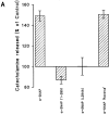
α-SNAP (1–285) and α-SNAP (L294A) are unable to stimulate catecholamine release from digitonin-permeabilized adrenal chromaffin cells but inhibit the α-SNAP stimulation of exocytosis. (A) Adrenal chromaffin cells were permeabilized for 45 min with permeabilization buffer and stimulated with 10 μM Ca2+ with or without 25 μg/ml of full-length α-SNAP, α-SNAP (1–285), α-SNAP (L294A), or the reverse mutation α-SNAP (A294L) for 30 min, and released catecholamine was assayed. The data were pooled from seven separate experiments, and the means were expressed as a percentage of the 10 μM Ca2+ control stimulation ± SEM. (B) Adrenal chromaffin cells were permeabilized for 20 min with permeabilization buffer, incubated with KGEP buffer with or without 25 μg/ml of recombinant α-SNAP (1–160), α-SNAP (1–285), or α-SNAP (L294A) for 25 min, and then subsequently stimulated with 10 μM Ca2+ with or without 25 μg/ml full-length α-SNAP for 30 min, and released catecholamine was assayed. The data was pooled from four separate experiments, and the means were expressed as a percentage of the 10 μM Ca2+ control stimulation ± SEM.
Figure 5
Figure 5
α-SNAP (1–285) and α-SNAP (L294A) are unable to stimulate catecholamine release from digitonin-permeabilized adrenal chromaffin cells but inhibit the α-SNAP stimulation of exocytosis. (A) Adrenal chromaffin cells were permeabilized for 45 min with permeabilization buffer and stimulated with 10 μM Ca2+ with or without 25 μg/ml of full-length α-SNAP, α-SNAP (1–285), α-SNAP (L294A), or the reverse mutation α-SNAP (A294L) for 30 min, and released catecholamine was assayed. The data were pooled from seven separate experiments, and the means were expressed as a percentage of the 10 μM Ca2+ control stimulation ± SEM. (B) Adrenal chromaffin cells were permeabilized for 20 min with permeabilization buffer, incubated with KGEP buffer with or without 25 μg/ml of recombinant α-SNAP (1–160), α-SNAP (1–285), or α-SNAP (L294A) for 25 min, and then subsequently stimulated with 10 μM Ca2+ with or without 25 μg/ml full-length α-SNAP for 30 min, and released catecholamine was assayed. The data was pooled from four separate experiments, and the means were expressed as a percentage of the 10 μM Ca2+ control stimulation ± SEM.
Figure 6
Figure 6
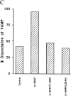
α-SNAP (1–285) and α-SNAP (L294A) associate with but are unable to support dissociation of the 20S complex. (A) A detergent extract of rat brain membrane proteins was incubated with 15 μg of NSF, 30 μg α-SNAPs for 30 min with 0.5 mM MgATP or MgATPγS as indicated at 4°C. Proteins were immunoprecipitated with an antisyntaxin antibody conjugated to protein G–Sepharose, and bound proteins were solubilized with SDS sample buffer and separated on a 12.5% polyacrylamide gel. Proteins were detected using specific antisera to NSF, α-SNAP, syntaxin, and VAMP. (B) Extracts were incubated without (control) or with added α-SNAP and with 15 μg NSF in the presence of 0.5 mM MgATP or MgATPγS as indicated. Endogenous α-SNAP in control incubations was sufficient to recruit exogenous NSF. (C) Extracts were incubated with NSF with no added α-SNAP (control) or with added α-SNAPs as indicated. The presence of VAMP in syntaxin immunoprecipitates was determined by immunoblotting, and the amount of VAMP dissociated in the presence of MgATP was calculated as a percentage of the amount of bound VAMP in MgATPγS incubations.The data shown are the mean values from two experiments.
Figure 6
Figure 6
α-SNAP (1–285) and α-SNAP (L294A) associate with but are unable to support dissociation of the 20S complex. (A) A detergent extract of rat brain membrane proteins was incubated with 15 μg of NSF, 30 μg α-SNAPs for 30 min with 0.5 mM MgATP or MgATPγS as indicated at 4°C. Proteins were immunoprecipitated with an antisyntaxin antibody conjugated to protein G–Sepharose, and bound proteins were solubilized with SDS sample buffer and separated on a 12.5% polyacrylamide gel. Proteins were detected using specific antisera to NSF, α-SNAP, syntaxin, and VAMP. (B) Extracts were incubated without (control) or with added α-SNAP and with 15 μg NSF in the presence of 0.5 mM MgATP or MgATPγS as indicated. Endogenous α-SNAP in control incubations was sufficient to recruit exogenous NSF. (C) Extracts were incubated with NSF with no added α-SNAP (control) or with added α-SNAPs as indicated. The presence of VAMP in syntaxin immunoprecipitates was determined by immunoblotting, and the amount of VAMP dissociated in the presence of MgATP was calculated as a percentage of the amount of bound VAMP in MgATPγS incubations.The data shown are the mean values from two experiments.

Similar articles

Cited by

References

    1. Barnard RJO, Morgan A, Burgoyne RD. Domains of α-SNAP required for the stimulation of exocytosis and for NSF binding and activation. Mol Biol Cell. 1996;7:693–701. - PMC - PubMed
    1. Beckers CJM, Block MR, Glick BS, Rothman JE, Balch WE. Vesicular transport between the endoplasmic reticulum and the Golgi stack requires the NEM-sensitive fusion protein. Nature. 1989;339:397–398. - PubMed
    1. Block MR, Glick BS, Wilcox CA, Wieland FT, Rothman JE. Purification of an N-ethylmaleimide-sensitive protein catalyzing vesicular transport. Proc Natl Acad Sci USA. 1988;85:7852–7856. - PMC - PubMed
    1. Burgoyne, R.D. 1992. Investigation of the intracellular regulators and components of the exocytotic pathway. In Neuromethods, Volume 20: Intracellular Messengers. A. Boulton, G. Baker, and C. Taylor, editors. Humana Press Inc., Totowa, NJ. 433–470.
    1. Burgoyne RD, Williams G. NSF and SNAP are present on adrenal chromaffin granules. FEBS Lett. 1997;414:349–352. - PubMed

Publication types