. 1997 Nov 17;139(4):907-16.
doi: 10.1083/jcb.139.4.907.

Differential localization of vesicular acetylcholine and monoamine transporters in PC12 cells but not CHO cells

Affiliations

Differential localization of vesicular acetylcholine and monoamine transporters in PC12 cells but not CHO cells

Y Liu et al. J Cell Biol. .

Abstract

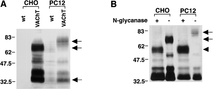
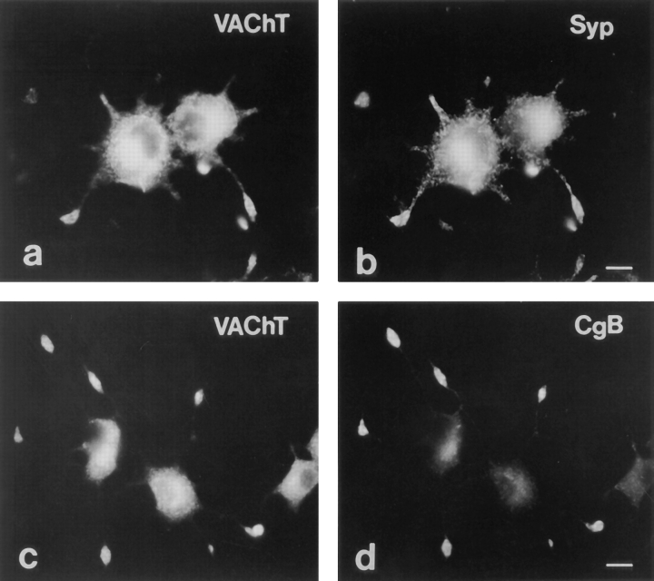
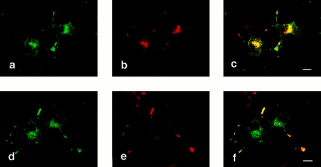
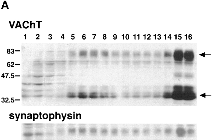
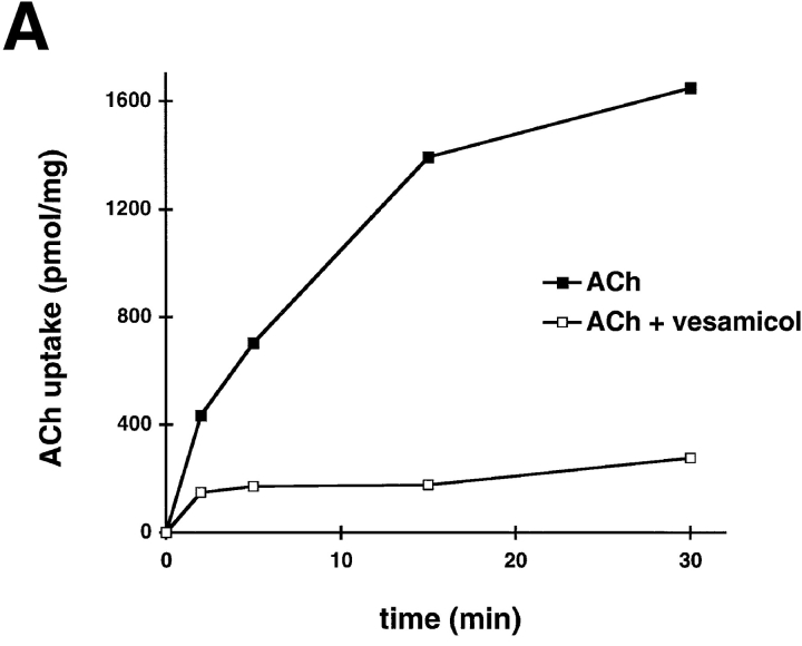
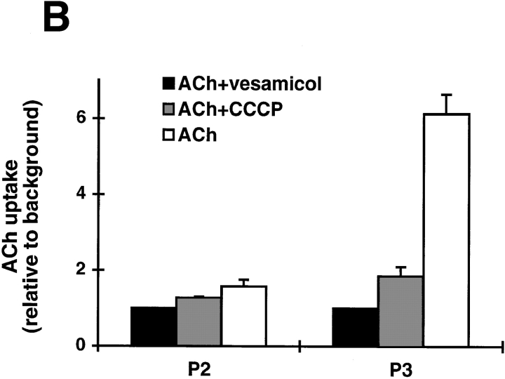
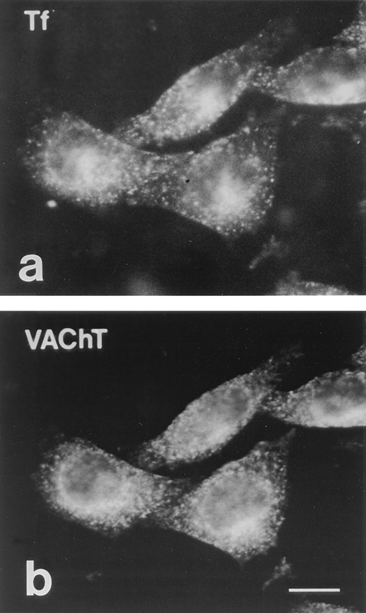
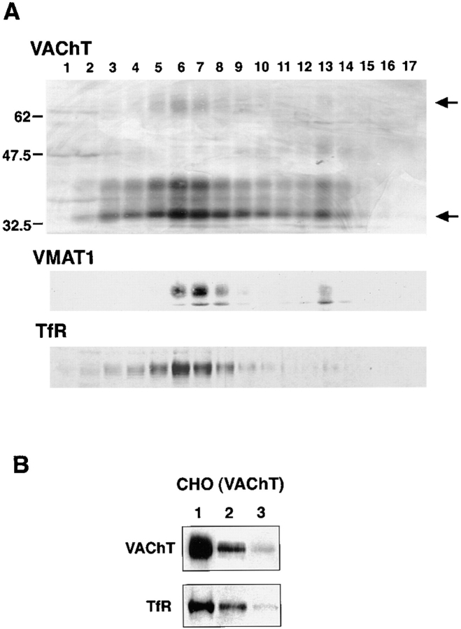
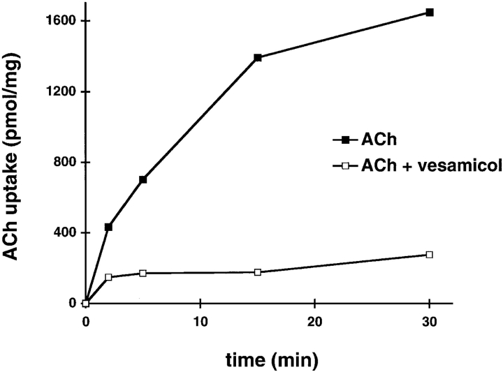
Previous studies have indicated that neuro-endocrine cells store monoamines and acetylcholine (ACh) in different secretory vesicles, suggesting that the transport proteins responsible for packaging these neurotransmitters sort to distinct vesicular compartments. Molecular cloning has recently demonstrated that the vesicular transporters for monoamines and ACh show strong sequence similarity, and studies of the vesicular monoamine transporters (VMATs) indicate preferential localization to large dense core vesicles (LDCVs) rather than synaptic-like microvesicles (SLMVs) in rat pheochromocytoma PC12 cells. We now report the localization of the closely related vesicular ACh transporter (VAChT). In PC12 cells, VAChT differs from the VMATs by immunofluorescence and fractionates almost exclusively to SLMVs and endosomes by equilibrium sedimentation. Immunoisolation further demonstrates colocalization with synaptophysin on SLMVs as well as other compartments. However, small amounts of VAChT also occur on LDCVs. Thus, VAChT differs in localization from the VMATs, which sort predominantly to LDCVs. In addition, we demonstrate ACh transport activity in stable PC12 transformants overexpressing VAChT. Since previous work has suggested that VAChT expression confers little if any transport activity in non-neural cells, we also determined its localization in transfected CHO fibroblasts. In CHO cells, VAChT localizes to the same endosomal compartment as the VMATs by immunofluorescence, density gradient fractionation, and immunoisolation with an antibody to the transferrin receptor. We have also detected ACh transport activity in the transfected CHO cells, indicating that localization to SLMVs is not required for function. In summary, VAChT differs in localization from the VMATs in PC12 cells but not CHO cells.


Figures

Figure 1
Figure 1
Western analysis of membranes from transfected CHO and PC12 cells. (A) An anti-peptide antibody recognizes VAChT in transfected CHO and PC12 cells. Western analysis of membranes prepared by differential centrifugation from CHO (left) and PC12 cells (right) shows that wild-type (wt) CHO cells express no immunoreactive VAChT, whereas a stable CHO transformant (VAChT) contains multiple immunoreactive forms. The two major species of ∼70 and ∼30 kD are indicated by the lower two arrows. Wild-type (wt) PC12 cells show several immunoreactive forms, most of which appear at higher levels in the stable transformant overexpressing rat VAChT. In PC12 cells, the two major species of ∼80 and ∼30 kD are indicated by the upper and lower arrows. Molecular masses in kD are shown to the left. (B) VAChT undergoes N-linked glycosylation in PC12 and CHO transformants. A postnuclear supernatant prepared from either PC12 cells (left) or CHO cells (right) was incubated overnight at 37°C in the presence (+) and absence (−) of N-glycanase. The arrows on the right indicate the undigested forms and the arrowhead the digested form of VAChT. VAChT thus undergoes N-linked glycosylation in both PC12 and CHO cells, and the extent of glycosylation differs in the two cell lines. Molecular masses (in kD) are indicated to the left.
Figure 2
Figure 2
VAChT colocalizes with synaptophysin in wild-type PC12 cells by immunofluorescence. Wild-type PC12 cells were stained with antibodies to VAChT (a and c), synaptophysin (b), and chromogranin B (d). For double immunofluorescence of the two sets of cells (a and b and c and d), the rabbit polyclonal antibody to VAChT was detected with a secondary antibody conjugated to fluorescein and the mouse monoclonal antibodies to synaptophysin and chromogranin B were detected with a secondary antibody conjugated to rhodamine. VAChT immunoreactivity occurs at both the tips of processes and in the cell body (a and c) and colocalizes with synaptophysin at these sites (b). In contrast, chromogranin B occurs predominantly at the tips of processes with very little at the cell body (d), a pattern clearly distinct from that of VAChT (c). Bars, 10 μm.
Figure 3
Figure 3
Colocalization of VAChT with synaptophysin in a stable PC12 cell transformant. A stable PC12 cell transformant overexpressing VAChT was immunostained for VAChT (a and d), synaptophysin (b), and chromogranin B (e). Using two sets of cells (a–c and d–f) for confocal laser microscopy, the rabbit polyclonal antibody to VAChT was detected with a secondary anti– rabbit antibody conjugated to Cy3 (a and d, green), and the mouse monoclonal antibody to synaptophysin was detected with a secondary anti–mouse antibody conjugated to Cy5 (b and e, red). The superimposed staining is shown in c and f. Similar to wild-type PC12 cells, VAChT (a) colocalizes with synaptophysin (b) at both the tips of processes and at a juxtanuclear region in the cell body (c, yellow). Despite the overexpression of VAChT in this cell line, VAChT immunoreactivity (d) appears quite distinct from chromogranin B (e), which occurs predominantly at the tips of processes and only at lower levels in the cell body (f, yellow). Bars, 10 μm.
Figure 4
Figure 4
VAChT localizes to SLMVs by velocity sedimentation through glycerol and immunoisolation. (A) A stable PC12 transformant overexpressing VAChT was disrupted by homogenization and the postnuclear supernatant subjected to velocity sedimentation through a gradient of 5 to 25% glycerol. Western analysis of the fractions shows that both high and low molecular weight forms of VAChT (arrows) appear at a position in the middle of the gradient that coincides with synaptophysin and corresponds to SLMVs. Larger amounts of both VAChT and synaptophysin also occur at the bottom of the gradient in fractions that include endosomes. Molecular mass markers (in kD) are indicated at the left. Fractions are numbered from the top of the gradient on the left to the bottom on the right. (B) A postnuclear supernatant was subjected to immunoisolation using Dynal beads coated with a monoclonal antibody to synaptophysin. Western analysis using the antibody to VAChT shows substantial immunoreactivity in the starting material (lane 1) and almost full recovery in the immunoisolated vesicles (lane 2). Immunoisolation with uncoated Dynal beads yields minimal VAChT immunoreactivity (lane 3). Immunostaining for synaptophysin (Syp) shows an identical pattern, confirming the colocalization of VAChT and synaptophysin. (C) Fraction 6 from the glycerol gradient was subjected to immunoisolation as described in B. Western analysis using the antibody to VAChT shows immunoreactivity in the starting material (lane 1) and essentially full recovery in the immunoisolated vesicles (lane 2). Control immunoisolation with uncoated Dynal beads yields no VAChT immunoreactivity (lane 3). Immunostaining for synaptophysin shows an identical pattern, confirming the colocalization of VAChT and synaptophysin on the same population of SLMVs.
Figure 4
Figure 4
VAChT localizes to SLMVs by velocity sedimentation through glycerol and immunoisolation. (A) A stable PC12 transformant overexpressing VAChT was disrupted by homogenization and the postnuclear supernatant subjected to velocity sedimentation through a gradient of 5 to 25% glycerol. Western analysis of the fractions shows that both high and low molecular weight forms of VAChT (arrows) appear at a position in the middle of the gradient that coincides with synaptophysin and corresponds to SLMVs. Larger amounts of both VAChT and synaptophysin also occur at the bottom of the gradient in fractions that include endosomes. Molecular mass markers (in kD) are indicated at the left. Fractions are numbered from the top of the gradient on the left to the bottom on the right. (B) A postnuclear supernatant was subjected to immunoisolation using Dynal beads coated with a monoclonal antibody to synaptophysin. Western analysis using the antibody to VAChT shows substantial immunoreactivity in the starting material (lane 1) and almost full recovery in the immunoisolated vesicles (lane 2). Immunoisolation with uncoated Dynal beads yields minimal VAChT immunoreactivity (lane 3). Immunostaining for synaptophysin (Syp) shows an identical pattern, confirming the colocalization of VAChT and synaptophysin. (C) Fraction 6 from the glycerol gradient was subjected to immunoisolation as described in B. Western analysis using the antibody to VAChT shows immunoreactivity in the starting material (lane 1) and essentially full recovery in the immunoisolated vesicles (lane 2). Control immunoisolation with uncoated Dynal beads yields no VAChT immunoreactivity (lane 3). Immunostaining for synaptophysin shows an identical pattern, confirming the colocalization of VAChT and synaptophysin on the same population of SLMVs.
Figure 5
Figure 5
VAChT predominates in light membrane fractions by equilibrium sedimentation through sucrose. A postnuclear supernatant from PC12 cells overexpressing VAChT was subjected to equilibrium sedimentation through a gradient of 0.6 to 1.6 M sucrose. Western analysis shows that VAChT occurs in light membrane fractions as a very broad peak that coincides with the peak for synaptophysin, a marker of SLMVs and transferrin receptor (TfR), a marker of endosomes. Secretogranin II indicates fractions containing LDCVs, which migrate as a peak distinct from VAChT and synaptophysin. However, the peak of VAChT immunoreactivity overlaps with that of secretogranin II. Molecular mass markers (in kD) are indicated on the left and high and low molecular weight forms of VAChT are indicated by arrows on the right. Fractions were collected from the top of the gradient (left) to the bottom (right).
Figure 6
Figure 6
VAChT occurs on LDCVs by two-step density gradient fractionation. PC12 cells stably overexpressing VAChT were incubated overnight in medium containing 0.25 μCi/ml [3H]norepinephrine, disrupted by homogenization, and a postnuclear supernatant subjected to two steps of density gradient fractionation. In the first step, the membranes were separated by velocity sedimentation through a gradient of 0.3 to 1.2 M sucrose. Radioactive fractions containing LDCVs in the middle of the gradient were then pooled and subjected to equilibrium sedimentation through a gradient of 0.6 to 1.6 M sucrose. Fractions from the second gradient show VAChT immunoreactivity that coincides with the LDCV marker secretogranin II as well as synaptophysin. However, some of the synaptophysin also occurs on slightly less dense vesicles that coincide with the endoplasmic reticulum marker ribophorin II. Molecular mass standards (in kD) are shown to the left and arrows indicating the two species of VAChT to the right. Fractions from the second gradient were collected from the top (left) to the bottom (right).
Figure 7
Figure 7
VAChT overexpression in PC12 cells confers vesicular ACh transport activity. (A) A population of light membrane vesicles (P3) from transfected PC12 cells (PC12[VAChT]; filled squares) demonstrates ACh transport activity substantially higher than in the same vesicles from untransfected cells (wt PC12; filled triangles). In addition, vesamicol inhibits the uptake by transfected cells (open squares) to a level lower than in untransfected cells, suggesting that endogenous PC12 cell VAChT has some transport activity. (B) Comparison of ACh uptake by different fractions from transfected PC12 cells. Membranes from a postnuclear supernatant collected by centrifugation at 27,000 g for 35 min (P2) show less vesamicol- and CCCP-sensitive ACh transport activity than membranes collected by centrifugation of the S2 at 200,000 g for 40 min (P3). ACh uptake is normalized to background accumulation in the presence of vesamicol for each of the membrane preparations. (C) Western analysis of equal amounts of protein from P2 and P3 fractions shows approximately equal amounts of VAChT and substantial amounts of transferrin receptor (TfR) in both fractions but considerably more of the LDCV marker secretogranin II (SgII) in P2 and more of the SLMV marker synaptophysin (Syp) in P3.
Figure 7
Figure 7
VAChT overexpression in PC12 cells confers vesicular ACh transport activity. (A) A population of light membrane vesicles (P3) from transfected PC12 cells (PC12[VAChT]; filled squares) demonstrates ACh transport activity substantially higher than in the same vesicles from untransfected cells (wt PC12; filled triangles). In addition, vesamicol inhibits the uptake by transfected cells (open squares) to a level lower than in untransfected cells, suggesting that endogenous PC12 cell VAChT has some transport activity. (B) Comparison of ACh uptake by different fractions from transfected PC12 cells. Membranes from a postnuclear supernatant collected by centrifugation at 27,000 g for 35 min (P2) show less vesamicol- and CCCP-sensitive ACh transport activity than membranes collected by centrifugation of the S2 at 200,000 g for 40 min (P3). ACh uptake is normalized to background accumulation in the presence of vesamicol for each of the membrane preparations. (C) Western analysis of equal amounts of protein from P2 and P3 fractions shows approximately equal amounts of VAChT and substantial amounts of transferrin receptor (TfR) in both fractions but considerably more of the LDCV marker secretogranin II (SgII) in P2 and more of the SLMV marker synaptophysin (Syp) in P3.
Figure 8
Figure 8
VAChT immunofluorescence colocalizes with transferrin receptors in transfected CHO cells. CHO cells expressing VAChT were preloaded with transferrin conjugated to Texas red. Double immunofluorescence of the same cells shows that the particulate cytoplasmic localization of internalized transferrin (a) coincides with the distribution of VAChT immunoreactivity (b). Thus, VAChT colocalizes with transferrin receptor in an endocytic compartment. Both VAChT and transferrin receptor also appear in a perinuclear compartment that occurs at the microtubule organizing center (36). Bar, 10 μm.
Figure 9
Figure 9
VAChT localizes to endosomes in CHO cells by density gradient fractionation and immunoisolation. (A) CHO cells expressing both VAChT and VMAT1 were homogenized and a postnuclear supernatant separated by equilibrium sedimentation through 0.6 to 1.6 M sucrose. Western analysis of the fractions shows comigration of VAChT with transferrin receptor (TfR), supporting the localization to endosomes indicated by immunofluorescence. Further, the peak of VAChT immunoreactivity coincides with that for VMAT1, suggesting that in contrast to PC12 cells, where they do not localize to the same membranes, VAChT and VMAT1 may colocalize in CHO cells. Molecular mass standards (in kD) are shown to the left and the two forms of VAChT indicated with arrows to the right. Fractions were collected from the top (left) to the bottom (right). (B) Fraction 7 from the sucrose gradient was subjected to immunoisolation using Dynal beads coated with a monoclonal antibody to the cytoplasmic domain of the transferrin receptor (H68.4). Western analysis using the antibody to VAChT shows immunoreactivity in the starting material (lane 1) and substantial but not complete recovery in the immunoisolated vesicles (lane 2). Immunoisolation with uncoated Dynal beads yields trace levels of VAChT immunoreactivity (lane 3), supporting the specificity of the immunoisolation. Immunostaining for transferrin receptor (TfR) shows a similar pattern of incomplete recovery. Densitometry indicates that at least 20% of the total VAChT in this light membrane fraction coexists with the transferrin receptor, presumably on endosomal vesicles.
Figure 10
Figure 10
VAChT expression in CHO cells confers ACh transport activity. A postnuclear supernatant prepared from CHO cells stably expressing VAChT demonstrates uptake of [3H]ACh (filled squares), which is sensitive to vesamicol (open squares). Membranes from untransfected cells do not exhibit vesamicol-dependent ACh transport (data not shown).

Similar articles

Cited by

References

    1. Alfonso A, Grundahl K, Duerr JS, Han H-P, Rand JB. The Caenorhabditis elegans unc-17 gene: a putative vesicular acetylcholine transporter. Science. 1993;261:617–619. - PubMed
    1. Anderson DC, King SC, Parsons SM. Proton gradient linkage to active uptake of 3H-acetylcholine by Torpedo electric organ synaptic vesicles. Biochemistry. 1982;21:3037–3043. - PubMed
    1. Bauerfeind R, Regnier-Vigouroux A, Flatmark T, Huttner WB. Selective storage of acetylcholine, but not catecholamines, in neuroendocrine synaptic-like microvesicles of early endosomal origin. Neuron. 1993;11:105–121. - PubMed
    1. Bejanin S, Cervini R, Mallet J, Berrard S. A unique gene organization for two cholinergic markers, choline acetyltransferase and a putative vesicular transporter of acetylcholine. J Biol Chem. 1994;269:21944–21947. - PubMed
    1. Blumberg D, Schweitzer ES. Vesamicol binding to subcellular membranes that are distinct from catecholaminergic vesicles in PC12 cells. J Neurochem. 1992;58:801–810. - PubMed

Publication types

MeSH terms