. 1997 Nov 17;139(4):975-83.
doi: 10.1083/jcb.139.4.975.

XMAP310: a Xenopus rescue-promoting factor localized to the mitotic spindle

Affiliations

XMAP310: a Xenopus rescue-promoting factor localized to the mitotic spindle

S S Andersen et al. J Cell Biol. .

Abstract

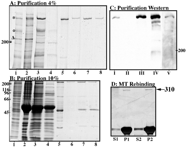
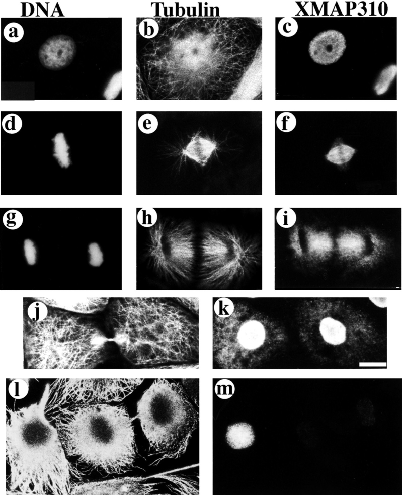
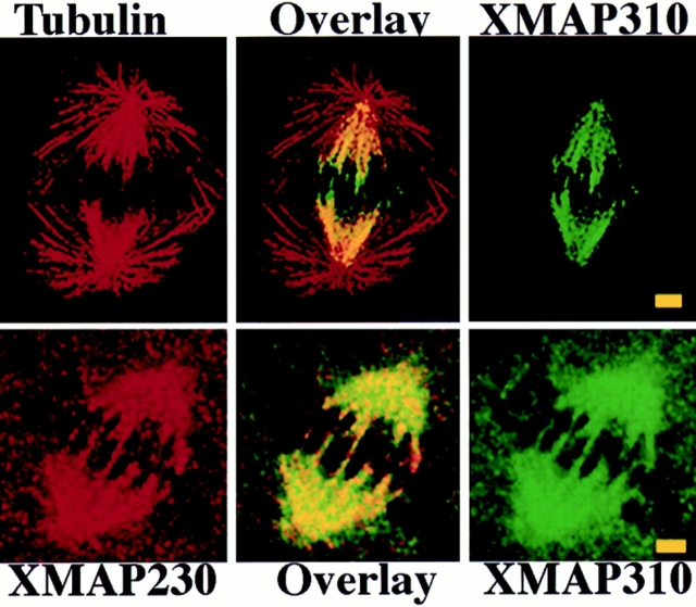
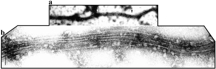
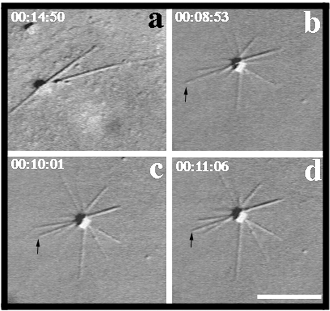
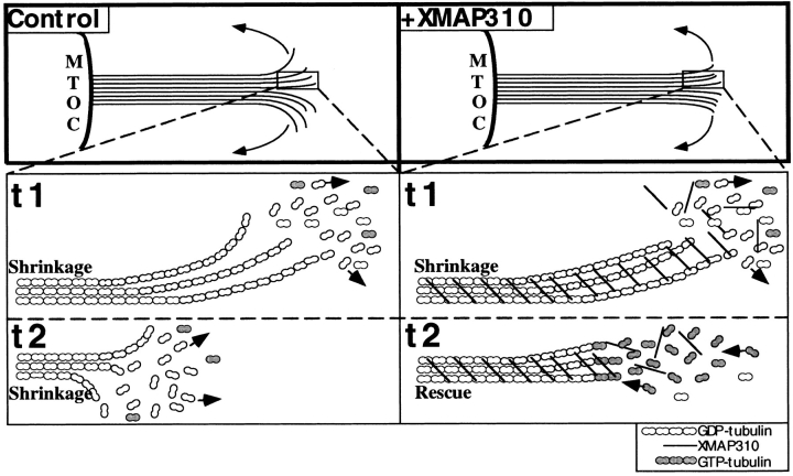
To understand the role of microtubule-associated proteins (MAPs) in the regulation of microtubule (MT) dynamics we have characterized MAPs prepared from Xenopus laevis eggs (Andersen, S.S.L., B. Buendia, J.E. Domínguez, A. Sawyer, and E. Karsenti. 1994. J. Cell Biol. 127:1289-1299). Here we report on the purification and characterization of a 310-kD MAP (XMAP310) that localizes to the nucleus in interphase and to mitotic spindle MTs in mitosis. XMAP310 is present in eggs, oocytes, a Xenopus tissue culture cell line, testis, and brain. We have purified XMAP310 to homogeneity from egg extracts. The purified protein cross-links pure MTs. Analysis of the effect of this protein on MT dynamics by time-lapse video microscopy has shown that it increases the rescue frequency 5-10-fold and decreases the shrinkage rate twofold. It has no effect on the growth rate or the catastrophe frequency. Microsequencing data suggest that XMAP230 and XMAP310 are novel MAPs. Although the three Xenopus MAPs characterized so far, XMAP215 (Vasquez, R.J., D.L. Gard, and L. Cassimeris. 1994. J. Cell Biol. 127:985-993), XMAP230, and XMAP310 are localized to the mitotic spindle, they have distinct effects on MT dynamics. While XMAP215 promotes rapid MT growth, XMAP230 decreases the catastrophe frequency and XMAP310 increases the rescue frequency. This may have important implications for the regulation of MT dynamics during spindle morphogenesis and chromosome segregation.


Figures

Figure 3
Figure 3
Purification of XMAP310. A and B show the different steps of the purification by Coomassie-stained 4% (A) and 10% (B) SDS-PAGE: lane 1, total MAPs eluted with salt (Andersen et al., 1994) from taxol stabilized MTs; lane 2, MT pellet of Ca2+ undepolymerized MTs; lane 3, Supernatant of Ca2+ depolymerized MTs/Sample loaded on the MonoS column; lane 4, Flow-through from the MonoS column; lane 5, pooled fractions from the MonoS column (the major 49-kD protein was determined as EF1-γ); lanes 6–8, fractions from the Superose 6 column. (C) Steps of the purification monitored with the Q4 mAb: lane I, start extract; lane II, extract after depletion with MTs; lane III, total MTs (V = 2 ml); lane IV, Ca2+-stable MT fraction (V = 500 μl); lane V, pooled fractions from the MonoS column (compare with A and B, lanes 5). (D) Purified XMAP310 rebinds to and pellets (P) with taxol stabilized MTs. Binding to MTs was for 10 min on ice. In lane 1 the MT concentration (in tubulin equivalents) was 2.5 μM and in 2 0.6 μM. However, the total amount of MTs used was the same in lanes 1 and 2 (S, supernatant after pelleting MTs).
Figure 1
Figure 1
XMAP310 localization in XL177 cells (Miller and Daniel, 1977) during the cell cycle. XL177 cells were stained with: Hoechst (a, d, g), a rabbit anti-tubulin pAb (b, e, h, j, l), and the Q4 mAb (c, f, i, k, m). Interphase (a–c), metaphase/early anaphase after pr-extraction with 0.5% TX-100 (d–f), late anaphase (g–i), late telophase without preextraction (j and k), late telophase with preextraction (l and m). Bar: (a–k) 10 μm; (l and m) 12.5 μm.
Figure 2
Figure 2
(Top) XMAP310 localization in the mitotic spindle: MTs stained with a rabbit anti-tublin pAb in the rhodamine channel (Tubulin), XMAP310 in the fluorescein channel (XMAP310) and in overlay (Overlay). (Bottom) XMAP230 and XMAP310 colocalize (Overlay) to the central region of the mitotic spindle in XL177 cells. Images were obtained by confocal microscopy. Bar, 2.5 μm.
Figure 4
Figure 4
Electron micrographs showing cross-linked MTs in bundles formed in the presence of XMAP310. After spontaneous assembly in the presence XMAP310, MTs were spotted onto carbon grids and visualized by negative stain and electron microscopy. (a) A long bundle consisting of three cross-linked MTs (b) magnification of the arrow-outlined area in a, note the uniform short spacing of the MTs. Bars: (a) 200 nm; (b) 60 nm.
Figure 5
Figure 5
MT dynamics visualized by video microscopy. MTs are nucleated by a centrosome (center of the MT asters). (a) MTs polymerized in the absence of XMAP310. (b–d) MTs polymerized with XMAP310. The MT indicated with an arrow experienced a catastrophe shortly after the frame shown in b, and shrank to the length shown in c where it was rescued and continued to grow, as shown in d. Time since transfer to 37°C is indicated by xx:xx:xx, which represents hours:minutes:seconds, respectively. Bar, 10 μm.
Figure 6
Figure 6
Model for how XMAP310 may promote rescues and MT cross-linking. (Top) Plane-projection of a depolymerizing (shrinking) MT in the absence (Control) or presence of XMAP310 (+XMAP310), with protofilaments (lines) peeling off the MT by out-wards curvature (arrows). Note the more blunt MT end in the presence of XMAP310 which could be promoted if cross-linking of protofilaments by XMAP310 prevents the outwards curvature of protofilaments. (Bottom) Magnifications of the boxed areas, showing part of the MTs at two different time points, t1 and t2. In the Control, the MT is shrinking both at t1 and t2, by loss of GDP-tubulin (arrows). In the presence of XMAP310 the MT is shrinking at t1. At t2 the MT has been rescued. A depolymerizing MT (t1) will meet resistance towards depolymerization due to the cross-linking of protofilaments/MTs, but XMAP310 does not dramatically prevent the loss of the tubulin dimers from the depolymerizing ends (little effect on vs). Eventually depolymerization of the MT is blocked long enough to maintain the depolymerizing MT-GDP-lattice in a straight conformation allowing new GTP-tubulin subunits to be added to the end, and the MT has been rescued (arrows indicate the direction of the flux of tubulin subunits).

References

    1. Andersen SSL, Buendia B, Domínguez JE, Sawyer A, Karsenti E. Effect on microtubule dynamics of XMAP230, a microtubule-associated protein present in Xenopus laeviseggs and dividing cells. J Cell Biol. 1994;127:1289–1299. - PMC - PubMed
    1. Andersen SSL, Ashford AJ, Tournebize R, Gavet O, Sobel A, Hyman AA, Karsenti E. Mitotic chromatin regulates phosphorylation of Stathmin/Op18. Nature. 1997;389:640–643. - PubMed
    1. Ashford, A.J., S.S.L. Andersen, and A.A. Hyman. 1998. Purification of bovine brain tubulin. In Cell Biology: A Laboratory Handbook. J.E. Celis, editor. Academic Press, San Diego. Vol. II. 205–212.
    1. Belmont LD, Mitchison TJ. Identification of a protein that interacts with tubulin dimers and increases the catastrophe rate of microtubules. Cell. 1996;84:623–631. - PubMed
    1. Belmont LD, Hyman AA, Sawin KE, Mitchison TJ. Real-time visualization of cell cycle-dependent changes in microtubule dynamics in cytoplasmic extracts. Cell. 1990;62:579–589. - PubMed

Publication types