. 1997 Nov 17;139(4):1025-32.
doi: 10.1083/jcb.139.4.1025.

Precocious mammary gland development in P-cadherin-deficient mice

Affiliations

Precocious mammary gland development in P-cadherin-deficient mice

G L Radice et al. J Cell Biol. .

Abstract

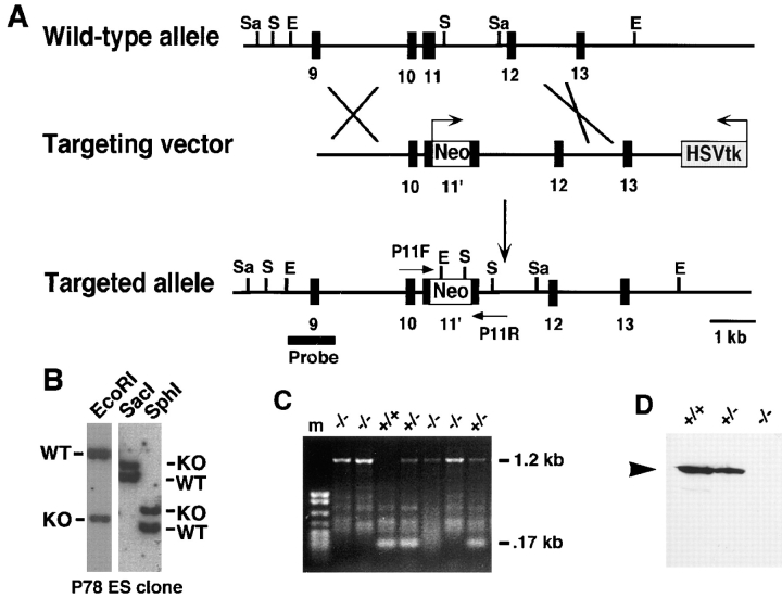
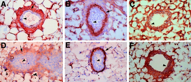
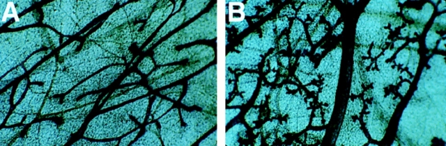
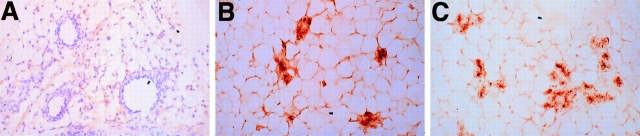
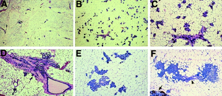
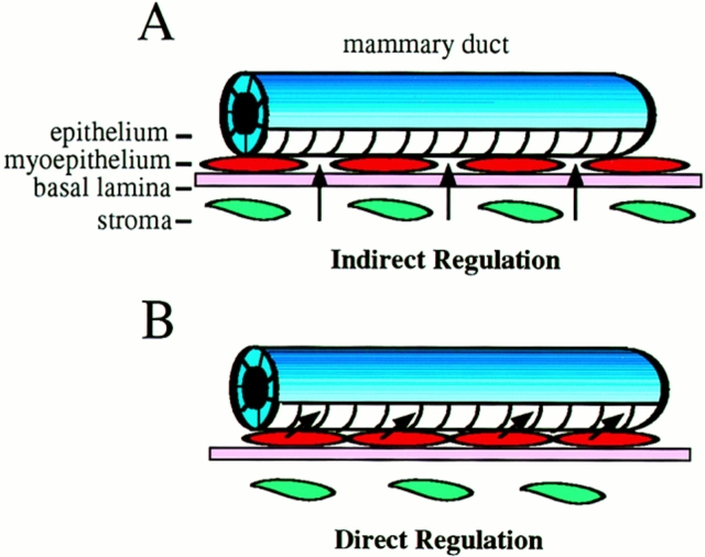
To investigate the functions of P-cadherin in vivo, we have mutated the gene encoding this cell adhesion receptor in mice. In contrast to E- and N-cadherin- deficient mice, mice homozygous for the P-cadherin mutation are viable. Although P-cadherin is expressed at high levels in the placenta, P-cadherin-null females are fertile. P-cadherin expression is localized to the myoepithelial cells surrounding the lumenal epithelial cells of the mammary gland. The role of the myoepithelium as a contractile tissue necessary for milk secretion is clear, but its function in the nonpregnant animal is unknown. The ability of the P-cadherin mutant female to nurse and maintain her litter indicates that the contractile function of the myoepithelium is not dependent on the cell adhesion molecule P-cadherin. The virgin P-cadherin-null females display precocious differentiation of the mammary gland. The alveolar-like buds in virgins resemble the glands of an early pregnant animal morphologically and biochemically (i.e., milk protein synthesis). The P-cadherin mutant mice develop hyperplasia and dysplasia of the mammary epithelium with age. In addition, abnormal lymphocyte infiltration was observed in the mammary glands of the mutant animals. These results indicate that P-cadherin-mediated adhesion and/or signals derived from cell-cell interactions are important determinants in negative growth control in the mammary gland. Furthermore, the loss of P-cadherin from the myoepithelium has uncovered a novel function for this tissue in maintaining the undifferentiated state of the underlying secretory epithelium.


Figures

Figure 1
Figure 1
Targeted disruption of the P-cadherin gene. (A) Schematic representation of the expected gene replacement at the P-cadherin locus. Exons are represented as closed boxes. The MC1-neomycin resistance cassette and MC1-thymidine kinase cassette are designated Neo and HSVtk, respectively; arrows indicate the orientation of the genes. The flanking probe used for screening ES cell clones and genotyping mice is shown (Probe). Restriction endonuclease sites are abbreviated as follows: E, EcoRI; S, SphI; Sa, SacI. The PCR primers, P11F and P11R, used for genotyping are shown. (B) Southern blot analysis of a targeted ES cell clone. The wild-type (WT) and knockout (KO) genomic fragments are indicated. (C) PCR blot analysis of P-cadherin intercross progeny indicating the genotypes. The wild-type, 0.17 kb, and mutant, 1.2 kb, PCR products are shown. (D) Western analysis of placental lysates from P-cadherin intercross progeny. The arrowhead indicates the 118-kD P-cadherin protein recognized by the mAb PCD-1. No P-cadherin protein was detected in the homozygous mice. kb, kilobases; m, markers.
Figure 2
Figure 2
Cadherin expression in wild-type and mutant mammary ducts. Mammary gland sections of wild-type (A–C) and mutant (D– F) animals were immunostained for P-cadherin (A and D), smooth muscle actin (B and E), and E-cadherin (C and F). P-cadherin is present in the myoepithelial cells surrounding the lumenal epithelial cells in the wild-type duct (A) and absent in the mutant duct (D). A diffuse nonspecific background staining was observed in the mutant (D), but the characteristic dark linear staining of the myoepithelial cells (A) was absent in the mutant. Myoepithelial cells are present in both the wild-type (B) and mutant (E) as shown by the smooth muscle actin staining. E-cadherin expression appears similar in wild-type (C) and mutant (F) ducts. The arrowheads indicate lumenal epithelial cells and arrows indicate myoepithelial cells. Original magnification 125×.
Figure 3
Figure 3
Structure of normal and mutant mammary glands in virgin female mice. Whole-gland stain of (A) wild-type and (B) mutant mammary gland from 10-wk-old virgin animals. P-cadherin–deficient virgin females display precocious alveolar differentiation. Original magnification 10×.
Figure 4
Figure 4
Expression of milk protein in P-cadherin–null virgin mice. Mammary gland sections of wild-type virgin (A) , mutant virgin (B), and wild-type pregnant day 14 (C) animals were immunostained with a polyclonal antibody against caseins. The P-cadherin–deficient virgin mammary gland express casein(s) similarly to a pregnant animal. The wild-type virgin gland was counterstained to distinguish the ducts. Original magnification 62×.
Figure 5
Figure 5
P-cadherin mutant mice develop focal hyperplasia and dysplasia with age. Histology of virgin mammary tissue from wild-type (A) and mutant (B–F) animals, 19–24 mo of age. The mutant mice (B) exhibit extensive alveolar differentiation compared with their control littermates (A). Higher magnification of the alveoli (C). P-cadherin mutant animals develop fibroblastic (D) and secretory hyperplasia (E) and exhibit extensive lymphocyte infiltration (F). The lymphocytes are concentrated around the ducts (arrowhead). A portion of the lymph node is shown (arrow). Original magnifications: (A and B) 12.5×; (C–F) 31×.
Figure 6
Figure 6
Possible models for regulation of mammary gland differentiation by P-cadherin. (A) The indirect regulation model is based on a cell adhesion defect. For example, inductive signals from the surrounding ECM (basal lamina) and/or stroma may more easily permeate a less cohesive myoepithelium leading to differentiation of the epithelium. (B) The direct regulation model is based on a P-cadherin–mediated cell signaling defect. For example, loss of P-cadherin may cause the myoepithelium to relay a positive signal(s), or alternatively, it may relieve an inhibitory signal(s) resulting in differentiation of the lumenal epithelium. The cell adhesion and cell signaling models are not mutually exclusive.

References

    1. Bchini O, Andres AC, Schubaur B, Mehtali M, LeMeur M, Lathe R, Gerlinger P. Precocious mammary gland development and milk protein synthesis in transgenic mice ubiquitously expressing human growth hormone. Endocrinology. 1991;128:539–546. - PubMed
    1. Behrens J, von Kries JP, Kuhl M, Bruhn L, Wedlich D, Grosschedl R, Birchmeier W. Functional interaction of β-catenin with the transcription factor LEF-1. Nature. 1996;382:638–642. - PubMed
    1. Birchmeier W, Behrens J. Cadherin expression in carcinomas: role in the formation of cell junctions and the prevention of invasiveness. Biochim Biophys Acta. 1994;1198:11–26. - PubMed
    1. Bradley, A. 1987. Production and analysis of chimeric mice. In Teratocarcinomas and Embryonic Stem Cells: A Practical Approach. E.J. Robertson, editor. IRL Press, Oxford. 113–151.
    1. Bussemakers MJG, Bokhoven AV, Voller M, Smit FP, Schalken JA. The genes for the calcium-dependent cell adhesion molecules P- and E-cadherin are tandemly arranged in the human genome. Biochem Biophys Res Com. 1994;203:1291–1294. - PubMed

Publication types