. 1997 Nov 17;186(10):1645-53.
doi: 10.1084/jem.186.10.1645.

Cytotoxic T lymphocyte antigen 4 (CTLA-4) interferes with extracellular signal-regulated kinase (ERK) and Jun NH2-terminal kinase (JNK) activation, but does not affect phosphorylation of T cell receptor zeta and ZAP70

Affiliations

Cytotoxic T lymphocyte antigen 4 (CTLA-4) interferes with extracellular signal-regulated kinase (ERK) and Jun NH2-terminal kinase (JNK) activation, but does not affect phosphorylation of T cell receptor zeta and ZAP70

C R Calvo et al. J Exp Med. .

Abstract

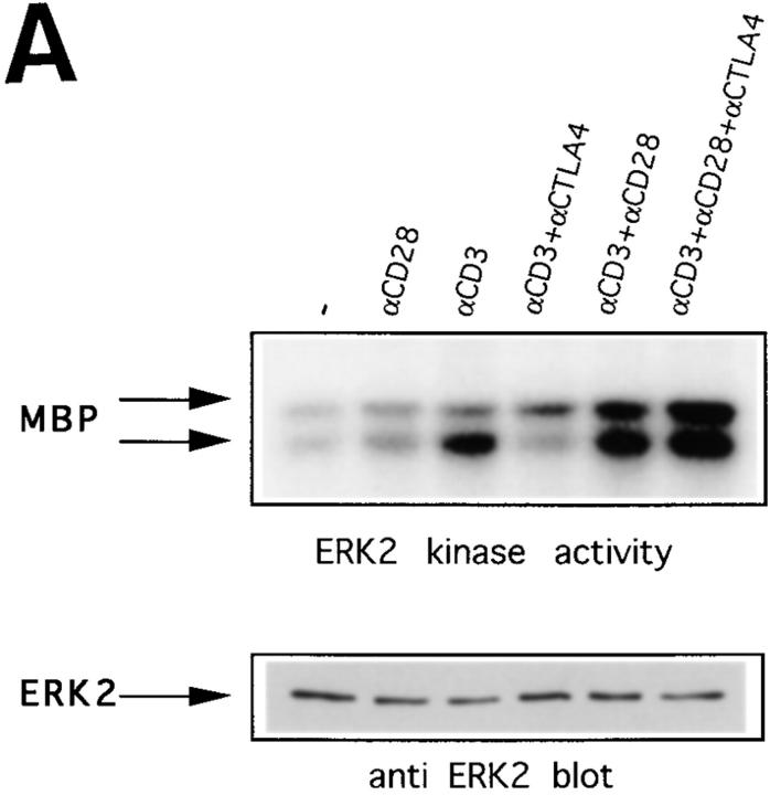
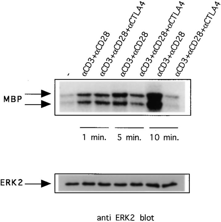
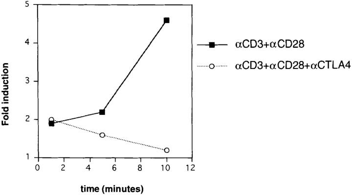
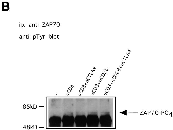
Cytotoxic T lymphocyte antigen 4 (CTLA-4) is an important regulator of T cell homeostasis. Ligation of this receptor leads to prominent downregulation of T cell proliferation, mainly as a consequence of interference with IL-2 production. We here report that CTLA-4 engagement strikingly selectively shuts off activation of downstream T cell receptor (TCR)/CD28 signaling events, i.e., activation of the microtubule-associated protein kinase (MAPKs) ERK and JNK. In sharp contrast, proximal TCR signaling events such as ZAP70 and TCR-zeta chain phosphorylation are not affected by CTLA-4 engagement on activated T cells. Since activation of the ERK and JNK kinases is required for stimulation of interleukin (IL)-2 transcription, these data provide a molecular explanation for the block in IL-2 production imposed by CTLA-4.


Figures

Figure 1
Figure 1
CTLA-4 inhibits ERK2 activity induced by anti-CD3 in preactivated T cells, but not in naive T cells. (A) 2-d preactivated T cells were coated on ice with the indicated combinations of anti-CD3, anti-CD28, and anti–CTLA-4. 1 min after addition of cross-linking anti– hamster antibody (10 μg/ml) in warm (37°C) medium the cells were lysed, ERK2 was immunoprecipitated, and in vitro kinase reactions were performed using MBP as substrate in the presence of γ-[32P]ATP. Indicated by arrows are the bands representing phosphorylated MBP (top). The same lysates were tested for equal protein abundance on immunoblot (bottom). (B) Naive purified T cells were rested for 5 h and coated with the different combinations of antibodies as indicated. Cells were lysed 1 or 5 min after addition of warm cross-linking antibody and in vitro kinase reactions were performed as in A.
Figure 1
Figure 1
CTLA-4 inhibits ERK2 activity induced by anti-CD3 in preactivated T cells, but not in naive T cells. (A) 2-d preactivated T cells were coated on ice with the indicated combinations of anti-CD3, anti-CD28, and anti–CTLA-4. 1 min after addition of cross-linking anti– hamster antibody (10 μg/ml) in warm (37°C) medium the cells were lysed, ERK2 was immunoprecipitated, and in vitro kinase reactions were performed using MBP as substrate in the presence of γ-[32P]ATP. Indicated by arrows are the bands representing phosphorylated MBP (top). The same lysates were tested for equal protein abundance on immunoblot (bottom). (B) Naive purified T cells were rested for 5 h and coated with the different combinations of antibodies as indicated. Cells were lysed 1 or 5 min after addition of warm cross-linking antibody and in vitro kinase reactions were performed as in A.
Figure 2
Figure 2
Combined triggering of CD3 and CD28 delays downregulation of ERK2 activity by CTLA-4. Preactivated T cells were coated with different combinations of antibodies as in Fig. 1 and lysed 1, 5, or 10 min after addition of warm cross-linking antibody. ERK2 was immunoprecipitated and in vitro kinase reactions were performed (top). The same lysates were tested for ERK2 protein abundance (middle). ERK2 activities were quantified using a phosphorimager and represented as relative values compared to ERK2 activities from unstimulated cells (unstimulated = 1; bottom).
Figure 2
Figure 2
Combined triggering of CD3 and CD28 delays downregulation of ERK2 activity by CTLA-4. Preactivated T cells were coated with different combinations of antibodies as in Fig. 1 and lysed 1, 5, or 10 min after addition of warm cross-linking antibody. ERK2 was immunoprecipitated and in vitro kinase reactions were performed (top). The same lysates were tested for ERK2 protein abundance (middle). ERK2 activities were quantified using a phosphorimager and represented as relative values compared to ERK2 activities from unstimulated cells (unstimulated = 1; bottom).
Figure 3
Figure 3
CTLA-4 reduces JNK activity induced by CD3 and CD28 triggering. Preactivated T cells were coated with different combinations of antibodies and lysed 10 min after addition of warm cross-linking antibody. Lysates were tested for JNK activity by precipitation with GST–c-jun precoupled to glutathione beads followed by in vitro kinase reactions as described in Materials and Methods. Phosphorylated GST–c-jun is indicated by the arrow (top). The same lysates were tested for JNK protein abundance on immunoblot (middle). JNK activities were quantified using a phosphorimager and represented as relative values compared to JNK activity from unstimulated cells (unstimulated = 1; bottom). Data for the 10-min time point are shown because no JNK activity could be measured at earlier times.
Figure 3
Figure 3
CTLA-4 reduces JNK activity induced by CD3 and CD28 triggering. Preactivated T cells were coated with different combinations of antibodies and lysed 10 min after addition of warm cross-linking antibody. Lysates were tested for JNK activity by precipitation with GST–c-jun precoupled to glutathione beads followed by in vitro kinase reactions as described in Materials and Methods. Phosphorylated GST–c-jun is indicated by the arrow (top). The same lysates were tested for JNK protein abundance on immunoblot (middle). JNK activities were quantified using a phosphorimager and represented as relative values compared to JNK activity from unstimulated cells (unstimulated = 1; bottom). Data for the 10-min time point are shown because no JNK activity could be measured at earlier times.
Figure 4
Figure 4
Anti-CD3–induced phosphorylation of TCR-ζ and ZAP70 is not affected by CTLA-4. (A) Preactivated T cells were coated with different combinations of antibodies and lysed 1, 5, or 10 min after addition of warm cross-linking antibody. TCR-ζ was immunoprecipitated and immunoblotted with antiphosphotyrosine antibody 4G10. Indicated with arrows are the p21 and p23 isoforms of tyrosine phosphorylated TCR-ζ. (B) Lysates prepared as in A from cells stimulated for 1 min were immunoprecipitated with anti-ZAP70 antibody and immunoblotted with antiphosphotyrosine antibody 4G10. Indicated with an arrow is phosphorylated ZAP70. Similar results were also obtained on lysates from 5 and 10 min–stimulated cells (not shown).
Figure 4
Figure 4
Anti-CD3–induced phosphorylation of TCR-ζ and ZAP70 is not affected by CTLA-4. (A) Preactivated T cells were coated with different combinations of antibodies and lysed 1, 5, or 10 min after addition of warm cross-linking antibody. TCR-ζ was immunoprecipitated and immunoblotted with antiphosphotyrosine antibody 4G10. Indicated with arrows are the p21 and p23 isoforms of tyrosine phosphorylated TCR-ζ. (B) Lysates prepared as in A from cells stimulated for 1 min were immunoprecipitated with anti-ZAP70 antibody and immunoblotted with antiphosphotyrosine antibody 4G10. Indicated with an arrow is phosphorylated ZAP70. Similar results were also obtained on lysates from 5 and 10 min–stimulated cells (not shown).

References

    1. Abbas AK. Die and let live: eliminating dangerous lymphocytes. Cell. 1996;84:655–658. - PubMed
    1. Zheng L, Fisher G, Miller RE, Peschon J, Lynch DH, Lenardo MJ. Induction of apoptosis in mature T cells by tumour necrosis. Nature (Lond) 1995;377:348–352. - PubMed
    1. Tivol EA, Schweitzer AN, Sharpe AH. Costimulation and autoimmunity. Curr Opin Immunol. 1996;8:822–830. - PubMed
    1. Lenschow DJ, Walunas TL, Bluestone JA. CD28/B7 system of T cell costimulation. Annu Rev Immunol. 1996;14:233–258. - PubMed
    1. Walunas TL, Lenschow DJ, Bakker CY, Linsley PS, Freeman GJ, Green JM, Thompson CB, Bluestone JA. CTLA-4 can function as a negative regulator of T cell activation. Immunity. 1994;1:405–413. - PubMed

Publication types

MeSH terms