. 1997 Dec 1;17(23):8937-44.
doi: 10.1523/JNEUROSCI.17-23-08937.1997.

Epsilon subunit-containing acetylcholine receptors in myotubes belong to the slowly degrading population

Affiliations

Epsilon subunit-containing acetylcholine receptors in myotubes belong to the slowly degrading population

C Sala et al. J Neurosci. .

Abstract

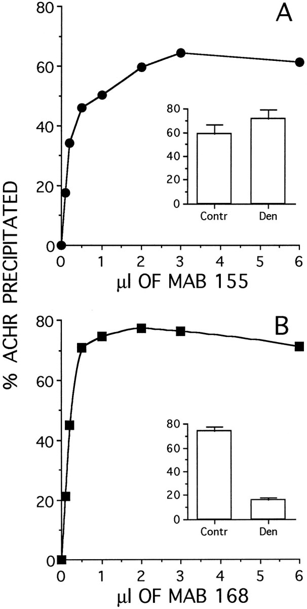
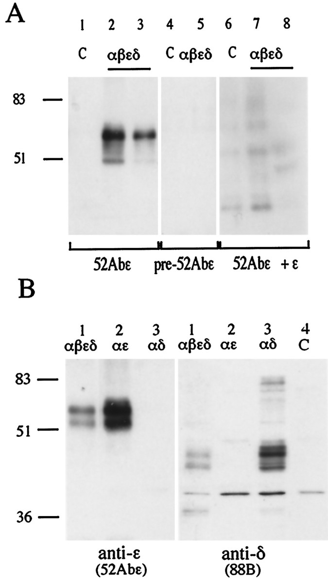
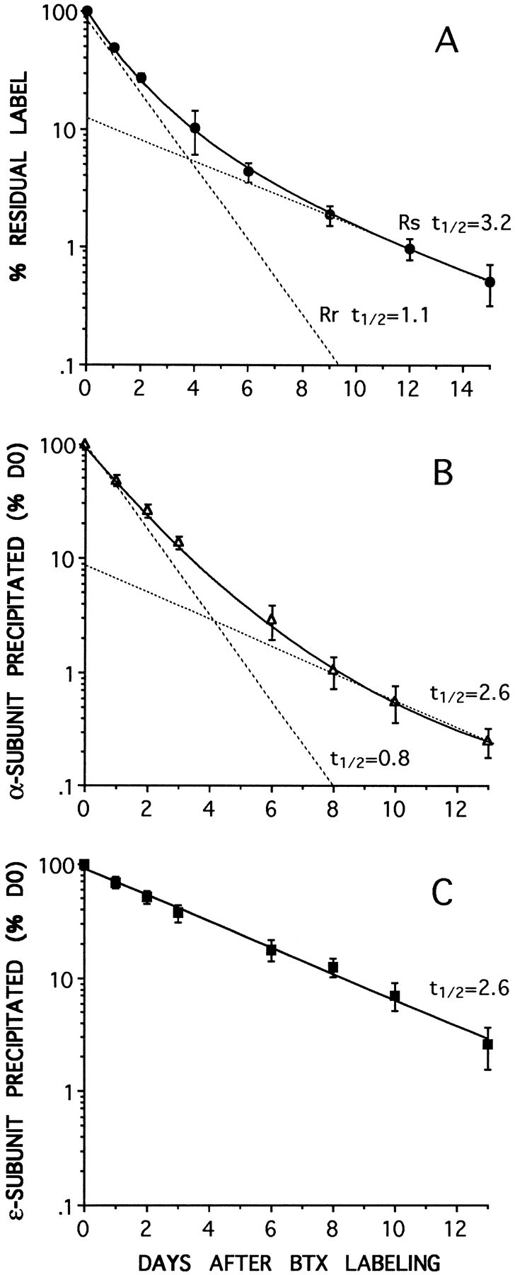
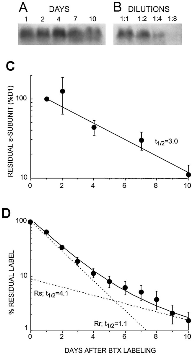
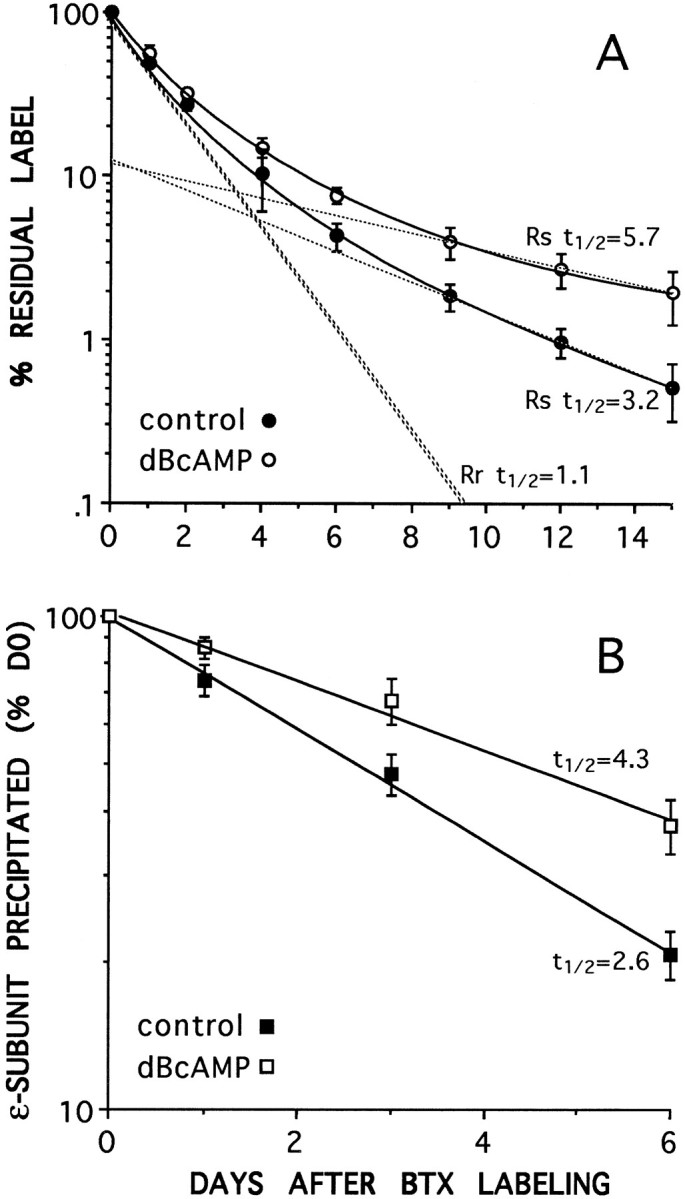
Two types of muscle acetylcholine receptors (AChRs) can be distinguished on the basis of their degradation rates and sensitivities to innervation, muscle activity, and agents elevating intracellular cAMP. The first type (Rs), is present in a stable form (degradation t1/2 = approximately 10 d) at the adult innervated neuromuscular junctions (NMJs). Rs can also exist in a less stable form (called accelerated Rs; t1/2 = approximately 3-5 d) at denervated NMJs and in aneurally cultured myotubes; agents that increase intracellular cAMP reversibly modulate Rs stability. The second type of AChR is a rapidly degrading receptor (Rr) expressed only in embryonic and noninnervated muscles. Rr can be stabilized by ATP and not by cAMP. This study tested the hypothesis that the degradation properties unique to the Rs are attributable to the presence of the epsilon subunit. Immunoprecipitation and Western blot analysis of AChRs extracted from rat muscle cells in tissue culture showed that AChRs recognized by antibodies against the epsilon subunit degraded as a single population with a half-life similar to that of the slow component, Rs, in these cells. In addition, as for Rs receptors in denervated NMJs and cultured muscle cell, the degradation rate of these epsilon-containing AChRs was stabilized by dibutyryl-cAMP. The data indicate that the epsilon-containing AChRs behave like Rs. Thus, the presence of the epsilon subunit is sufficient for selecting an AChR molecule to the Rs pool.


Figures

Fig. 1.
Fig. 1.
Efficiency of immunoprecipitation of adult AChR by the anti-α subunit mAb 155 (A) or the anti-ε subunit mAb 168 (B). Innervated soleus muscles were labeled in vitro with 125I-BTX, extracted with 2% Triton X-100, and immunoprecipitated with mAbs 155 and 168 (see Materials and Methods). The amount of immunoprecipitated radioactivity (AChR) was expressed as the percentage of total activity in the reaction mixture. The insets compare the maximum percent immunoprecipitated from innervated control (Contr) and 7 d denervated (Den) soleus muscle at saturating concentrations of antibodies.
Fig. 2.
Fig. 2.
The anti-ε 52Abε polyclonal antibody specifically recognizes the ε AChR subunit on Western blots.A, HEK 293 kidney fibroblast cells were transfected with cDNAs coding for the αβεδ subunits (2:1:1:1) of rat AChR (lanes 2, 3, 5, 7, 8) or left as untreated controls (lanes 1, 4, 6). The 52Abε antibody recognized a major band at molecular weight 60 kDa in the ε-containing AChR-transfected cells (lanes 2, 3) but not in the control nontransfected cells (lane 1). No bands were seen with preimmune serum (lanes 4, 5) or after preabsorption of the 52Abε antibody with the ε peptide (lanes 6–8). Numbers on theleft correspond to molecular mass standards (in kilodaltons). B, Samples obtained from HEK 293 cells transfected with αβεδ (2:1:1:1), αε (2:1), or αδ (2:1) cDNAs were incubated with either the anti-ε subunit 52Abε antibody or the anti-γ/δ subunit mAb 88B. The 52Abε antibody recognized a major bands of 60 kDa in the cells transfected with αβεδ and αε but none in the cells transfected with αδ. mAb 88B specifically immunodecorated the lanes loaded with membrane preparations obtained from the cells transfected with αβεδ and αδ but not from cells transfected with αε or the nontransfected control. (C).
Fig. 3.
Fig. 3.
AChR degradation rates measured by either125I-BTX release (A) or by subunit-specific immunoprecipitation with anti-α (B) or anti-ε (C) subunit mAbs. At each of the time points data are expressed as a percentage of the radioactivity immunoprecipitated from the cells at the time of labeling with 125I-BTX (day 0). Biphasic degradation curves for total AChRs are obtained both by 125I-BTX release (A) or by anti-α subunit mAb 155 immunoprecipitation (B). In each case the best fit to the data (solid curve) consists of the sum of two components (dashed lines): a slow component (Rs), with at½ value of ∼3 d (3.2 and 2.6 d), and a fast component (Rr), with at½ of ∼1 d (1.1 and 0.8 d). The receptor immunoprecipitated by the anti-ε subunit mAb 168 (C) degrades as a single exponential (solid line) with at½ of 2.6 d, similar to the slow components in A and B. No fast component is seen. The values are the means ± SD of at least three different experiments. The data in B andC were obtained from the same sets of cells.
Fig. 4.
Fig. 4.
Western blot analysis of ε-AChR degradation in cultured muscle cells compared with total AChR degradation measured by125I-BTX release. A, Sample of Western blots at different times after BTX labeling, showing the ∼60 kDa immunogenic band recognized by the anti-ε subunit antibody 52Abε (as in Fig. 2). B, Successive dilutions of antigen established linearity of the Western blot response. C, A plot of the density of the anti-ε subunit bands, decreasing with time after labeling, gives a single exponential decay with a half-life of ∼3.0 d (n = 3 experiments), similar to the slow components in Figure 3, A and B. No fast component is seen. D, Residual label from parallel plates assayed by 125I-BTX release gives a double-exponential fit for total AChR (as in Fig. 3A,B), revealing two AChR populations with slow, Rs, and fast, Rr, components having t½ values of 4.1 and 1.1 d, respectively.
Fig. 5.
Fig. 5.
Dibutyryl-cAMP (dBcAMP) slows the degradation rate of the slow component, Rs, when measured by125I-BTX release (A) and of the material immunoprecipitated by the anti-ε subunit mAb 168 (B). Incubation in dB-cAMP increases thet½ of Rs (from 3.2 to 5.8 d) and that of the ε-subunit (from 2.7 to 4.3 d) but does not affect the t½ of Rr, which remains at 1.1 d (A). Dashed lines inA give the two component exponential decays for Rs and Rr (see Materials and Methods), which when summed give the best fit to the experimental data (solid lines) for each condition. The two exponentials for Rr overlap for control and dB-cAMP-treated cells.

Similar articles

Cited by

References

    1. Andreose J, Xu R, Lømo T, Salpeter MM. Degradation of two AChR populations at rat neuromuscular junction: regulation by electrical stimulation. J Neurosci. 1993;13:3433–3438. - PMC - PubMed
    1. Blount P, Merlie JP. Characterization of an adult muscle acetylcholine receptor subunit by expression in fibroblasts. J Biol Chem. 1991;266:14692–14696. - PubMed
    1. Brehm P, Yeh E, Patrick J, Kidokoro Y. Metabolism of acetylcholine receptors on embryonic amphibian muscle. J Neurosci. 1983;3:101–107. - PMC - PubMed
    1. Brenner HR, Sakmann B. Neurotrophic control of channel properties at neuromuscular synapses of rat muscle. J Physiol (Lond) 1983;337:159–171. - PMC - PubMed
    1. Brenner HR, Witzemann V, Sakmann B. Imprinting of acetylcholine receptor messenger RNA accumulation in mammalian neuromuscular synapses. Nature. 1990;344:544–547. - PubMed

Publication types

MeSH terms

LinkOut - more resources