. 1997 Dec 1;17(23):8975-83.
doi: 10.1523/JNEUROSCI.17-23-08975.1997.

Cyclin dependent kinase inhibitors and dominant negative cyclin dependent kinase 4 and 6 promote survival of NGF-deprived sympathetic neurons

Affiliations

Cyclin dependent kinase inhibitors and dominant negative cyclin dependent kinase 4 and 6 promote survival of NGF-deprived sympathetic neurons

D S Park et al. J Neurosci. .

Abstract

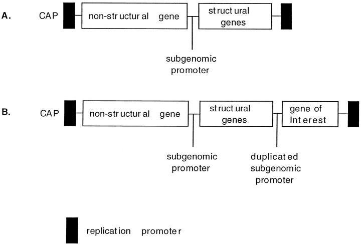
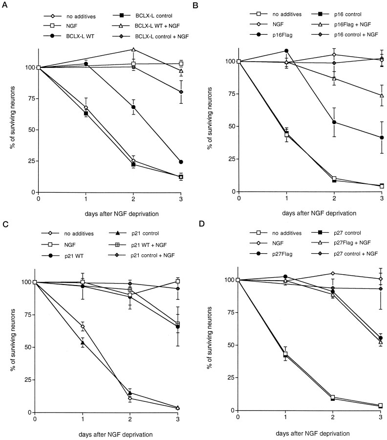
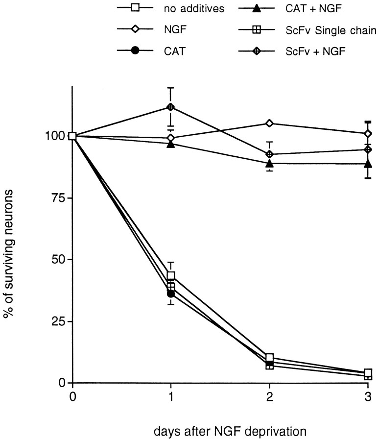
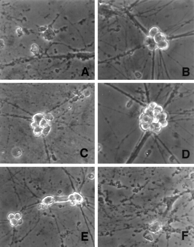
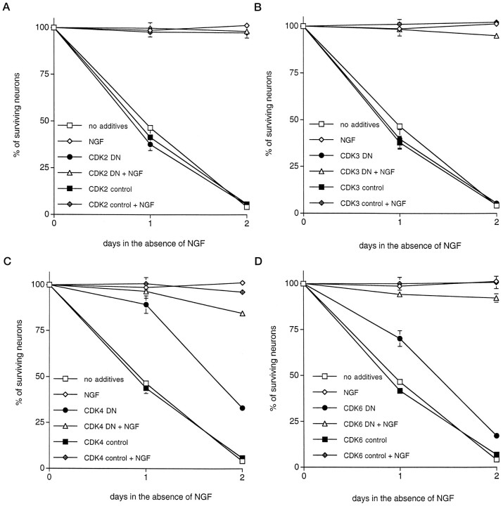
Neuronal apoptosis plays a critical role in both normal development and disease. However, the precise molecular events controlling neuronal apoptosis are not well understood. Previously, we hypothesized that cell cycle regulatory molecules function in controlling the apoptotic pathways of trophic factor-deprived neurons. To test this hypothesis, we used the RNA alphavirus Sindbis to express three known cyclin dependent kinase inhibitors (CKIs), p16(ink4), p21(waf/cip), and p27(kip1), and dominant negative mutant forms of four known G1 cyclin dependent kinases (CDKs), Cdk2, Cdk3, Cdk4, and Cdk6, in primary cultured rat superior cervical ganglion sympathetic neurons. We demonstrate that expression of each of the CKIs protects the postmitotic cultured neurons from apoptotic death evoked by withdrawal of NGF. In addition, we show that expression of dominant negative forms of Cdk4 or Cdk6, but not Cdk2 or Cdk3, protects NGF-deprived sympathetic neurons from death. Such findings suggest the participation of several CDKs and their cognate cyclins in a neuronal apoptotic pathway.


Figures

Fig. 1.
Fig. 1.
Diagram of the double subgenomic promoter containing Sindbis virus vector. A, Wild-type Sindbis vector. B, Sindbis vector with duplicated subgenomic promoter.
Fig. 2.
Fig. 2.
Expression of CDK inhibitors andBcl-xL using the Sindbis viral delivery system promotes survival of primary cultured sympathetic neurons deprived of trophic support. Each data point is the mean ± SEM of three samples and is expressed relative to the number of neurons present in each well at the time of NGF withdrawal. Control viruses for each vector were generated by removal of the start codon and, in some cases, by introduction of a premature stop site.Flag denotes that the wild-type (WT) protein has a flag epitope attached to the C terminal. The values of p derived from Student’st tests comparing the wild-type or flagged protein with the control-stop virus at days 1 and 2 are given. A–D, Effects of overexpression of Bcl-xL(p < 0.01) (A), p16 (p < 0.05) (B), p21 (p < 0.005) (C), and p27 (p < 0.05) (D) and respective controls on the time course of survival of sympathetic neurons after withdrawal of NGF. Similar results were obtained in at least three independent experiments.
Fig. 3.
Fig. 3.
Sindbis virus-mediated expression of non-cell cycle-related control proteins, CAT and anScFv single-chain antibody control, fails to promote survival of primary cultured sympathetic neurons deprived of trophic support. Each data point is the mean ± SEM of three samples and is expressed relative to the number of neurons present in each well at the time of NGF withdrawal.
Fig. 4.
Fig. 4.
Expression of FLAG-tagged p27 in sympathetic neurons grown in the presence of NGF and infected by an engineered Sindbis virus. A, B, Immunofluorescence staining with an antibody directed against the FLAG epitope of neurons in culture infected with Sindbis virus expressing p27-FLAG (A) or containing the p27 control sequence (B). Arrows indicate the locations of individual neurons. Neurons were stained 2 d after infection.
Fig. 5.
Fig. 5.
Phase contrast micrographs of rat sympathetic neurons cultured in NGF-free medium in the presence of the following additives for 2 d: (A) no additives, (B) NGF, (C) p16 Sindbis virus, (D) p21 Sindbis virus, (E) p27 Sindbis virus, and (F) noncoding control Sindbis virus. Magnification, 375×.
Fig. 6.
Fig. 6.
Expression of dominant negative Cdk4 and Cdk6 but not Cdk2 and Cdk3 promotes survival of primary cultured sympathetic neurons deprived of trophic support. Each data point is the mean ± SEM of three samples and is expressed relative to the number of neurons present in each well at the time of NGF withdrawal. Control viruses for each vector were generated by removal of the start codon and, in some cases, by introduction of a premature stop site.DN denotes dominant negative. The values ofp derived from Student’s t tests comparing the wild-type protein with the control-stop virus at days 1 and 2 are given. A–D, Effects of overexpression of DN Cdk2 (p > 0.05) (A), DN Cdk3 (p > 0.05) (B), DN Cdk4 (p < 0.01) (C), and DN Cdk6 (p < 0.025) (D) and respective controls on the time course of survival of sympathetic neurons after withdrawal of NGF. Similar results were obtained in at least three independent experiments.

Similar articles

Cited by

References

    1. Batistatou A, Greene LA. Internucleosomal DNA cleavage and neuronal cell survival/death. J Cell Biol. 1993;122:523–532. - PMC - PubMed
    1. Batistatou A, Merry DE, Korsmeyer SJ, Greene LA. Bcl-2 affects survival, but not neuronal differentiation of PC12 cells. J Neurosci. 1993;13:4422–4428. - PMC - PubMed
    1. Berry DE, Lu Y, Schimdt B, Falon PG, O’Connell C, Hu S-X, Xu H-J, Blanck G. Retinoblastoma protein inhibits IFN induced apoptosis. Oncogene. 1996;12:1809–1819. - PubMed
    1. Brooks SF, Gibson LA, Rubin LL. Apoptosis induced by NGF-withdrawal from differentiated PC12 cells involves activation of P34cdc2 kinase. Soc Neurosci Abstr. 1993;19:885.
    1. Chellepan SP, Hiebert S, Mudryj M, Horowitz JM, Nevins JR. The E2F transcription factor is a cellular target for the RB protein. Cell. 1991;65:1053–1061. - PubMed

Publication types

MeSH terms

Substances

LinkOut - more resources