Comparative Study
. 1997 Dec 1;17(23):9060-7.
doi: 10.1523/JNEUROSCI.17-23-09060.1997.

Pyruvate protects neurons against hydrogen peroxide-induced toxicity

Affiliations
Comparative Study

Pyruvate protects neurons against hydrogen peroxide-induced toxicity

S Desagher et al. J Neurosci. .

Abstract

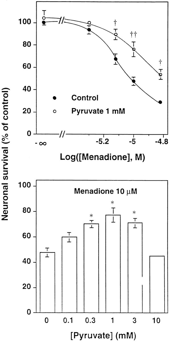
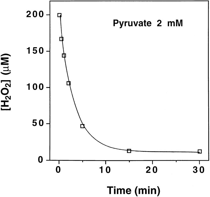
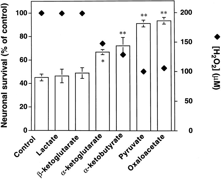
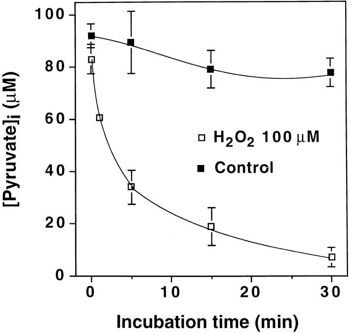
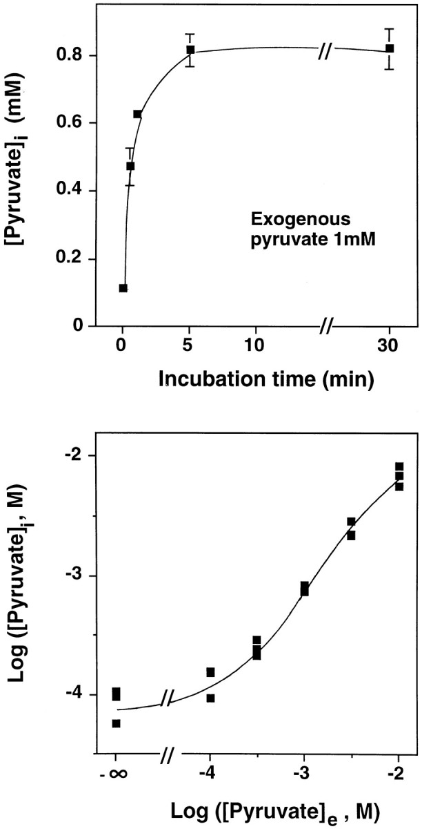
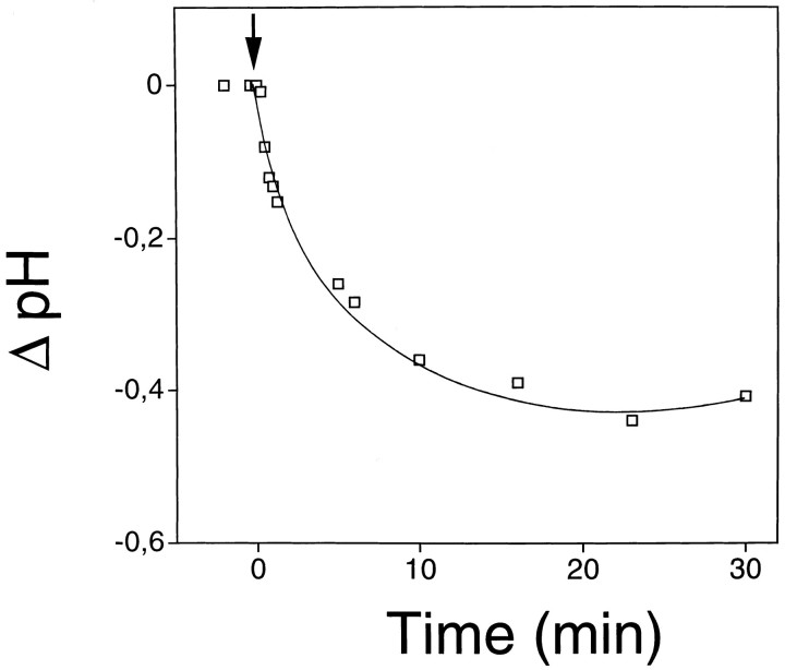
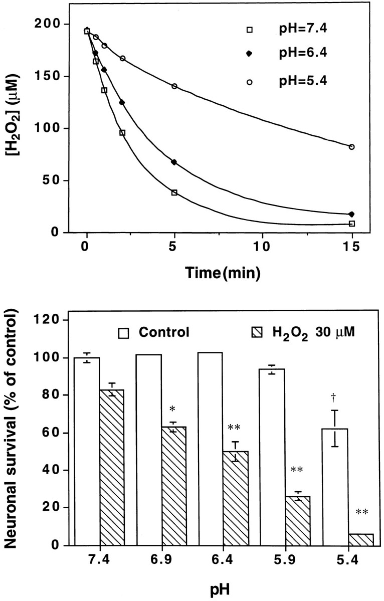
Hydrogen peroxide (H2O2) is suspected to be involved in numerous brain pathologies such as neurodegenerative diseases or in acute injury such as ischemia or trauma. In this study, we examined the ability of pyruvate to improve the survival of cultured striatal neurons exposed for 30 min to H2O2, as estimated 24 hr later by the 3-[4,5-dimethylthiazol-2-yl]-2,5-diphenyltetrazoliumbromide assay. Pyruvate strongly protected neurons against both H2O2 added to the external medium and H2O2 endogenously produced through the redox cycling of the experimental quinone menadione. The neuroprotective effect of pyruvate appeared to result rather from the ability of alpha-ketoacids to undergo nonenzymatic decarboxylation in the presence of H2O2 than from an improvement of energy metabolism. Indeed, several other alpha-ketoacids, including alpha-ketobutyrate, which is not an energy substrate, reproduced the neuroprotective effect of pyruvate. In contrast, lactate, a neuronal energy substrate, did not protect neurons from H2O2. Optimal neuroprotection was achieved with relatively low concentrations of pyruvate (</=1 mM), whereas at high concentration (10 mM) pyruvate was ineffective. This paradox could result from the cytosolic acidification induced by the cotransport of pyruvate and protons into neurons. Indeed, cytosolic acidification both enhanced the H2O2-induced neurotoxicity and decreased the rate of pyruvate decarboxylation by H2O2. Together, these results indicate that pyruvate efficiently protects neurons against both exogenous and endogenous H2O2. Its low toxicity and its capacity to cross the blood-brain barrier open a new therapeutic perspective in brain pathologies in which H2O2 is involved.


Figures

Fig. 1.
Fig. 1.
Pyruvate protects neurons from exogenous H2O2-induced toxicity. Primary cultures of striatal neurons were preincubated in Krebs’ bicarbonate buffer at 37°C for 30 min with 2 mm (top) or increasing concentrations (bottom) of sodium pyruvate and then further incubated for 30 min with increasing concentrations of H2O2 (top) or with 200 μm H2O2 (bottom) in the presence or absence of the indicated concentration of pyruvate. Pyruvate and H2O2 were simultaneously applied to the cells. Neuronal survival was estimated 24 hr later by the MTT colorimetric assay. Results are expressed as the percentage of surviving neurons compared with control cultures. Data are the mean ± SEM of three independent experiments, each performed on triplicate wells. When not visible, the sizes of the error bars are less than those of the symbols. p < 0.001; significantly different from the corresponding values determined in the absence of pyruvate (ANOVA followed by Bonferroni’s test). *p < 0.05; **p < 0.01; significantly different from the value obtained in the absence of pyruvate (ANOVA followed by Dunnett’s test).
Fig. 2.
Fig. 2.
Pyruvate partly protects neurons from menadione-induced toxicity. Primary cultures of neurons were incubated in Krebs’ bicarbonate buffer at 37°C for 1 hr with increasing concentrations of menadione in the absence or the presence of 1 mm sodium pyruvate (top) or with 10 μm menadione in the presence of increasing concentrations of pyruvate (bottom). Cells were then washed and further incubated for 30 min with or without pyruvate and replaced into the initial culture medium supplemented with the corresponding concentrations of pyruvate. Neuronal survival was estimated 24 hr later. Results are expressed as the percentage of surviving neurons compared with control cultures not treated with menadione. Data are the mean ± SEM of three independent experiments, each performed on triplicate wells. p < 0.01;††p < 0.001; significantly different from the corresponding values determined in the absence of pyruvate (ANOVA followed by Bonferroni’s test). *p < 0.01; significantly different from the value obtained in the absence of pyruvate (ANOVA followed by Dunnett’s test).
Fig. 3.
Fig. 3.
Kinetics of the reaction of H2O2 and pyruvate in the absence of cells. Pyruvate (2 mm) and H2O2 (200 μm) were mixed in Krebs’ bicarbonate buffer at 37°C in the absence of cells. The residual concentrations of H2O2 were determined at indicated times as described in Materials and Methods. Data are the mean ± SEM of three independent experiments each performed in triplicate. The error bars are not visible, because they are smaller than the symbols.
Fig. 4.
Fig. 4.
H2O2-scavenging capacities and neuroprotecting properties of various α-ketoacids. A 200 μm concentration of H2O2 was incubated with 2 mm sodium lactate, β-ketoglutarate, α-ketoglutarate, α-ketobutyrate, pyruvate, or oxaloacetate in Krebs’ bicarbonate buffer for 2 min at 37°C in the absence of cells. The residual concentration of H2O2(filled symbols) was determined in each experimental condition. The error bars are not visible, because they are smaller than the symbols. In a separate set of experiments, cultured neurons were preincubated for 30 min with a 2 mmconcentration of each compound and further incubated for 30 min with 200 μm H2O2 in their presence or absence. Neuronal survival was estimated 24 hr later. Results are expressed as the percentage of living neurons compared with cultures not treated with H2O2. Data are the mean ± SEM of three independent experiments each performed in triplicate. *p < 0.05; **p < 0.01; significantly different from the control value (ANOVA followed by Dunnett’s test).
Fig. 5.
Fig. 5.
Exogenous H2O2 decreased intracellular pyruvate. Striatal neurons were incubated in Krebs’ bicarbonate buffer with or without 100 μmH2O2 for indicated times. Then, neuronal cultures were washed with 500 IU/ml catalase for 30 sec. Cells were subsequently treated as indicated in Materials and Methods. The cytosolic neuronal volume and the residual intracellular pyruvate were measured as described in Materials and Methods. Data are the mean ± SEM of three independent experiments, each performed in triplicate.
Fig. 6.
Fig. 6.
Pyruvate uptake by striatal neurons. Primary cultures of striatal neurons were incubated in Krebs’ bicarbonate buffer at 37°C with 1 mm sodium pyruvate for the indicated times (top) or with increasing concentrations of pyruvate ([Pyruvate]e) for 10 min (bottom). Neurons were treated, and intracellular concentrations of pyruvate ([Pyruvate]i) were determined as described in Materials and Methods. Top, Data are the mean ± SEM of two independent experiments, each performed in triplicate. Bottom, Individual results of three independent experiments, also performed in triplicate.
Fig. 7.
Fig. 7.
Cytosolic acidification by 10 mmsodium pyruvate. Cultured striatal neurons, previously loaded with carboxy-SNARF-1, were perfused for 30 min with 10 mm sodium pyruvate (arrow) in Krebs’ bicarbonate buffer at a constant extracellular pH of 7.4. The exposure to pyruvate resulted in a long-lasting decrease of the 580:640 nm fluorescence ratio, determined as described in Materials and Methods. After pyruvate removal, the ratio increased, returning to its resting value by the end of a 25 min washout (data not shown). Each point is the mean from 14 cells. Two other independent experiments gave similar results.
Fig. 8.
Fig. 8.
Influence of pH on the neuroprotective effect of pyruvate. Top, Kinetics of H2O2degradation by pyruvate in acid solutions. Pyruvate (2 mm) and H2O2 (200 μm) were incubated at 37°C in the absence of cells in HEPES-buffered salt solutions adjusted to different pH for increasing times. The residual concentrations of H2O2 were determined as described in Materials and Methods. Data are the mean ± SEM of three independent experiments, each performed in triplicate. The error bars are not visible, because they are smaller than the symbols.Bottom, Neurotoxic effects of H2O2 in acid solutions. Cultured neurons were preincubated for 15 min and then incubated for 30 min with or without 30 μm H2O2 in HEPES-buffered salt solutions adjusted to different pH. Neuronal survival was estimated 24 hr later. Results are expressed as the percentage of living neurons compared with control cultures incubated at pH 7.4 in the absence of H2O2. Data are the mean ± SEM of three independent experiments, each performed in triplicate.p < 0.01; significantly different from the control value; *p < 0.05; **p < 0.01; significantly different from the value obtained in the presence of H2O2 at pH 7.4 (ANOVA followed by Dunnett’s test).

Similar articles

Cited by

References

    1. Andrae U, Singh J, Ziegler-Skylakakis K. Pyruvate and related α-ketoacids protect mammalian cells in culture against hydrogen peroxide-induced cytotoxicity. Toxicol Lett. 1985;28:93–98. - PubMed
    1. Bakker EP, Van Dam K. The movement of monocarboxylic acids across phospholipid membranes: evidence for an exchange diffusion between pyruvate and other monocarboxylate ions. Biochim Biophys Acta. 1974;339:285–289. - PubMed
    1. Beal MF. Does impairment of energy metabolism result in excitotoxic neuronal death in neurodegenerative illnesses? Ann Neurol. 1992;31:119–130. - PubMed
    1. Berridge MV, Tan AS. Characterization of the cellular reduction of 3-(4,5-dimethylthiazol-2-yl)-2,5-diphenyltetrazolium bromide (MTT): subcellular localization, substrate dependence, and involvement of mitochondrial electron transport in MTT reduction. Arch Biochem Biophys. 1993;303:474–482. - PubMed
    1. Bradford MM. A rapid and sensitive method for the quantitation of microgram quantities of protein utilizing the principle of protein dye binding. Anal Biochem. 1976;72:248–254. - PubMed

Publication types

LinkOut - more resources