Comparative Study
. 1997 Dec 1;17(23):9095-103.
doi: 10.1523/JNEUROSCI.17-23-09095.1997.

Phosphodiesterase I, a novel adhesion molecule and/or cytokine involved in oligodendrocyte function

Affiliations
Comparative Study

Phosphodiesterase I, a novel adhesion molecule and/or cytokine involved in oligodendrocyte function

B Fuss et al. J Neurosci. .

Abstract

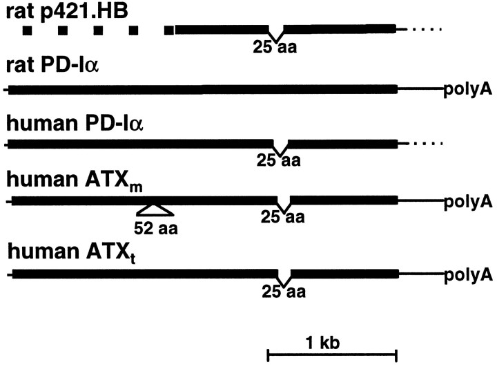
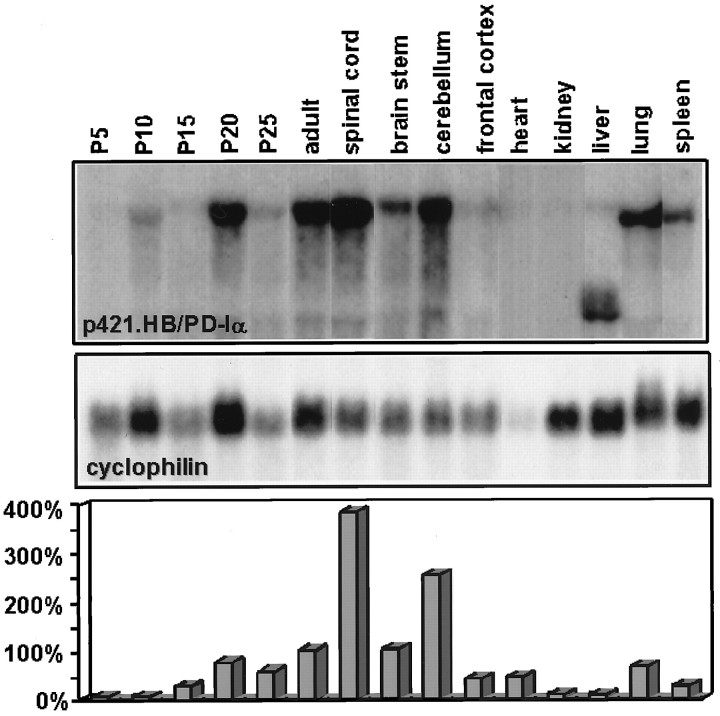
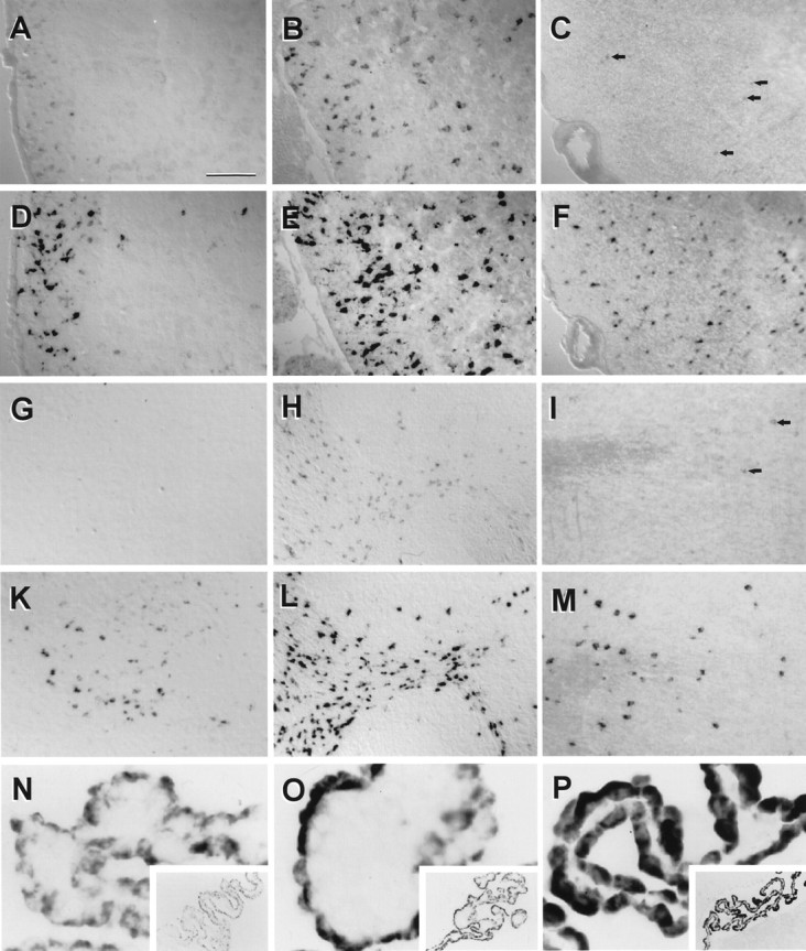
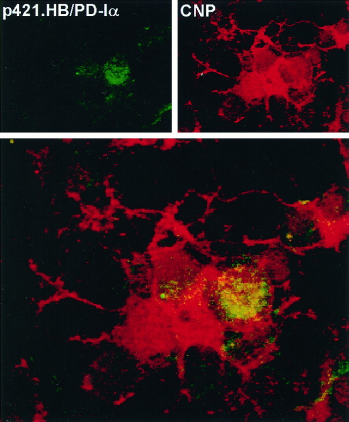
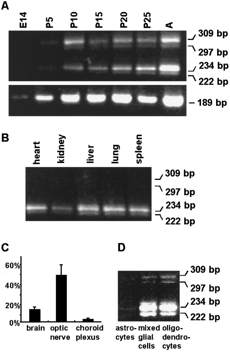
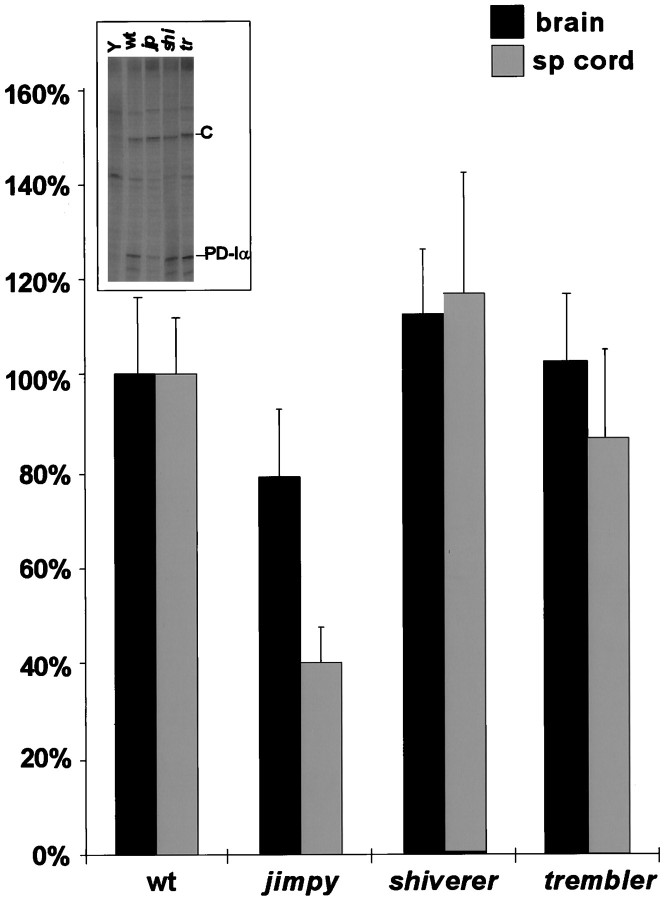
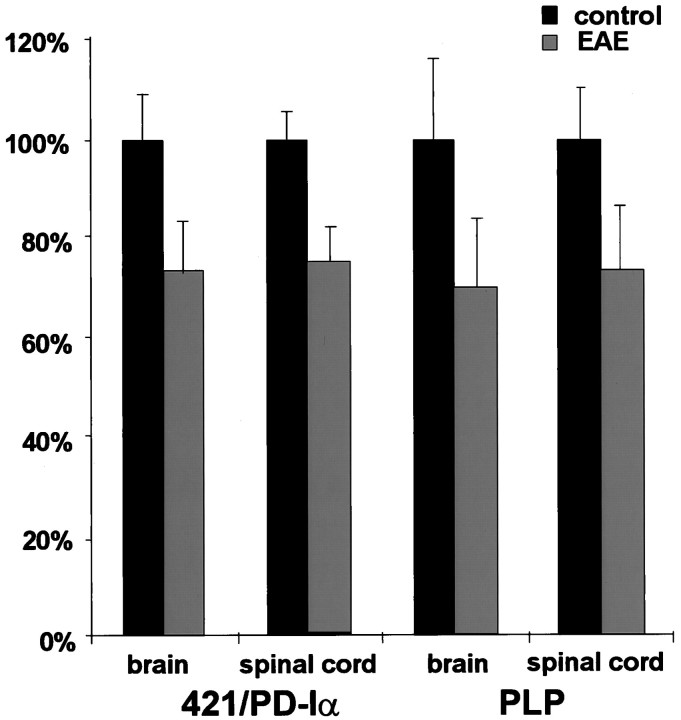
One of the more complex developmental processes occurring postnatally in the CNS is the formation of the myelin sheath by oligodendrocytes. To examine the molecular events that take place during myelination, we isolated oligodendrocyte-derived cDNA clones, one of which (p421.HB) represents a putative alternatively spliced isoform of rat brain-specific phosphodiesterase I (PD-Ialpha) and a species homolog of the human cytokine autotaxin. Analysis of the structural composition of the p421.HB/PD-Ialpha protein suggests a transmembrane-bound ectoenzyme, which, in addition to the phosphodiesterase-active site contains presumed cell recognition and Ca2+-binding domains. Consequently, it may be involved in extracellular signaling events. Expression of p421.HB/PD-Ialpha is enriched in brain and spinal cord, where its mRNA can be detected in oligodendrocytes and in cells of the choroid plexus. Expression in the brain increases during development with an intermediate peak of expression around the time of active myelination and maximal expression in the adult. We have identified four presumably alternatively spliced isoforms, two of which appear to be CNS-specific. Decreased levels of p421.HB/PD-Ialpha mRNA in the dysmyelinating mouse mutant jimpy, but not shiverer, suggest a role for p421.HB/PD-Ialpha during active myelination and/or late stages of oligodendrocyte differentiation. Furthermore, p421.HB/PD-Ialpha mRNA levels were reduced in the CNS at onset of clinical symptoms in experimental autoimmune encephalomyelitis. These data together implicate the importance of p421.HB/PD-Ialpha in oligodendrocyte function, possibly through cell-cell and/or cell-extracellular matrix recognition.


Figures

Fig. 1.
Fig. 1.
Comparison of PD-Iα/ATX cDNA clones. Sequence analysis revealed identity of the partial cDNA clone p421.HB with the C-terminal coding region of rat PD-Iα plus 3′-untranslated sequences. However, in the cDNA clone, p421.HB sequences coding for amino acids 596–615 (25 aa) of rat PD-Iα are missing. In addition, human PD-Iα is identical to human teratocarcinoma-derived autotaxin (ATXt), whereas human melanoma-derived autotaxin (ATXm) has an additional 52 amino acid (52 aa) insertion. Coding regions are represented bythick lines; 5′- and 3′-untranslated sequences are represented by thin lines.
Fig. 2.
Fig. 2.
Northern blot analysis of mRNA prepared from different adult rat tissues and rat brain of different developmental stages [postnatal days 5 (P5), 10 (P10), 15 (P15), 20 (P20) and 25 (P25) and adult] using the p421.HB cDNA clone (Fig. 1) and a cyclophilin-specifc cDNA probe. Ten micrograms of total RNA were separated on a 1.2% formaldehyde gel and hybridized with 32P-labeled DNA fragments specific for p421.HB/PD-Iα and cyclophilin. For quantification Northern blots were analyzed by phosphorimaging techniques, and mRNA levels were normalized to cyclophilin. For the diagram at thebottom the mRNA level in adult brain was set to 100%.
Fig. 3.
Fig. 3.
Localization of p421.HB/PD-Iα mRNA (A–C, G–I, N–P) in comparison with PLP mRNA (D–F, K–M) in the ventral horn of the spinal cord (A–F), cerebellum (G–M), and choroid plexus of the lateral ventricle (N–P) of postnatal day 4 (A, D, G, K, N) and 21 (B, E, H, L, O) and adult (C, F, I, M, P) rats. p421.HB/PD-Iα mRNA-positive cells were observed in white matter areas in a distribution similar to PLP mRNA-positive cells (compare A with D, B with E, C with F, G with K, and H with L, I with M), although p421.HB/PD-Iα mRNA expression appears to be lower than the one of PLP. In contrast to PLP, at postnatal day 4 no p421.HB/PD-Iα mRNA-positive cells were detectable at the base of the cerebellum (compare G withK). In the spinal cord, however, p421. HB/PD-Iα mRNA-positive cells were visible more restricted to the area close to the outer surface (A) than PLP mRNA-positive cells (D). In the adult, only very few p421.HB/PD-Iα mRNA-positive cells were detectable, which were localized in and close to white matter tracts (arrows inC, I), where PLP-positive cells can also be detected (compare C with F,I with M). Additional strong p421.HB/PD-Iα mRNA-positive signals were detectable in cells of the choroid plexus, with increasing levels of expression with age (N–P). Sections hybridized with sense cRNA probes showed no labeling. Scale bars (in A): A, B, D, E, 100 μm; C, F, 210 μm; G, H, 110 μm; H, L, 130 μm; I, M, 200 μm; N–P, 20 μm; N–P,insets, 410 μm.
Fig. 4.
Fig. 4.
Analysis of p421.HB/PD-Iα mRNA expression in cultured oligodendrocytes by confocal imaging. The expression of p421.HB/PD-Iα mRNA by oligodendrocytes could be directly demonstrated by combined in situ hybridization and immunocytochemistry. In 8-d-old cultures of panned oligodendrocytes cells positive for both 421.HB/PD-Iα mRNA (fluorescein,green) and CNP (Texas Red, red) could be detected, although not all of the CNP-positive cells were positive for p421.HB/PD-Iα mRNA. Controls hybridized with sense cRNA probes were negative.
Fig. 5.
Fig. 5.
Characterization of p421.HB/PD-Iα isoforms. The existence of alternatively spliced isoforms of p421.HB/PD-Iα could be demonstrated by RT-PCR using two primer pairs: one, in which both oligonucleotides flanked the proposed alternatively spliced 75 bp (Fig.1, 25 aa) sequence (A, top panel, B, D), and a second one in which one oligonucleotide flanked the 75 bp region, and the other one was located within this 75 region (A, bottom panel). RNA was derived from rat brains of different developmental ages [A, embryonic day 14 (E14), postnatal days 5 (P5), 10 (P10), 15 (P15), 20 (P20), and 25 (P25), and adult (A)], different adult rat tissues (B), different CNS regions of adult rats (C), and from cells in culture (D). The amplification products were analyzed on 3.5% agarose gels (ethidium bromide-stained gels are shown in A, B, D). For quantification (C) scanned images were used and analyzed with the NIH-Image software. Percentages of the two longer isoforms (309 bp, 297 bp), including the alternatively spliced 75 bp exon [coding for amino acids 551–557 (AETGKFRGSKHENKKNLNGSVEPRK) in rat PD-Iα], are given in C (total p421.HB/PD-Iα expression = 100%). The molecular weights of the fragments obtained are marked at the right margins in base pairs.
Fig. 6.
Fig. 6.
Characterization of p421.HB/PD-Iα mRNA levels in dysmyelinating mouse mutants [jimpy(jp), shiverer(shi), and trembler(tr)]. Levels of p421.HB/PD-Iα mRNA present in brain and spinal cord of 21-d-old dysmyelinating mutant mice were compared with wild-type (wt) levels by RPA. Ten micrograms of total RNA were used for hybridization with 32P-labeled cRNA probes for p421.HB and cyclophilin. Protected fragments were analyzed in 6% denaturing acrylamide gels by phosphorimaging techniques (the inset shows a representative example of the separation of protected fragments; C, protected cyclophilin band; PD-Iα, protected p421.HB/PD-Iα band). Three independent experiments using RNA of at least two animals each were used for statistical analysis. Amounts of protected p421.HB/PD-Iα fragments were standardized by the cyclophilin values obtained in the same hybridization reaction. Wild-type mRNA levels were set to 100%, and mutant levels were adjusted accordingly. Error bars represent SD.
Fig. 7.
Fig. 7.
Characterization of p421.HB/PD-Iα and PLP mRNA levels at onset of clinical symptoms in EAE. Levels of p421.HB/PD-Iα and PLP mRNA expressed in brain and spinal cord of EAE (PLP peptide-injected) mice were compared with normal (control, BSA-injected) levels by RPA. Tissues for RNA preparation were taken from EAE mice at onset of clinical symptoms and of time-matched control animals. Ten micrograms of total RNA of each animal were used for hybridization with 32P-labeled cRNA probes for p421.HB and PLP, each in combination with cyclophilin for normalization. Protected fragments were analyzed in 6% denaturing acrylamide gels by phosphorimaging techniques. For statistical analysis four pairs of samples (EAE vs control) were used, and levels of p421.HB/PD-Iα and PLP mRNA were normalized to cyclophilin values. Control mRNA levels were set to 100%, and EAE mRNA levels were calculated accordingly. Error bars represent SD.

Similar articles

Cited by

References

    1. Agresti C, D’Urso D, Levi G. Reversible inhibitory effects of interferon-γ and tumor necrosis factor-α on oligodendroglial lineage cell proliferation and differentiation in vitro. Eur J Neurosci. 1996;8:1106–1116. - PubMed
    1. Altschul SF, Gish W, Miller W, Myers EW, Lipman DJ. Basic local alignment search tool. J Mol Biol. 1990;215:403–410. - PubMed
    1. Baba H, Fuss B, Watson JB, Zane LT, Macklin WB. Identification of novel mRNAs expressed in oligodendrocytes. Neurochem Res. 1994;19:1091–1099. - PubMed
    1. Babu YS, Bugg CE, Cook WJ. Structure of calmodulin refined 2.2 Å resolution. J Mol Biol. 1988;204:191–204. - PubMed
    1. Barres BA, Hart IK, Coles HSR, Burne JF, Voyvodic JT, Richardson WD, Raff MC. Cell Death and control of cell survival in the oligodendrocyte lineage. Cell. 1992;70:31–46. - PubMed

Publication types

MeSH terms