. 1997 Dec 1;17(23):9157-64.
doi: 10.1523/JNEUROSCI.17-23-09157.1997.

Delayed reduction of ischemic brain injury and neurological deficits in mice lacking the inducible nitric oxide synthase gene

Affiliations

Delayed reduction of ischemic brain injury and neurological deficits in mice lacking the inducible nitric oxide synthase gene

C Iadecola et al. J Neurosci. .

Abstract

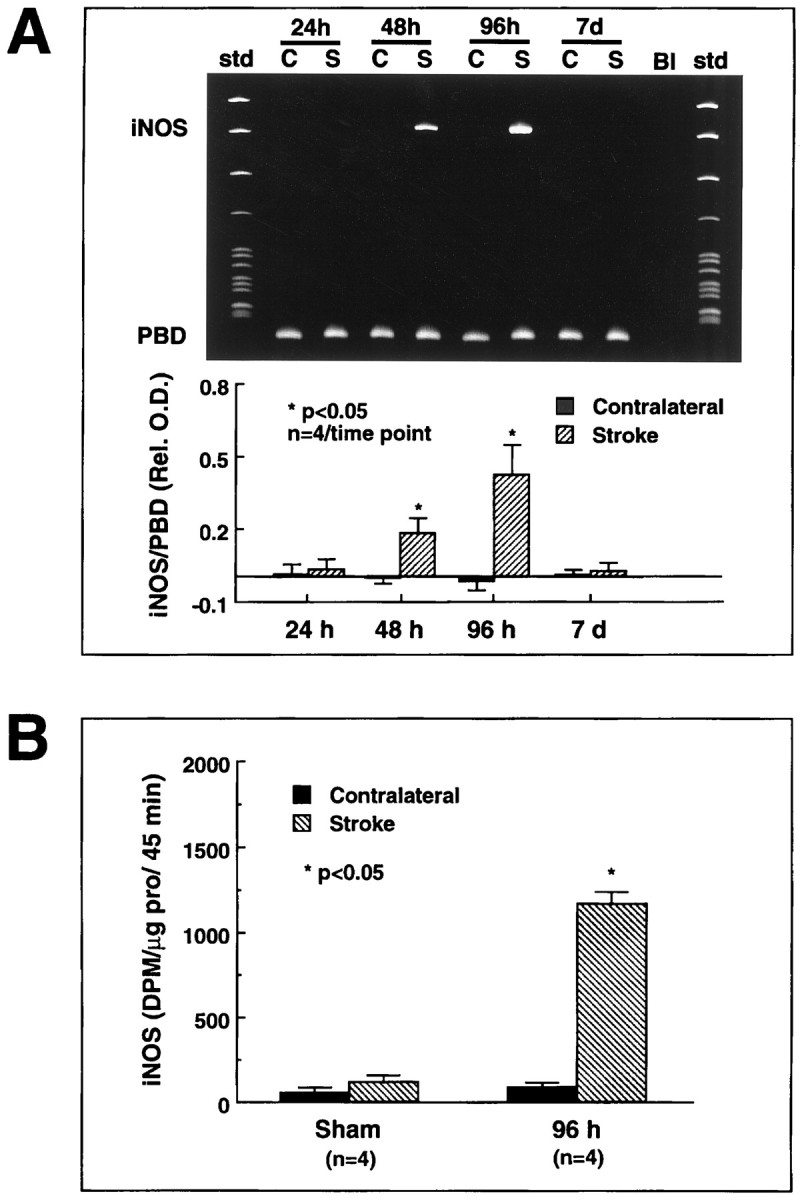
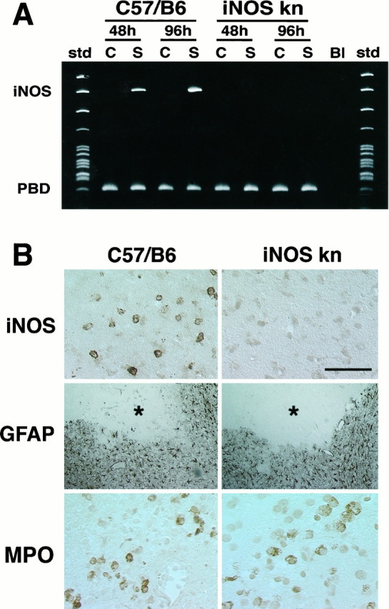
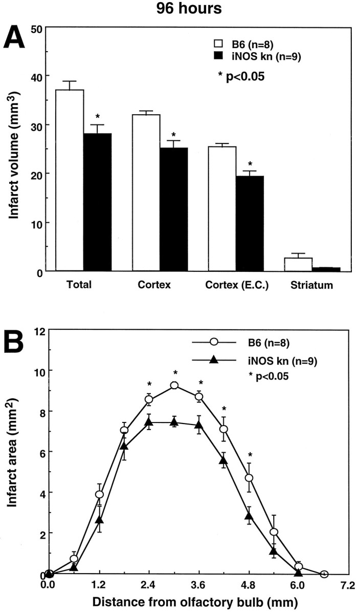
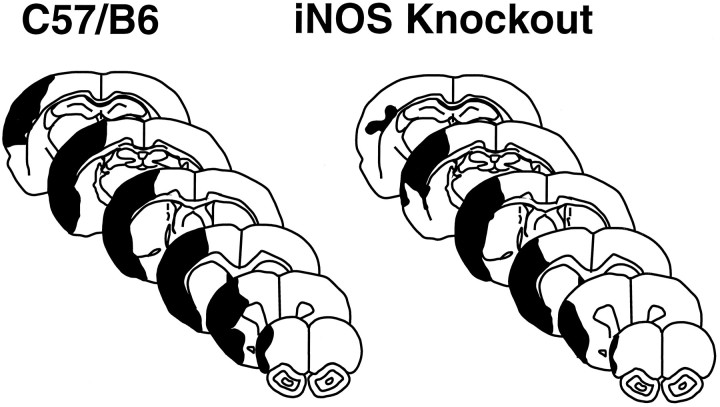
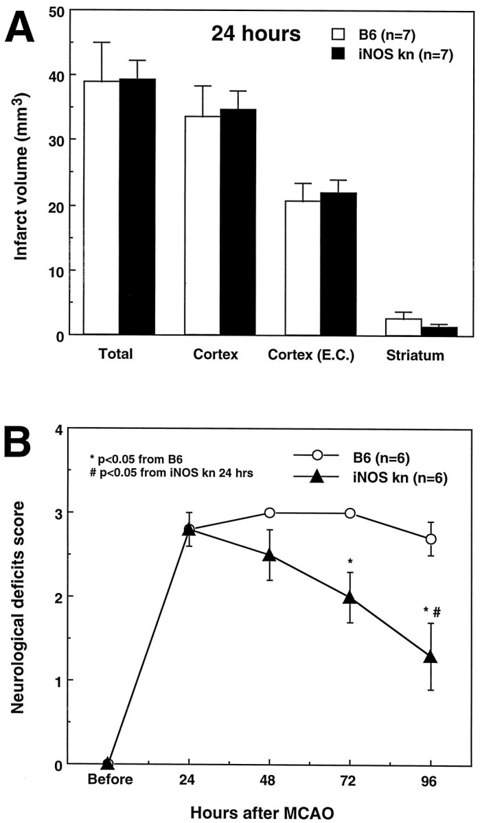
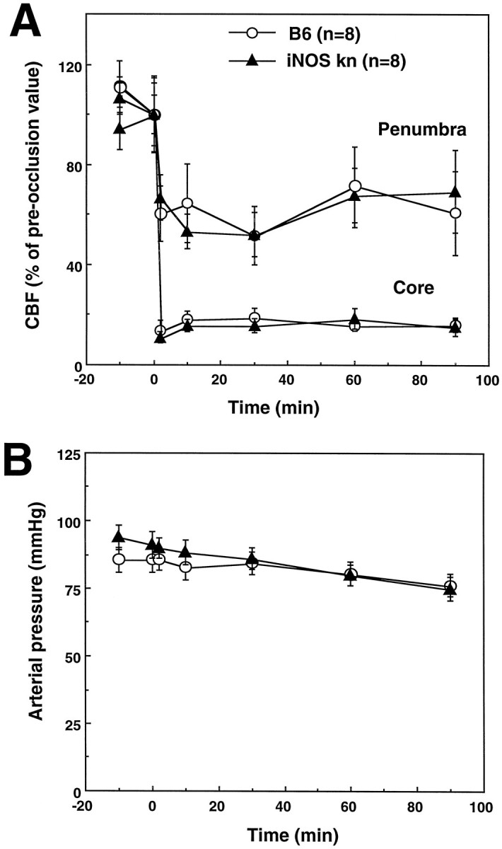
Inducible nitric oxide synthase (iNOS), an enzyme that produces toxic amounts of nitric oxide, is expressed in a number of brain pathologies, including cerebral ischemia. We used mice with a null mutation of the iNOS gene to study the role of iNOS in ischemic brain damage. Focal cerebral ischemia was produced by occlusion of the middle cerebral artery (MCA). In wild-type mice, iNOS mRNA expression in the post-ischemic brain begun between 24 and 48 hr peaked at 96 hr and subsided 7 d after MCA occlusion. iNOS mRNA induction was associated with expression of iNOS protein and enzymatic activity. In contrast, mice lacking the iNOS gene did not express iNOS message or protein after MCA occlusion. The infarct and the motor deficits produced by MCA occlusion were smaller in iNOS knockouts than in wild-type mice (p < 0.05). Such reduction in ischemic damage and neurological deficits was observed 96 hr after ischemia but not at 24 hr, when iNOS is not yet expressed in wild-type mice. The decreased susceptibility to cerebral ischemia in iNOS knockouts could not be attributed to differences in the degree of ischemia or vascular reactivity between wild-type and knockout mice. These findings indicate that iNOS expression is one of the factors contributing to the expansion of the brain damage that occurs in the post-ischemic period. iNOS inhibition may provide a novel therapeutic strategy targeted specifically at the secondary progression of ischemic brain injury.


Figures

Fig. 1.
Fig. 1.
Expression of iNOS message and enzymatic activity after cerebral ischemia in B6 mice. A, Expression of iNOS message after focal cerebral ischemia. iNOS mRNA was determined by RT-PCR. A strong iNOS signal is observed at 48 and 96 hr after ischemia (top panel). The bottom panelshows the time course of the ratio of the optical density of the iNOS band by the density of the band corresponding to porphobilinogen deaminase (PBD), a housekeeping gene used for normalization. Significant elevation is observed at 48 and 96 hr after ischemia (p < 0.05; ANOVA and Tukey’s test). No expression is observed at 24 hr or 7 d (p > 0.05). B, iNOS enzymatic activity after cerebral ischemia. iNOS activity was measured using the citrulline conversion assay of Bredt and Snyder modified for detection of calcium-independent arginine-to-citrulline conversion, i.e., iNOS activity (Ross and Iadecola, 1996). Virtually no iNOS activity was observed in sham-operated mice. Substantial iNOS activity developed in the ischemic region 96 hr after MCA occlusion. The 96 hr time point was chosen because at this time iNOS message is maximal. These findings suggest that cerebral ischemia leads to expression of iNOS message and enzymatic activity in the injured brain.
Fig. 2.
Fig. 2.
A, iNOS message detected by RT-PCR in wild-type mice (C57/B6) and iNOS knockouts at 48 and 96 hr after ischemia. No iNOS signal is detected in the ischemic brain at either time points. B, Immunoreactivity foriNOS, glial fibrillary acidic protein (GFAP), or myeloperoxidase (MPO) in iNOS null mice and controls (C57/B6) 96 hr after MCA occlusion. Cerebral ischemia is followed by expression of iNOS immunoreactivity in C57/B6 mice but not in iNOS knockouts (top panels). Immunoreactivity for the astrocytic marker GFAP (middle panels) is comparable between iNOS null mice and wild-type controls. The asterisk indicates the infarcted area. GFAP expression occurs at the border of the lesion. To detect infiltrating inflammatory cells in the post-ischemic brain, immunocytochemistry for MPO, an enzyme expressed in polymorphonuclear cells infiltrating the ischemic brain, was performed. Infiltrating myeloperoxidase-positive cells are observed both in iNOS null mice and in controls (bottom panels). Scale bar (shown intop right panel in B): topand bottom panels, 50 μm; middle panel, 250 μm.
Fig. 3.
Fig. 3.
A, Infarct volume in wild-type controls (C57/B6) and iNOS knockouts killed 96 hr after occlusion of the MCA. Infarct size is smaller in iNOS knockouts (p < 0.01; t test). The difference persists after correction for ischemic swelling [Cortex (E.C.)]. B, Distribution of the infarcted areas at different rostrocaudal levels in wild-type controls (C57/B6) and iNOS knockouts. The area of infarction is smaller in the iNOS knockouts at all rostrocaudal levels. Values for iNOS null mice and controls were compared at each rostrocaudal level by the unpairedt test.
Fig. 4.
Fig. 4.
Representative brain sections illustrating the distribution of the ischemic damage in wild-type mice (C57/B6) and iNOS knockouts 96 hr after MCA occlusion. The area of infarction involves the cerebral cortex almost exclusively. Note that the infarct is smaller in the knockouts and that the area spared from infarction involves the peripheral regions of the infarct. This peripheral region represents the so-called ischemic penumbra.
Fig. 5.
Fig. 5.
A, Infarct volume in wild-type controls (C57/B6) and iNOS knockouts killed 24 hr after occlusion of the MCA. Data were corrected for ischemic swelling [Cortex (E.C.)]. Infarct volume is not different between wild-type mice and iNOS knockouts (p > 0.05;t test). B, Neurological deficits in wild-type mice (C57/B6) and iNOS knockouts. Mice were examined before induction of ischemia and at 24 hr intervals until the time they were killed 96 hr after MCA occlusion. Higher neurological scores indicate more severe deficits. Neurological scores were not different 24 hr after MCA occlusion (p > 0.05; Mann–Whitney U test). However, although the scores remained stable in wild-type mice during the following 96 hr (p > 0.05; Kruskal–Wallis ANOVA), they improved markedly in the iNOS knockouts (p< 0.05 from wild-type mice at 96 hr and p < 0.05 from 24 hr).
Fig. 6.
Fig. 6.
A, Effect of MCA occlusion on CBF in iNOS knockouts and wild-type controls (C57/B6). The reduction in CBF produced by MCA occlusion is similar in the two groups both in the center of the ischemic territory (Core) and at the periphery (Penumbra). B, Arterial pressure of the mice before and after MCA occlusion. Arterial pressure did not differ between controls and iNOS knockouts (p > 0.05).

Similar articles

Cited by

References

    1. Adamson DC, Wildemann B, Sasaki M, Glass JD, McArthur JC, Christov VI, Dawson TM, Dawson VL. Immunologic NO synthase: elevation in severe AIDS dementia and induction by HIV-1 gp41. Science. 1996;274:1917–1921. - PubMed
    1. Astrup J, Symon L, Branston NM, Lassen NA. Cortical evoked potential and extracellular K+ and H+ at critical levels of brain ischemia. Stroke. 1977;8:51–57. - PubMed
    1. Bagasra O, Michaels FH, Zheng Y-M, Bobroski LE, Spitsin SV, Fu F, Tawadros R, Koprowski H. Activation of the inducible form of nitric oxide synthase in the brains of patients with multiple sclerosis. Proc Natl Acad Sci USA. 1995;92:12041–12045. - PMC - PubMed
    1. Baird AE, Benfield A, Schlaug G, Siewert B, Lovblad KO, Edelman RR, Warach S. Enlargement of human cerebral ischemic lesion volumes measured by diffusion-weighted magnetic resonance imaging. Ann Neurol. 1997;41:581–589. - PubMed
    1. Baron J-C, von Kummer R, del Zoppo GJ. Treatment of acute ischemic stroke. Challenging the concept of a rigid and universal time window. Stroke. 1995;26:2219–2221. - PubMed

Publication types

LinkOut - more resources