Comparative Study
. 1997 Dec 1;17(23):9183-93.
doi: 10.1523/JNEUROSCI.17-23-09183.1997.

Neuronal and non-neuronal collapsin-1 binding sites in developing chick are distinct from other semaphorin binding sites

Affiliations
Comparative Study

Neuronal and non-neuronal collapsin-1 binding sites in developing chick are distinct from other semaphorin binding sites

T Takahashi et al. J Neurosci. .

Abstract

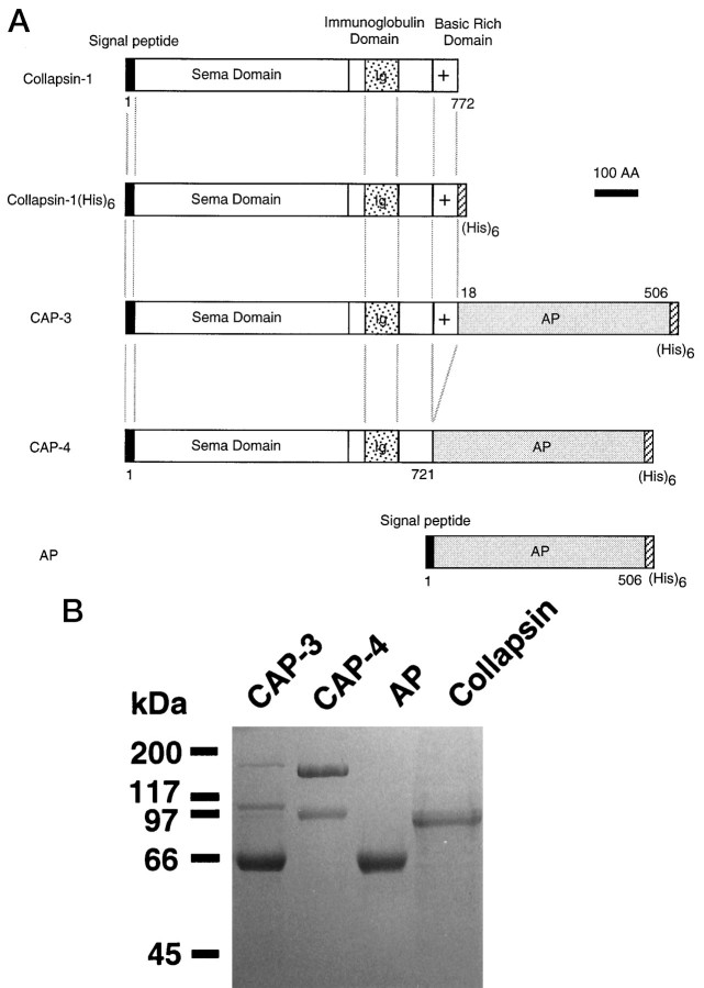
The collapsin and semaphorin family of extracellular proteins contributes to axonal path finding by repulsing axons and collapsing growth cones. To explore the mechanism of collapsin-1 action, we expressed and purified a truncated collapsin-1-alkaline phosphatase fusion protein (CAP-4). This protein retains biological activity as a DRG growth cone collapsing agent and saturably binds to DRG neurons with low nanomolar affinity. Specific CAP-4 binding sites are present on DRG neurons, sympathetic neurons, and motoneurons, but not on retinal, cortical, or brainstem neurons. Outside the nervous system, high levels of CAP-4 binding sites are present in the mesenchyme surrounding major blood vessels and developing bone and in lung. These sites provide a substrate for the collapsin-1-dependent patterning of non-neuronal tissues perturbed in sema III (-/-) mice. The staining patterns for mouse semaphorin D/III and chick collapsin-1 fusion proteins are indistinguishable from one another but quite separate from that for semaphorin B and M-semaphorin F fusion proteins. These data imply that a family of high-affinity semaphorin binding sites similar in complexity to the semaphorin ligand family exists.


Figures

Fig. 1.
Fig. 1.
Purification of a collapsin-1–alkaline phosphatase fusion protein. A, Schematic illustrating the structure of recombinant proteins. Because a full-length collapsin-1–alkaline phosphatase fusion protein (CAP-3) was degraded in the basic rich region at the C terminal of collapsin-1, a truncated form of collapsin-1 lacking the basic rich region at the C terminal was fused with alkaline phosphatase (CAP-4). B, SDS–PAGE of purified recombinant proteins. Fusion protein preparations (2 μg) were analyzed by 10% SDS–PAGE. The CAP-3 preparation contains a significant fraction of degraded 66 kDa protein similar in molecular weight to alkaline phosphatase (AP) and a second degraded protein species of 110 kDa. Only a minor portion of theCAP-3 is full-length 165 kDa fusion protein. Although the CAP-4 preparation contains a minor amount of degraded 97 kDa protein, the majority of protein is full-length 160 kDa fusion protein. Collapsin-1-His6 exhibits the expected full-length 95 kDa size.
Fig. 2.
Fig. 2.
Growth cone collapse activity of collapsin-1-His6 and CAP-4. Growth cone collapse of chick E7 DRG explants (A), dissociated chick E7 sympathetic neurons (B), and chick E7 retinal explants (C) was determined with various concentrations of CAP-4 (closed circle) and collapsin-1-His6 (Col-His; open circle) as indicated. The EC50 values of collapsin-1-His6 and CAP-4 are 50 pm and 5 nm, respectively, for both DRG and sympathetic neurons. The SEM from three to five determinations is shown.
Fig. 3.
Fig. 3.
Saturable staining of chick E7 DRG neurons by CAP-4. Dissociated cultures of chick E7 DRG neurons were stained with 10 nm CAP-4 (top left), with 10 nm CAP-4 in the presence of 50 nmcollapsin-1-His6 (top right), or with 10 nm human placental alkaline phosphatase (bottom right). CAP-4 stained a subpopulation of DRG neurons. The CAP-4 staining was abolished in the presence of excess collapsin-1-His6. Human placental alkaline phosphatase did not stain DRG neurons. The growth cones of methanol-fixed DRG neurons were stained with 10 nm CAP-4 (bottom left). Scale bar: bottom left, 30 μm; rightand top left, 65 μm.
Fig. 4.
Fig. 4.
Quantitation of CAP-4 binding to chick sympathetic neurons. A, Saturation of CAP-4 binding to sympathetic neurons. Dissociated cultures of chick E7 sympathetic neurons were stained with various amount of CAP-4 (Total; open circle) or CAP-4 plus 100 nmcollapsin-1-His6 (Nonspecific; closed circle). Bound alkaline phosphatase activity was quantitated after the incubation with p-nitrophenyl phosphate by measuring the optical density at 415 nm. Specific CAP-4 binding was calculated by subtracting nonspecific from total binding (Specific; square). B, Scatchard analysis of specific CAP-4 binding to sympathetic neurons. Scatchard analysis of specific CAP-4 binding from A is shown. The calculated KD is 3.3 nm, and the Bmax is 1 nmol of CAP-4 bound per well. C, Inhibition of CAP-4 binding by collapsin-1. The binding of 4 nm CAP-4 to chick sympathetic neurons in the presence of the indicated concentrations of collapsin-1-His6 was quantitated as described in Materials and Methods. The IC50 is 500 pm, which corresponds to an apparent Ki of 250 pm assuming competitive inhibition and aKD for CAP-4 of 3.3 nm. Error bars indicate SEM for three to five determinations inA–C.
Fig. 5.
Fig. 5.
CAP staining of different neuronal populations. Chick E7 sympathetic neurons were stained with 10 nm CAP-4 (Ch Sym, CAP-4), and this staining was abolished in the presence of 50 nm collapsin-1-His6(Ch Sym, CAP-4 + Col). Rat E15 DRG neurons were also stained with 10 nm CAP-4 (Rat DRG, CAP-4), and labeling was abolished by the addition of 50 nm collapsin-1-His6 (Rat DRG, CAP-4 + Col). Although large diameter cells in rat E15 motoneuron-enriched cultures were stained with 10 nm CAP-4 (Rat MN, CAP-4), rat cortical neurons (Rat Cortex, CAP-4) and rat brainstem neurons (Rat BS, CAP-4) were not stained. Scale bar, 50 μm.
Fig. 6.
Fig. 6.
CAP-4 staining of E10 DRG neuronal subpopulations.A, Dissociated chick E10 DRG neurons were cultured in either NGF or NT-3 for 48 hr and then stained with 3 nmCAP-4 as described in Materials and Methods. Note the prominent staining of the NGF-dependent neurons and the absence of staining of NT-3-dependent cells. Scale bar, 50 μm. B, Dissociated chick E10 DRG neurons were cultured in both NGF and NT-3 for 48 hr and then stained with CAP-4 and anti-trkC antibody as described in Materials and Methods. The percentage of CAP-4-binding-positive neurons also exhibiting trkC immunoreactivity is low compared with the fraction of CAP-4-negative neurons that are trkC immunoreactive. The percentage of E10 DRG neurons stained by CAP-4 was 50 ± 2%. The SEM from three separate experiments is shown.
Fig. 7.
Fig. 7.
In situ localization of CAP-4 binding sites in developing chick nervous system. Transverse thoracic vibratome sections (dorsal surface at top) of chick E7 and E12 embryos were stained with 10 nm CAP-4. DRGand the motoneurons (MN) of the ventral horn of the spinal cord were stained in E7 embryos (top panel). In E12 embryos, staining of the ventral horn was minimal, but the ventral midline of the spinal cord was stained (bottom panel; arrow). This ventral midline staining was not detectable in E7 specimens (middle panel; arrow). Staining of mesenchyme at the surface of the vertebral bodies was prominent in E7 preparations (middle panel). Scale bar: top andmiddle panels, 60 μm; bottom panel, 120 μm.
Fig. 9.
Fig. 9.
Differential localization of semaphorin binding sites. A, Purification of semaphorin B, D, and M-F fusion proteins. Fusion proteins (2 μg) were analyzed by 10% SDS–PAGE. Semaphorin B extracellular domain–alkaline phosphatase fusion protein (Sema B) is purified in its full-length form of 150 kDa. The truncated semaphorin D–alkaline phosphatase fusion protein preparation (Sema D) contains the predicted protein of 165 kDa and a degraded protein of 97 kDa. The M-semaphorin F extracellular domain–alkaline phosphatase fusion protein (M-Sema F) preparation exhibits protein staining at the predicted size of 150 kDa and a minor fraction of a degraded protein at 66 kDa. B, Localization of semaphorin–alkaline phosphatase binding sites. Cryostat sections of E9 chick embryo were stained with CAP-4, Sema B–AP, Sema D–AP and M-Sema F–AP (10 nm). Although CAP-4 andSema D–AP stained the mesenchyme around the aorta in transverse thoracic sections, Sema B–AP andM-Sema F–AP did not. Coronal sections containing the midline of the dorsal midbrain and transverse sections of the nasal retina were stained by Sema B–AP and M-Sema F–AP but not by CAP-4 or Sema D–AP. The dorsal surface is at the top of each section. Scale bar, 100 μm.
Fig. 8.
Fig. 8.
Localization of CAP-4 binding sites in extraneuronal sites. Transverse thoracic sections (dorsal surface attop) of the chick embryo were stained with 10 nm CAP-4. Low magnification images of sections from E9 embryos reveal intense mesenchymal staining (CAP-4) that is blocked by the addition of 50 nm collapsin-1-His6 (CAP-4 + Col-His). The positions of the spinal cord (sc), the aorta (★), the limb (l), and the heart (h) are indicated. The mesenchyme around the aorta was stained with 10 nm CAP-4 (Aorta, CAP-4) in the E9 chick embryo. This staining was blocked in the presence of 50 nm collapsin-1-His6(Aorta, CAP-4 + Col-His). The lung parenchyma was stained with 10 nm CAP-4 (Lung, CAP-4). The histological border of lung tissue is indicated by the asterisks. In the limb, the mesenchyme surrounding bone was stained with 10 nm CAP-4 (Limb, CAP-4). Scale bar: two top panels, 800 μm; two bottom left panels, 100 μm; two bottom right panels, 200 μm.

Similar articles

Cited by

References

    1. Behar O, Golden JA, Mashimo H, Schoen FJ, Fishman MC. Semaphorin III is needed for normal patterning and growth of nerves, bones and heart. Nature. 1996;383:525–528. - PubMed
    1. Byk T, Dobransky T, Cifuentes-Diaz C, Sobel A. Identification and molecular characterization of unc-33-like phosphoprotein (Ulip), a putative mammalian homologue of the axonal guidance-associated unc-33 gene product. J Neurosci. 1996;16:688–701. - PMC - PubMed
    1. Camu W, Henderson CE. Purification of embryonic rat motoneuron by panning on a monoclonal antibody to the low-affinity NGF receptor. J Neurosci Methods. 1992;44:59–70. - PubMed
    1. Chen H-J, Flanagan JG. Identification and cloning of ELF-1, a developmentally expressed ligand for the Mek-4 and Sek receptor tyrosine kinases. Cell. 1994;79:157–168. - PubMed
    1. Chen H-J, Nakamoto M, Bergemann AD, Flanagan JG. Complementary gradients in expression and binding of ELF-1 and Mek4 in development of the topographic retinotectal projection map. Cell. 1995;82:371–381. - PubMed

Publication types

MeSH terms

LinkOut - more resources