Comparative Study
. 1997 Dec 1;17(23):9204-11.
doi: 10.1523/JNEUROSCI.17-23-09204.1997.

Birthdate and cell marker analysis of scrambler: a novel mutation affecting cortical development with a reeler-like phenotype

Affiliations
Comparative Study

Birthdate and cell marker analysis of scrambler: a novel mutation affecting cortical development with a reeler-like phenotype

J L González et al. J Neurosci. .

Abstract

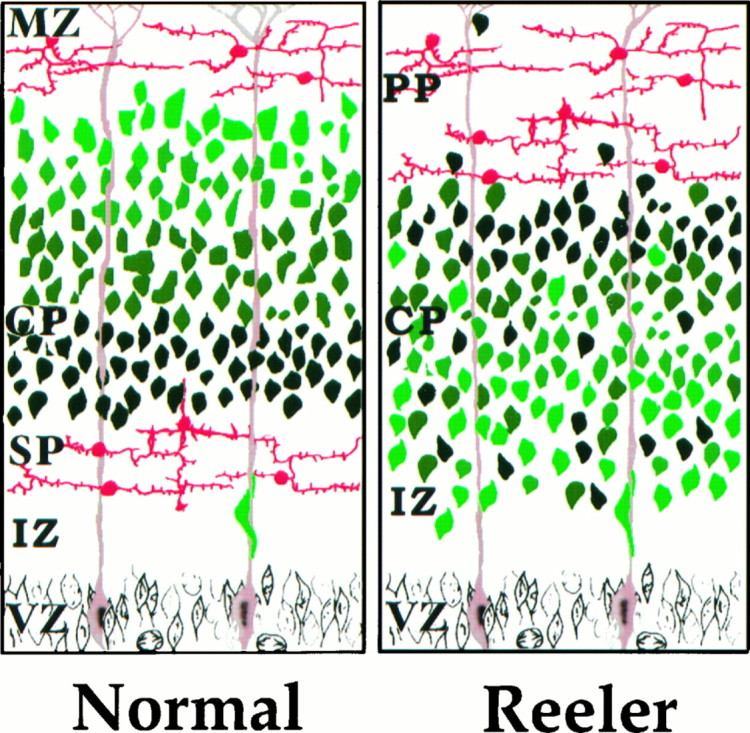
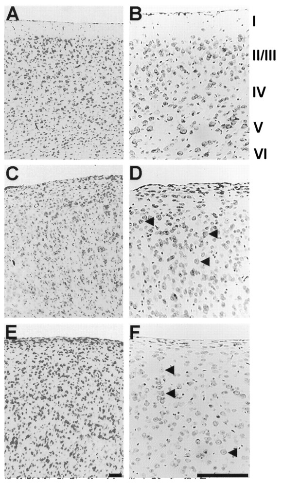
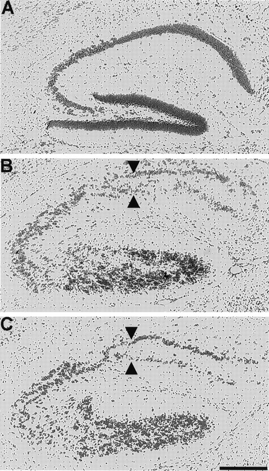
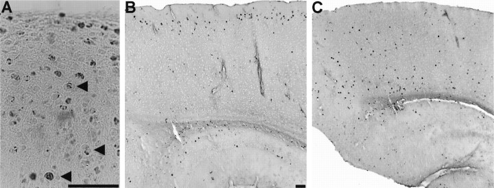
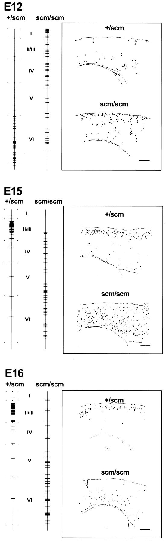
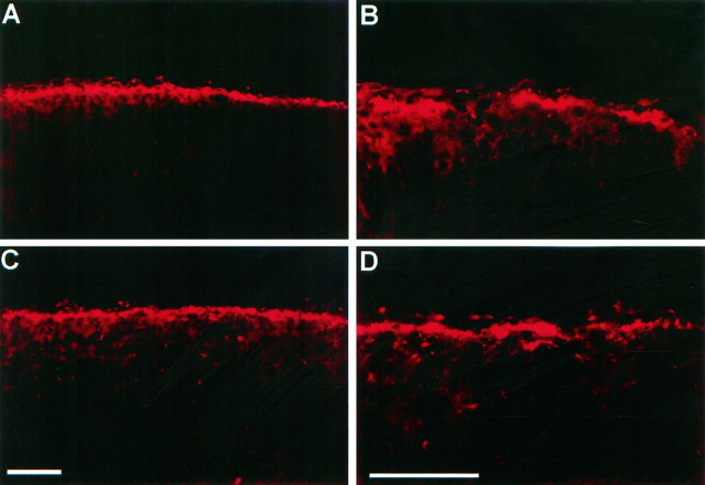
The reeler mutation in mice produces an especially well characterized disorder, with systematically abnormal migration of cerebral cortical neurons. The reeler gene encodes a large protein, termed Reelin, that in the cortex is synthesized and secreted exclusively in the Cajal-Retzius neurons of the cortical marginal zone (D'Arcangelo et al., 1995). In reeler mutant mice, loss of Reelin protein is associated with a systematic loss of the normal, "inside-out" sequence of neurogenesis in the cortex: neurons are formed in the normal sequence but become localized in the cortex in a somewhat inverted, although relatively disorganized "outside-in" pattern. Here we show that the scrambler mutant mouse exhibits a loss of lamination in the cortex and hippocampus that is indistinguishable from that seen in the reeler mouse. We use BrdU birthdating studies to show that scrambler cortex shows a somewhat inverted "outside-in" sequence of birthdates for cortical neurons that is similar to that previously described in reeler cortex. Finally, we perform staining with the CR-50 monoclonal antibody (Ogawa et al., 1995), which recognizes the Reelin protein (D'Arcangelo et al., 1997). We show that Reelin immunoreactivity is present in the scrambler cortex in a normal pattern, suggesting that Reelin is synthesized and released normally. Our data suggest that scrambler is a mutation in the same gene pathway as the reeler gene (Relnrl) and is most likely downstream of Relnrl.


Figures

Fig. 1.
Fig. 1.
Summary of defects in reeler neocortical development. This schematic diagram of neocortical development illustrates the major defects in the reeler mouse cortex. First, the correspondence of age and position of neurons is roughly reversed in reeler neocortex relative to normal. In the normal cortex, the oldest cortical plate (CP) neurons (dark green) lie deeper in the cortex than cortical plate neurons formed later in development (light green). In the reeler cortex, older CP neurons (dark green) tend to settle in the more superficial aspect of the cortical plate zone than younger CP neurons (light green). Second, the layering of neurons by age is not as well preserved in reeler mice as in normal. Third, the preplate (PP) is not split into subplate (SP) and marginal zone (MZ) in reeler neocortex. Whereas the preplate layer of neurons (red) is split into two layers by migrating CP neurons in the normal cortex, in the reeler cortex these neurons remain as a single layer in the superficial aspect of the brain. The intermediate zone (IZ) and ventricular zone (VZ) appear normal in reeler mice.
Fig. 2.
Fig. 2.
Histological appearance of the normal, reeler, and scrambler cortex. In A and B, photomicrographs of cresyl violet-stained, sagittal sections illustrate the typical six-layered structure of the normal cortex (although layersII/III are not sharply distinct). The sagittal sections shown in C and D demonstrate the characteristic lamination abnormality in the reeler mouse cortex.E and F show sagittal sections from the cortex of the scrambler mouse. As in reeler, the layers are not clearly distinct, and the cortex exhibits a lower overall level of cytoarchitectonic organization. The marginal zone (layer I), as defined by a low abundance of cell nuclei, is not well formed. Large pyramidal neurons can be seen through all layers of the cortex (arrowheads in D, F) and are somewhat more common at more superficial locations, as in reeler. Scale bars, 400 μm.
Fig. 3.
Fig. 3.
Histological appearance of the normal, reeler, and scrambler hippocampus. The sagittal section shown in Aexemplifies the lamination pattern observed in the normal mouse hippocampus. The sagittal section shown in B illustrates the abnormal lamination of the reeler hippocampus. Note the almost complete duplication of the pyramidal cell layer (arrowheads) and overall level of disorganization. (For a more detailed description of abnormalities in reeler hippocampus, seeStanfield and Cowan, 1979a,.) In C, a sagittal section from the scrambler cortex shows a disruption and partial duplication of the pyramidal cell layer (arrowheads) quite similar to that observed in the hippocampus of the reeler mouse. Scale bar, 400 μm.
Fig. 4.
Fig. 4.
Patterns of BrdU labeling in normal and scrambler mouse cortex. A shows large neurons inscm/scm cortex labeled with BrdU at E12 (arrowheads), indicating that the largest cortical neurons (characteristic of layer V), although abnormally positioned, are still among the older neurons in the cortex. In Band C, labeling is illustrated in sagittal sections taken from animals that received BrdU at E16 and were analyzed as adults. B shows +/scm neocortex. Note the neat organization of the labeled neurons at the superficial aspect of the cortex in an area corresponding to layers II/III. Cshows scm/scm neocortex labeled with BrdU on E16. Note the much wider distribution of labeled neurons, with the majority located in the deeper half of the cortex. This observation closely matches results from similar studies inRelnrl/Relnrlcortex (Caviness, 1982). Scale bars, 100 μm.
Fig. 5.
Fig. 5.
Summary patterns of BrdU labeling after injection at E12, E15, and E16. The positions of BrdU-labeled cells are represented by horizontal lines drawn on avertical line extending the thickness of the cortex from pia to white matter. Each tick mark on thevertical line represents a single BrdU-labeled neuron. Each line combines the vertical position of all BrdU-labeled neurons from the camera lucida drawing of a region of the parietal cortex, lying dorsal to the hippocampus, of a single sagittal section. (See Materials and Methods for details.) The approximate boundaries of each layer are indicated for normal cortex. For each labeling age, samples of camera lucida drawings from BrdU-labeled normal and scm/scm brain sections are displayed in the insets. Scale bars, 400 μm.
Fig. 6.
Fig. 6.
CR-50 staining in the scrambler cortex.A and B show CR-50 immunocytochemistry on a sagittal section of normal P0 cortex at low and high magnifications, respectively. C and D show CR-50 immunocytochemistry on a sagittal section of P0 cortex from ascm/scm littermate at similar magnifications. Scale bars, 100 μm.

Similar articles

Cited by

References

    1. Allendoerfer KL, Shatz CJ. The subplate, a transient neocortical structure: its role in the development of connections between thalamus and cortex. Annu Rev Neurosci. 1994;17:185–218. - PubMed
    1. Caviness VS., Jr . Reeler mutant mouse: a genetic experiment in developing mammalian cortex. In: Cowan WM, Ferrendelli JA, editors. Approaches to the cell biology of neurons, Vol 2. Society for Neuroscience; Bethesda, MD: 1977. pp. 27–46.
    1. Caviness VS., Jr Neocortical histogenesis in normal and reeler mice: a developmental study based upon 3H-thymidine autoradiography. Brain Res. 1982;256:293–302. - PubMed
    1. Caviness VS, Jr, Korde MG. Monoaminergic afferents to the neocortex: a developmental histofluorescence study in normal and Reeler mouse embryos. Brain Res. 1981;209:1–9. - PubMed
    1. Caviness VS, Jr, Crandall JE, Edwards MA. The reeler malformation: implications for neocortical histogenesis. In: Peters A, Jones EG, editors. Cerebral neocortex. Plenum; New York: 1988. pp. 59–89.

Publication types

MeSH terms

LinkOut - more resources