Comparative Study
. 1997 Dec 1;17(23):9233-47.
doi: 10.1523/JNEUROSCI.17-23-09233.1997.

Subcortical input to the smooth and saccadic eye movement subregions of the frontal eye field in Cebus monkey

Affiliations
Comparative Study

Subcortical input to the smooth and saccadic eye movement subregions of the frontal eye field in Cebus monkey

Tian et al. J Neurosci. .

Abstract

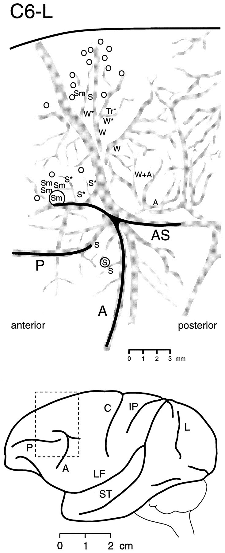
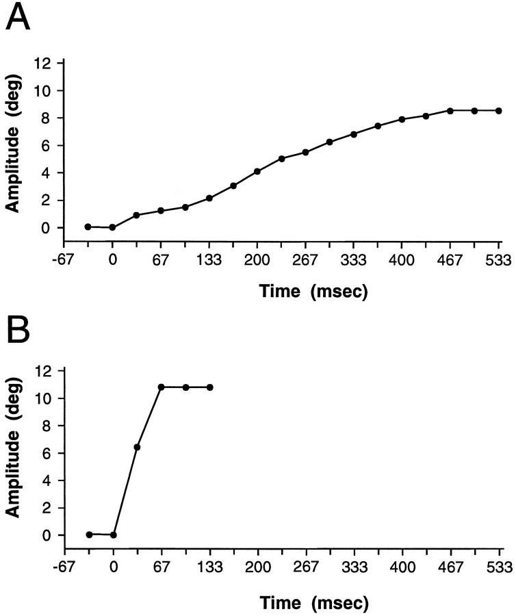
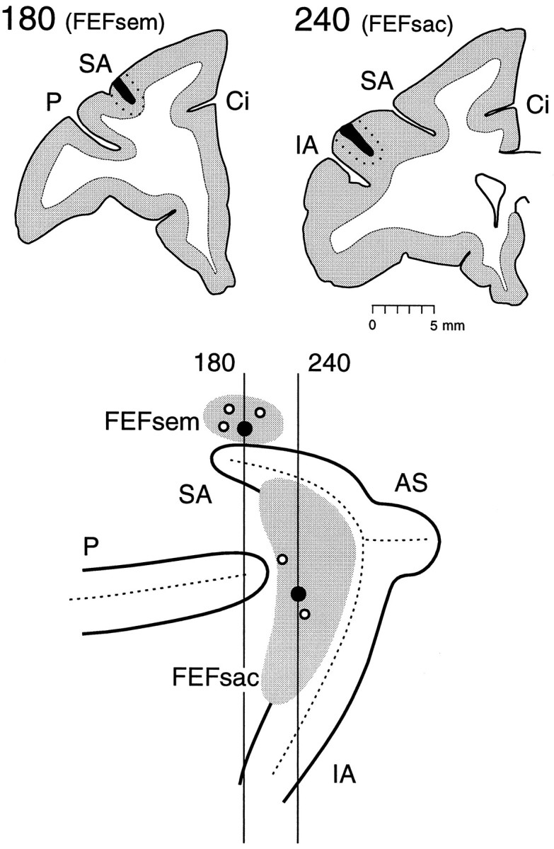
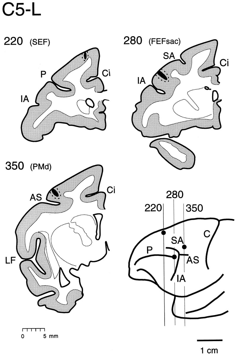
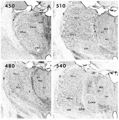
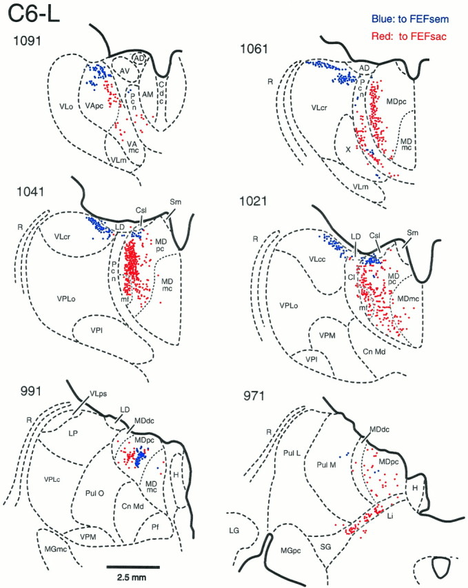
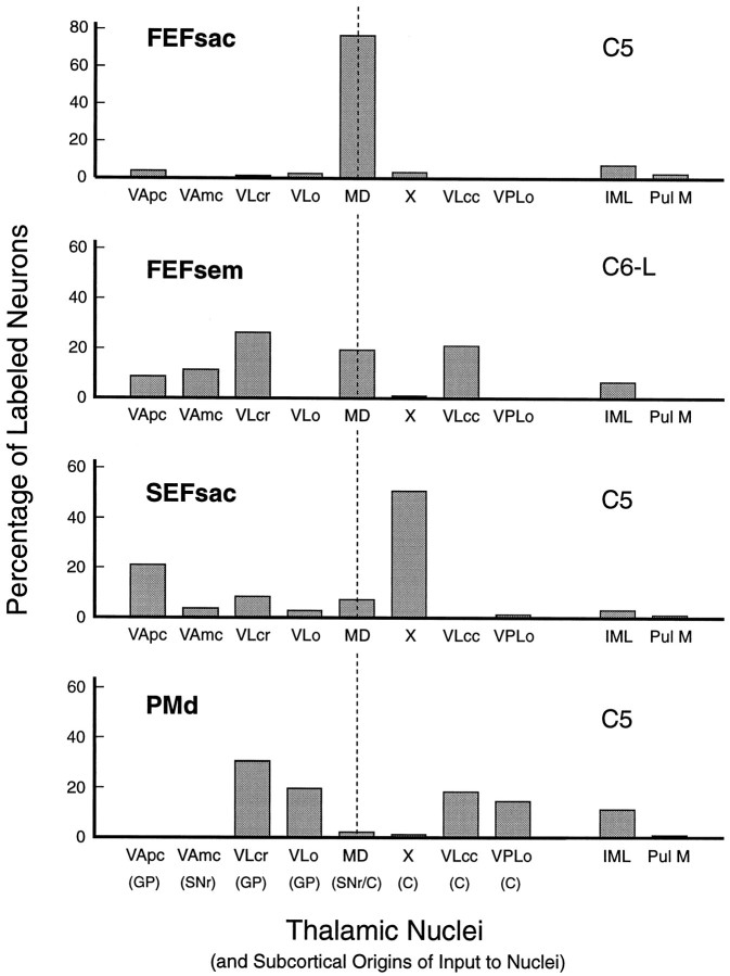
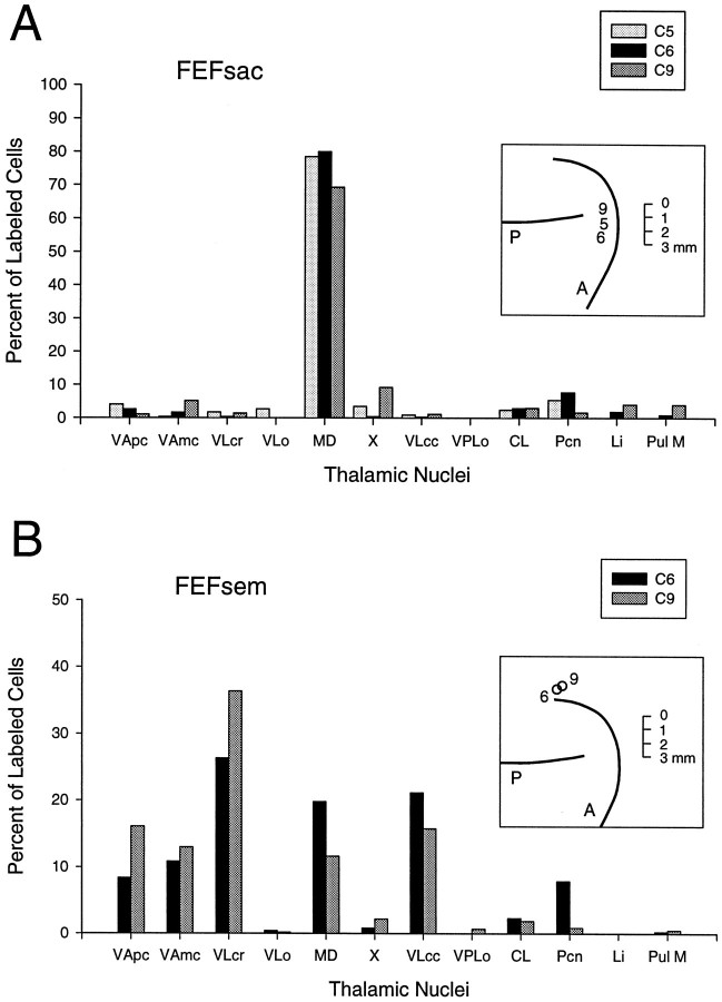
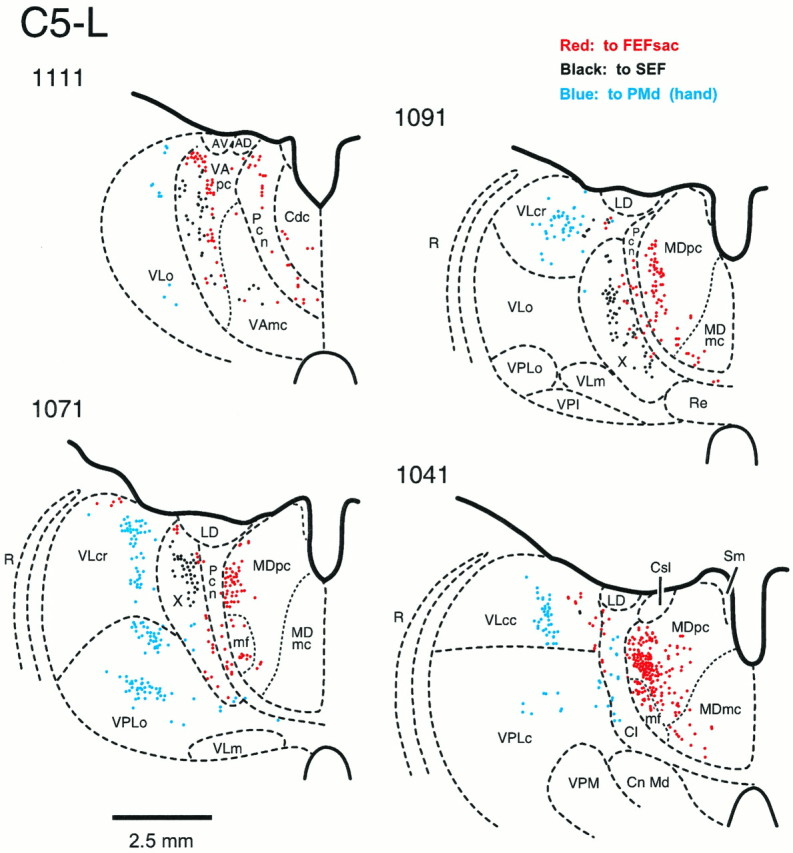
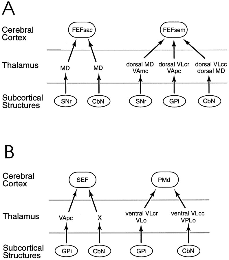
We have recently identified two functional subregions in the frontal eye field (FEF) of the Cebus monkey, a smooth eye movement subregion (FEFsem) and a saccadic subregion (FEFsac). The thalamic input to these two subregions was studied and quantified to gain more information about the influence of the cerebellum and basal ganglia on the oculomotor control mechanisms of the cerebral cortex. A recent study using transneuronal transport of virus demonstrated that there are neurons in the basal ganglia and cerebellum that project to the FEFsac with only a single intervening synapse (Lunch et al., 1994). In the present study, we concentrated on the thalamic input to the FEFsem to define possible basal ganglia-thalamus-cortex and cerebellum-thalamus-cortex channels of information flow to the FEFsem. We localized the functional subregions using low threshold microstimulation, and retrogradely transported fluorescent tracers were then placed into the FEFsem and FEFsac. The neurons that project to the FEFsem are distributed in (1) the rostral portion of the ventral lateral nucleus, pars caudalis, (2) the caudal portion of the ventral lateral nucleus, pars caudalis, (3) the mediodorsal nucleus, (4) the ventral anterior nucleus, pars parvocellularis, and (5) the ventral anterior nucleus, pars magnocellularis. In contrast, the large majority of neurons that project to the FEFsac are located in the paralaminar region of the mediodorsal nucleus. The FEFsac and FEFsem thus each receive neural input from both basal ganglia-receiving and cerebellar-receiving cell groups in the thalamus, but each receives input from a unique combination of thalamic nuclei.


Figures

Fig. 3.
Fig. 3.
Top, A typical microstimulation map within a region including the frontal eye field, the supplementary eye field, and the dorsal premotor cortex of monkey C6, left hemisphere (C6-L). The map was reconstructed by projecting and tracing a photograph made through the operating microscope of the cortical blood vessels (gray) and then plotting the position of each electrode penetration from individual electrode placement photographs. The locations where movements were elicited at thresholds <50 μA are indicated by letters: S, saccade; Sm, smooth eye movement; Tr, trunk muscle contraction; W, wrist flexion or dorsiflexion; W+A, wrist dorsiflexion evoked superficially in cortex plus forearm pronation and abduction evoked deeper in cortex; A, supination of forearm; and O, no response. Anasterisk indicates responses evoked at high threshold (>50 μA); a circle around a stimulation point indicates the location of a tracer injection (see also Fig. 4).Bottom, A lateral view of the hemisphere. Thedotted rectangle indicates the location of the microstimulation map. A, Arcuate sulcus;AS, arcuate spur; C, central sulcus;IP, intraparietal sulcus; L, lunate sulcus; LF, lateral fissure; P, principal sulcus; ST, superior temporal sulcus.
Fig. 1.
Fig. 1.
Typical time–amplitude trajectories of eye movements in a Telazol-anesthetized Cebus monkey (C6) are illustrated for smooth (A) and saccadic (B) eye movements. Measurements of eye position were made using video-editing equipment, manually frame by frame. [Modified from Tian and Lynch (1996a) with permission.]
Fig. 4.
Fig. 4.
Top, Injection sites in the smooth eye movement subregion of the FEF (section180) and in the saccadic subregion of theFEF (section 240) in monkey C6 on coronal sections. Single placements of different fluorescent tracers are within the gray matter of each functional subregion of the FEF.Bottom, A lateral view of the left hemisphere of monkey C6. The dotted lines indicate the fundi of sulci; theheavy lines indicate the shoulders of sulci.AS, Arcuate spur; Ci, cingulate sulcus;FEFsac, saccadic subregion of the FEF;FEFsem, smooth eye movement subregion of the FEF;IA, inferior arcuate sulcus; P, principal sulcus; SA, superior arcuate sulcus.
Fig. 5.
Fig. 5.
Injection sites of the FEFsac(section 280), the SEF (section220), and the hand area of the PMd(section 350) on coronal sections of monkey C5 (C5-L). Single placements of different fluorescent tracers are within the gray matter of each functional subregion of the FEF. AS, Arcuate spur;C, central sulcus; Ci, cingulate sulcus;FEFsac, saccadic subregion of the FEF; IA, inferior arcuate sulcus; LF, lateral fissure; P, principal sulcus; PMd, dorsal part of premotor cortex;SA, superior arcuate sulcus; SEF, supplementary eye field.
Fig. 2.
Fig. 2.
Macrophotographs of cresyl violet-stained sections from the thalamus of Cebus monkey C9. Section450 is at the most rostral level; 540 is at the most caudal level. See Results for descriptions of nuclei and cytoarchitectural features. AD, Anterior dorsalis;AM, anterior medialis; AV, anterior ventralis; CL, central lateral nucleus; Cn Md, centrum medianum; LD, lateralis dorsalis;MD, medialis dorsalis; MDmc ormc, medialis dorsalis, pars magnocellularis;MDpc or pc, medialis dorsalis, pars parvocellularis; PCN, paracentral nucleus;R, reticular nucleus; TMT, mammillothalamic tract; VAmc, ventralis anterior, pars magnocellularis; VApc, ventralis anterior, pars parvocellularis; VLcc, caudal portion of ventralis lateralis, pars caudalis; VLcr, rostral portion of ventralis lateralis, pars caudalis; VLo, ventralis lateralis, pars oralis; VPI, ventralis posterior inferior; VPLo, ventralis posterior lateralis, pars oralis; VPM, ventralis posterior medialis;X, area X in the ventral lateral complex.
Fig. 6.
Fig. 6.
The origin of thalamic inputs to theFEFsem (blue dots) andFEFsac (red dots) in the left hemisphere of monkey C6 on coronal sections (C6-L). Section1091 is at the most rostral level, and section971 is at the most caudal level. The fluorescent tracers FB and DY were injected into the FEFsac and theFEFsem, respectively (see Table 1 and Fig. 4). A total of 31 sections at 250 μm intervals were plotted. AD, Anterior dorsalis; AM, anterior medialis;AV, anterior ventralis; Cdc, centralis densocellularis; Cl, central lateral nucleus; Cn Md, centrum medianum; Csl, centralis superior lateralis; H, habenula; LD, lateralis dorsalis; LG, lateral geniculate nucleus;Li, nucleus limitans; LP, lateralis posterior; MD, medialis dorsalis; MDdc, medialis dorsalis, pars densocellularis; MDmc, medialis dorsalis, pars magnocellularis; MDmf ormf, medialis dorsalis, pars multiformis;MDpc, medialis dorsalis, pars parvocellularis;MGmc, medial geniculate nucleus, pars magnocellularis;MGpc, medial geniculate nucleus, pars parvocellularis;Pcn, paracentral nucleus; Pf, parafascicularis; Pul L, pulvinaris lateralis;Pul M, pulvinaris medialis; Pul O, pulvinaris oralis; R, reticular nucleus; SG, suprageniculate nucleus;Sm, stria medullaris thalami; VAmc, ventralis anterior, pars magnocellularis; VApc, ventralis anterior, pars parvocellularis; VLc, ventralis lateralis, pars caudalis; VLcc, caudal portion ofVLc; VLcr, rostral portion ofVLc; VLm, ventralis lateralis, pars medialis; VLo, ventralis lateralis, pars oralis;VLps, ventralis lateralis, pars postrema;VPI, ventralis posterior inferior; VPLc, ventralis posterior lateralis, pars caudalis; VPLo, ventralis posterior lateralis, pars oralis; VPM, ventralis posterior medialis; X, area Xin the ventral lateral complex.
Fig. 8.
Fig. 8.
Quantitative comparison of distribution patterns of labeled neurons in thalamic nuclei from the four cortical injection sites. Labeled neurons were counted within the cytoarchitectural boundaries of 31 sections spaced at 250 μm intervals in monkeys C5 and C6. Each graph illustrates the percentage, in each nucleus, of the total number of neurons labeled by that particular injection. The thalamic nuclei are arranged on the x-axis so that regions that receive input from the internal segment of the globus pallidus and the pars reticulata of the substantia nigra are on theleft of the vertical dashed line and the nuclear regions that receive input from the cerebellar nuclei are on the right of the dashed line. The medial dorsal nucleus receives input from both the basal ganglia and the cerebellum. Intralaminar nuclei (Pcn and Cl), indicated byIML, and the Pul M are not included in the basal ganglia versus cerebellum distribution dichotomy. A total of 5692 neurons were labeled by the FEFsac injection, 2876 by the FEFsem injection, 460 by theSEFsac injection, and 1288 by the PMdinjection. C, Cerebellum, predominantly via the dentate nucleus; FEFsac, saccadic subregion of the frontal eye field; FEFsem, smooth eye movement subregion of the frontal eye field; GP, globus pallidus;IML, intralaminar nuclei; MD, medialis dorsalis; PMd, hand region of the dorsal premotor cortex; Pul M, pulvinaris medialis;SEFsac, saccadic subregion of the supplementary eye field; SNr, substantia nigra pars reticularis;VAmc, ventralis anterior, pars magnocellularis;VApc, ventralis anterior, pars parvocellularis;VLcc, caudal portion of ventralis lateralis, pars caudalis; VLcr, rostral portion of ventralis lateralis, pars caudalis; VLo, ventralis lateralis, pars oralis;VPLo, ventralis posterior lateralis, pars oralis;X, area X of ventral lateral complex.
Fig. 9.
Fig. 9.
A, Comparison of the thalamic distributions of FEFsac-labeled neurons in three monkeys. B, Comparison of the thalamic distributions ofFEFsem-labeled neurons in two monkeys. Each graph illustrates the percentage, in each nucleus, of the total number of neurons labeled by that particular injection. In theinset in A, the numbers(5, 6, and 9) show the relative positions of the threeFEFsac injection sites on a standardized drawing of the arcuate sulcus region for monkeys C5, C6, and C9, respectively. Tracers were DY in C5, FB in C6, and FB in C9. In the inset inB, the two circles indicate the relative positions of the FEFsem injections in monkeys C6 and C9. Tracers were DY in C6 and FR in C9. The thalamic nuclei are arranged on the x-axis so that regions that receive input from the internal segment of the globus pallidus and the pars reticulata of the substantia nigra are on the left of theMD and the nuclear regions that receive input from the cerebellar nuclei are on the right of theMD. The medial dorsal nucleus receives input from both the basal ganglia and the cerebellum. A, Arcuate sulcus;CL, central lateral nucleus; Li, nucleus limitans; MD, medialis dorsalis; P, principal sulcus; Pcn, paracentral nucleus; Pul M, pulvinaris medialis; VAmc, ventralis anterior, pars magnocellularis; VApc, ventralis anterior, pars parvocellularis; VLcc, caudal portion of ventralis lateralis, pars caudalis; VLcr, rostral portion of ventralis lateralis, pars caudalis; VLo, ventralis lateralis, pars oralis; VPLo, ventralis posterior lateralis, pars oralis; X, areaX in the ventral lateral complex.
Fig. 7.
Fig. 7.
The origin of thalamic inputs to theFEFsac, SEF, and PMd in the left hemisphere of monkey C5 on coronal sections (C5-L) (see Table 1 for fluorescent tracers used in these injections). Section 1111 is at the most rostral level, and section 1041 is at the most caudal level. A total of 31 sections at 250 μm intervals were plotted. Red dots indicate the neurons labeled from an injection site in theFEFsac; black dots indicate the neurons labeled from an injection site in the SEF; andlight blue dots indicate the neurons labeled from an injection site of the PMd (hand). AD, Anterior dorsalis; AV, anterior ventralis;Cdc, centralis densocellularis; Cl, central lateral nucleus; Cn Md, centrum medianum;Csl, centralis superior lateralis; LD, lateralis dorsalis; MDmc, medialis dorsalis, pars magnocellularis; mf, medialis dorsalis, pars multiformis; MDpc, medialis dorsalis, pars parvocellularis; Pcn, paracentral nucleus;R, reticular nucleus; Re, nucleus of reuniens; Sm, stria medullaris thalami;VAmc, ventralis anterior, pars magnocellularis;VApc, ventralis anterior, pars parvocellularis;VLc, ventralis lateralis, pars caudalis;VLcc, caudal portion of VLc;VLcr, rostral portion of VLc;VLm, ventralis lateralis, pars medialis;VLo, ventralis lateralis, pars oralis;VPI, ventralis posterior inferior; VPLc, ventralis posterior lateralis, pars caudalis; VPLo, ventralis posterior lateralis, pars oralis; VPM, ventralis posterior medialis; X, area Xin the ventral lateral complex.
Fig. 10.
Fig. 10.
Summary diagram of GPi- andSNr-thalamocortical and cerebellothalamocortical connection patterns. A, Putative circuits from basal ganglia and cerebellum through thalamic nuclei to theFEFsac and FEFsem. B, Putative circuits from basal ganglia and cerebellum through thalamic nuclei to the SEF and PMd. Each of the functional areas in the cerebral cortex receives a major neural input from both a basal ganglia-receiving and a cerebellar-receiving cell group in the thalamus. It is proposed that some neurons from the basal ganglia and cerebellar nuclei synapse on thalamic neurons that, in turn, project to the cortical eye fields. However, this specific connectivity has thus far been confirmed with transneuronal transport experiments only in the case of the FEFsac (Lynch et al., 1994). The terms “dorsal” and “ventral” are used with theVLcr and VLcc nuclei to emphasize the fact that even though both the FEFsem and thePMd receive input from these two nuclei, the respective pathways originate in separate subregions of these nuclei. Similarly, the term “dorsal MD” is used to emphasize that theMD projection to the FEFsem originates in the dorsal-most portion of paralaminar MD, whereas theMD projection to the FEFsac originates relatively more ventrally in paralaminar MD.CbN, Cerebellar nuclei; FEF, frontal eye field; FEFsac, saccadic subregion of theFEF; FEFsem, smooth eye movement subregion of the FEF; GPi, internal globus pallidus; MD, medialis dorsalis;PMd, dorsal premotor cortex; SEF, supplementary eye field; SNr, substantia nigra, pars reticulata; VAmc, ventralis anterior, pars magnocellularis; VApc, ventralis anterior, pars parvocellularis; VLcc, caudal portion of ventralis lateralis, pars caudalis; VLcr, rostral portion of ventralis lateralis, pars caudalis; VLo, ventralis lateralis, pars oralis; VPLo, ventralis posterior lateralis, pars oralis; X, area X in the ventral lateral complex.

Similar articles

Cited by

References

    1. Alexander GE, DeLong MR, Strick PL. Parallel organization of functionally segregated circuits linking basal ganglia and cortex. Annu Rev Neurosci. 1986;9:357–381. - PubMed
    1. Alexander GE, Crutcher MD, DeLong M. Basal ganglia-thalamocortical circuits: parallel substrates for motor, oculomotor, “prefrontal” and “limbic” functions. In: Uylings HBM, Van Eden CG, Bruin JPC, Corner MA, Feenstra MGP, editors. Progress in brain research, Vol 85. Elsevier; New York: 1990. pp. 119–146. - PubMed
    1. Asanuma C, Thach WT, Jones EG. Cytoarchitectonic delineation of the ventral lateral thalamic region in the monkey. Brain Res. 1983a;286:219–235. - PubMed
    1. Asanuma C, Thach WT, Jones EG. Distribution of cerebellar terminations in the ventral lateral thalamic region of the monkey. Brain Res. 1983b;286:237–265. - PubMed
    1. Asanuma C, Thach WT, Jones EG. Anatomical evidence for segregated focal groupings of efferent cells and their terminal ramifications in the cerebellothalamic pathway of the monkey. Brain Res Rev. 1983c;5:267–297. - PubMed

Publication types

MeSH terms

Substances

LinkOut - more resources