. 1997 Dec 1;17(23):9285-97.
doi: 10.1523/JNEUROSCI.17-23-09285.1997.

Dissociable forms of inhibitory control within prefrontal cortex with an analog of the Wisconsin Card Sort Test: restriction to novel situations and independence from "on-line" processing

Affiliations

Dissociable forms of inhibitory control within prefrontal cortex with an analog of the Wisconsin Card Sort Test: restriction to novel situations and independence from "on-line" processing

R Dias et al. J Neurosci. .

Abstract

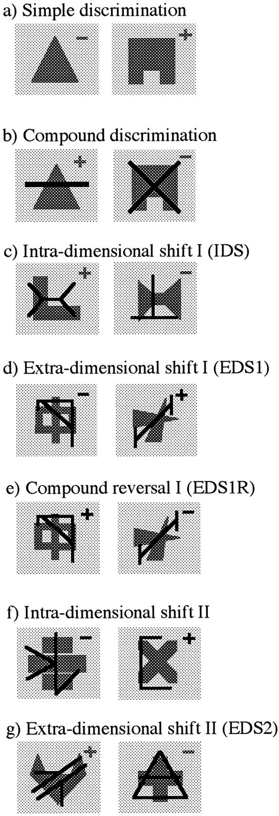
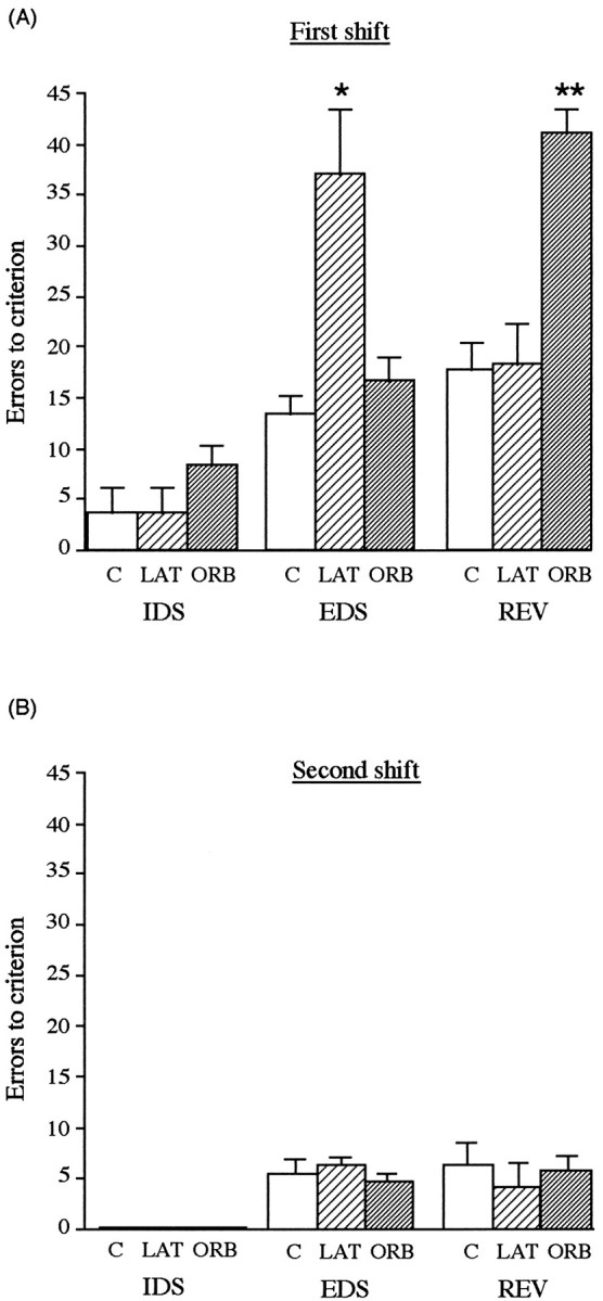
Attentional set-shifting and discrimination reversal are sensitive to prefrontal damage in the marmoset in a manner qualitatively similar to that seen in man and Old World monkeys, respectively (Dias et al., 1996b). Preliminary findings have demonstrated that although lateral but not orbital prefrontal cortex is the critical locus in shifting an attentional set between perceptual dimensions, orbital but not lateral prefrontal cortex is the critical locus in reversing a stimulus-reward association within a particular perceptual dimension (Dias et al., 1996a). The present study presents this analysis in full and extends the results in three main ways by demonstrating that (1) mechanisms of inhibitory control and "on-line" processing are independent within the prefrontal cortex, (2) impairments in inhibitory control induced by prefrontal damage are restricted to novel situations, and (3) those prefrontal areas involved in the suppression of previously established response sets are not involved in the acquisition of such response sets. These findings suggest that inhibitory control is a general process that operates across functionally distinct regions within the prefrontal cortex. Although damage to lateral prefrontal cortex causes a loss of inhibitory control in attentional selection, damage to orbitofrontal cortex causes a loss of inhibitory control in affective processing. These findings provide an explanation for the apparent discrepancy between human and nonhuman primate studies in which disinhibition as measured on the Wisconsin Card Sort Test is associated with dorsolateral prefrontal damage, whereas disinhibition as measured on discrimination reversal is associated with orbitofrontal damage.


Figures

Fig. 1.
Fig. 1.
The shape and line exemplars used for the various stages of the attentional set-shifting paradigm. In this example the dimension of “shape” is relevant in all the discriminations except that requiring an EDS (d) and reversal (e) and subsequent IDS II (f). On any one trial of a compound discrimination, a shape exemplar may be paired with one or other of the line exemplars. Correct and incorrect choices are indicated by + and −, respectively. Gray typefacespecifies that “shape” is the relevant dimension, whereasblack typeface specifies that “line” is the relevant dimension.
Fig. 2.
Fig. 2.
A, Mean number of errors (±SEM) to reach criterion on a visual discrimination that requires an IDS (third discrimination of the series of three that were presented postoperatively), an EDS, and a reversal, EDSR, in monkeys that received excitotoxic lesions of either the lateral (LAT) (n = 3) (pale hatched bars) or orbital prefrontal cortex (ORB) (n = 3) (dark hatched bars) or a sham operation (n = 3) (open bars). B, Mean number of errors (±SEM) to reach criterion on subsequent visual discriminations that require an intradimensional shift (IDS), an extradimensional shift (EDS), and a reversal (REV). * Lateral prefrontal lesioned group differed significantly from controls and orbital prefrontal lesioned group; p < 0.001. ** Orbital prefrontal lesioned group differed significantly from controls and lateral prefrontal lesioned group; p < 0.001.
Fig. 3.
Fig. 3.
Mean cumulative errors made over the first session of the reversal (EDSR) by monkeys that received excitotoxic lesions of either the lateral (LAT) (n = 3) (filled triangles) or orbital prefrontal cortex (ORB) (n = 3) (filled circles) or a sham operation (n = 3) (open circles). Horizontal line ata and b indicates the level of performance significantly different from chance (p < 0.05) on 16 trials, i.e., 12 or more incorrect responses, and 20 trials, i.e., 15 or more incorrect responses, respectively.
Fig. 4.
Fig. 4.
Schematic diagrams of a series of coronal sections through the frontal lobe illustrating the site of the lesion of the orbital (a) and lateral (b) prefrontal cortices. The three different types of shading represent the area of tissue that was damaged in all three marmosets (black shading), in two of the three marmosets (dark stippling), and in one marmoset only (pale stippling) after an orbital (a) or a lateral lesion (b). Orbital prefrontal cortex corresponds to areas 10–13 (marked orb on the sections), and lateral prefrontal cortex corresponds to area 9 (markedlat on the sections), as defined by Brodmann (1909) in his description of the frontal cortex in the marmoset.m, Medial prefrontal cortex; pm, premotor cortex; cn, caudate nucleus; p, putamen).
Fig. 5.
Fig. 5.
Low (a, c) and high (b, d) power photomicrographs of cresyl violet-stained coronal sections through an intermediate level of the frontal pole taken from a representative marmoset from both the orbital (a, b) and lateral (c, d) lesioned groups. The extensive cell loss in the orbital (ORB) prefrontal cortex ina is in stark contrast to the dense layering of neurons seen in the same region in c. Similarly, the almost total loss of neurons in the lateral prefrontal cortex (LAT) in c is in marked contrast to the dense layering of neurons seen in the same region ina. The stars mark the same locations in each pair of low and high power photomicrographs (e.g.,a and b, c andd); large arrowheads in band d mark the boundaries between the cortex and white matter; small arrowheads in a mark the lateral and medial boundaries of the orbital prefrontal lesion;small arrowheads in c mark the medial and dorsal boundaries of the lateral prefrontal lesion. Scale bars:a, c, 1 mm; b, d, 400 μm. M, Medial prefrontal cortex; PM, premotor cortex.
Fig. 6.
Fig. 6.
Mean number of errors (±SEM) made by sham-operated controls (open bars) (n = 3), lateral prefrontal lesioned marmosets (pale hatched bars) (n = 3), and orbital prefrontal lesioned marmosets (dark hatched bars) (n = 3) to reach criterion on a series of five IDSs.

Similar articles

Cited by

References

    1. Amaral DG, Price JL, Pitkanen A, Carmichael ST. The primate amygdala. In: Aggleton JP, editor. The amygdala. Wiley; New York: 1992. pp. 1–66.
    1. Brodmann K. Vergleichende lokalisationslehre der großhirnrinde in ihren prinzipierdargestellt auf grund des zellenbaues. Verlag Johan Ambrosius Barth; Leipzig: 1909.
    1. Butter CM. Impairments in selective attention to visual stimuli in monkeys with inferotemporal and lateral striate lesions. Brain Res. 1969;12:374–383. - PubMed
    1. Cicerone KD, Lazar RM. Effects of frontal lobe lesions on hypothesis sampling during concept formation. Neuropsychologia. 1983;21:513–524. - PubMed
    1. Courtney SM, Ungerleider LG, Keil K, Haxby JV. Object and spatial visual working memory activate separate neural systems in human cortex. Cereb Cortex. 1996;6:39–49. - PubMed

Publication types

MeSH terms