Comparative Study
. 1997 Dec 1;17(23):9298-307.
doi: 10.1523/JNEUROSCI.17-23-09298.1997.

Selective synaptic distribution of kainate receptor subunits in the two plexiform layers of the rat retina

Affiliations
Comparative Study

Selective synaptic distribution of kainate receptor subunits in the two plexiform layers of the rat retina

J H Brandstätter et al. J Neurosci. .

Abstract

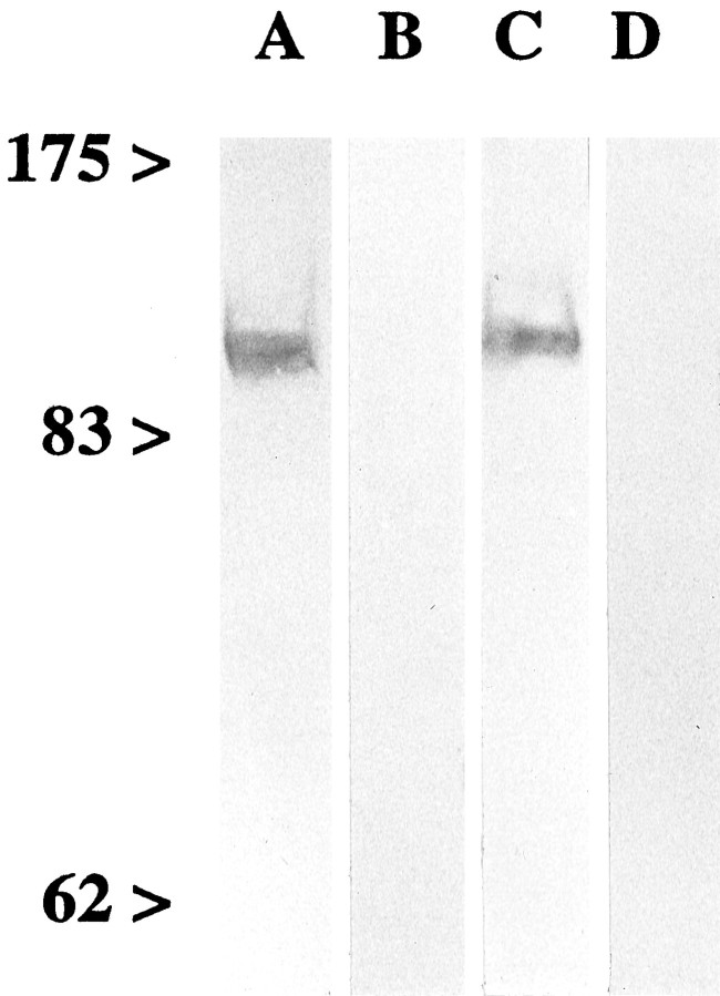
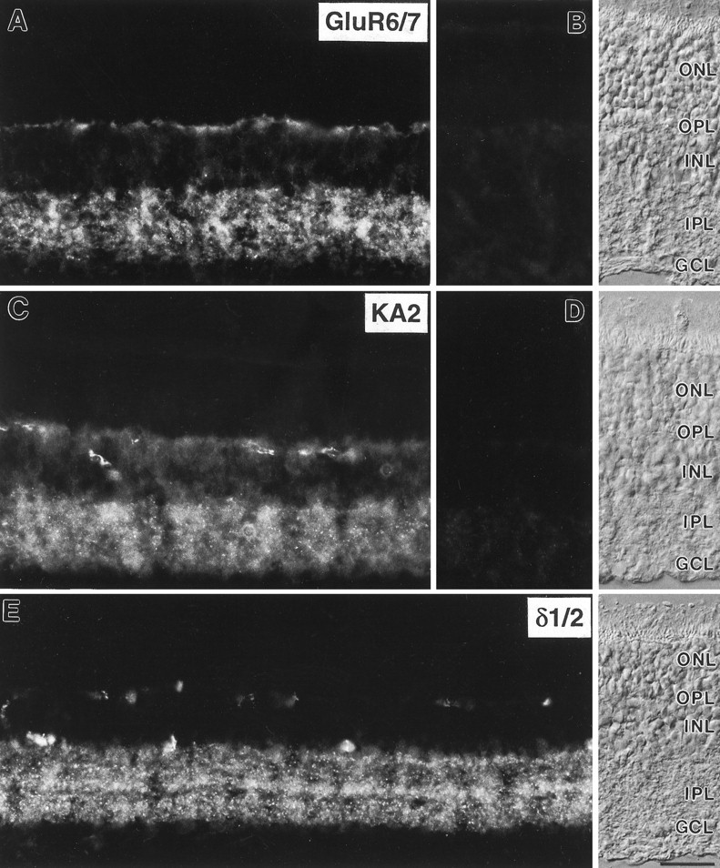
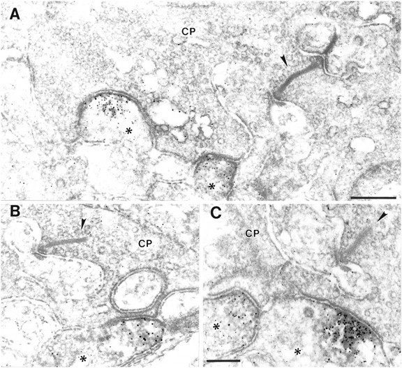
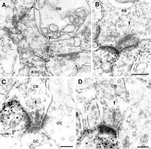
The synaptic localization of the kainate receptor subunits GluR6/7 and KA2 and of the ionotropic glutamate receptor subunits delta1/2 was studied in the rat retina using receptor-specific antisera. GluR6/7 and KA2 were present in both synaptic layers of the retina: the inner plexiform layer (IPL) and the outer plexiform layer (OPL). The localization of delta1/2 was restricted to the IPL. Detailed ultrastructural examination showed that in the OPL GluR6/7 was localized in horizontal cell processes postsynaptic to both rod spherules and cone pedicles. It was always only one of the two invaginating horizontal cell processes at the photoreceptor synapses labeled for GluR6/7. KA2 in the OPL was found only postsynaptic to cone pedicles and never postsynaptic to rod spherules. The KA2-labeled processes made flat contacts with the cone pedicles, suggesting they are the dendrites of OFF bipolar cells. In the IPL the different receptor subunits were localized postsynaptically to ribbon synapses of both rod and cone bipolar cells. As a rule, only one of the two postsynaptic elements at the bipolar cell dyad was stained for each of the receptor subunits examined. The selective and heterogeneous distribution of these receptors at the ribbon synapses of the OPL and IPL suggests a high degree of differential processing of the glutamatergic signals.


Figures

Fig. 1.
Fig. 1.
Specificity of the antisera against GluR6/7 and KA2. Membrane proteins of rat retina (80 μg/lane) were separated on a 7.5% SDS-PAGE gel and transferred onto nylon membrane.Numbers and arrowheads indicate the position and weight (kDa) of the marker bands. A,C, The antiserum against GluR6/7 (A) and KA2 (C) labeled a protein band at ∼110 and 120 kDa, respectively. B,D, Labeling was prevented by coincubation of the antiserum with the antigenic peptide, shown in B for GluR6/7 and in D for KA2.
Fig. 2.
Fig. 2.
Micrographs of vertical cryostat sections of rat retina immunostained with the antisera against GluR6/7, KA2, and δ1/2. A, Diffuse and punctate labeling for GluR6/7 was present in both synaptic layers, the OPL and theIPL. B, Preadsorption of the anti-GluR6/7 antiserum with the immunogen resulted in a complete loss of specific immunoreactivity.C, Diffuse and punctate labeling for KA2 was also present in both plexiform layers of the rat retina. In addition, somata in the inner nuclear layer showed weak KA2 immunostaining.D, No specific immunolabel was detected after preadsorption of the anti-KA2 antiserum with the immunogen.E, Punctate labeling for δ1/2 was present in several distinct strata in the IPL. Stained profiles in theOPL are unspecifically labeled blood vessels. The retinal layers are shown with Nomarski optics. ONL, Outer nuclear layer; OPL, outer plexiform layer;INL, inner nuclear layer; IPL, inner plexiform layer; GCL, ganglion cell layer. Scale bar, 25 μm.
Fig. 3.
Fig. 3.
High-power electron micrographs showing the ultrastructural localization of GluR6/7 immunoreactivity in the OPL.A, B, GluR6/7 staining in the OPL was found in horizontal cell processes (asterisk) postsynaptic to rod spherules (RS), and (C, D) in horizontal cell processes (asterisk) postsynaptic to cone pedicles (CP). Note in each case that only one of the two lateral horizontal cell processes at the photoreceptor synapses is labeled for GluR6/7. The presynaptic ribbon in the terminals of the photoreceptor cells is marked with an arrowhead. Scale bars: 0.2 μm (shown in D for C, D).
Fig. 4.
Fig. 4.
High-power electron micrographs showing the ultrastructural localization of KA2 immunoreactivity in the OPL.A–C, KA2 staining in the OPL was found in dendrites of OFF-cone bipolar cells (asterisk) that made flat contacts with the cone pedicles (CP) and were never associated with the synaptic complex of the CP. The presynaptic ribbon in the CPs is marked with anarrowhead. Scale bars (shown in A forA, B): 0.3 μm; C, 0.2 μm.
Fig. 5.
Fig. 5.
High-power electron micrographs showing the ultrastructural localization of GluR6/7 immunoreactivity at synapses in the IPL. GluR6/7 staining in the IPL was detected in the processes of amacrine and ganglion cells (asterisk) postsynaptic to OFF-cone (A; CB), ON-cone (B; CB), and rod bipolar cells (C; RB). Note in each case that only one of the two postsynaptic elements at the bipolar cell dyad is labeled for GluR6/7. The presynaptic ribbon in the terminals of the bipolar cells in A and B is marked with anarrowhead. GC, Ganglion cell. Scale bars, 0.1 μm.
Fig. 6.
Fig. 6.
High-power electron micrographs showing the ultrastructural localization of KA2 immunoreactivity at synapses in the IPL. KA2 staining in the IPL was detected in the processes of amacrine and ganglion cells (asterisk) postsynaptic to OFF-cone (A; CB), ON-cone (B;CB), and rod bipolar cells (C;RB). Note in each case that only one of the two postsynaptic elements at the bipolar cell dyad is labeled for KA2. The presynaptic ribbon in the terminals of the bipolar cells is marked with an arrowhead. Scale bar (shown in C forA–C): 0.1 μm.
Fig. 7.
Fig. 7.
High-power electron micrographs showing the ultrastructural localization of δ1/2 immunoreactivity at synapses in the IPL. A, B, δ1/2 staining was found in amacrine cell processes postsynaptic to rod bipolar cell (RB) ribbon synapses. In A, the labeled amacrine cell was identified as an AII amacrine cell. The second amacrine cell at the bipolar dyad in A, making a reciprocal synapse onto the bipolar terminal (arrow), is not labeled. In B, the labeled amacrine cell process (asterisk) postsynaptic to the rod bipolar cell (RB) synapse could not be identified. C,D, δ1/2 staining was also present in processes of amacrine and ganglion cells (asterisk) postsynaptic to OFF-cone (C; CB) and ON-cone (D; CB) bipolar cell synapses. Note at all bipolar cell synapses shown in A–D that only one of the two postsynaptic elements is labeled for δ1/2. GC, Ganglion cell. The presynaptic ribbon in the terminals of the bipolar cells is marked with an arrowhead. Scale bars (shown inB for A, B): 0.2 μm;C, D, 0.1 μm.

Similar articles

Cited by

References

    1. Araki K, Meguro H, Kushiya E, Takayama C, Inoue Y, Mishina M. Selective expression of the glutamate receptor channel delta 2 subunit in cerebellar Purkinje cells. Biochem Biophys Res Commun. 1993;197:1267–1276. - PubMed
    1. Bettler B, Egebjerg J, Sharma G, Pecht G, Hermans-Borgmeyer I, Moll C, Stevens CF, Heinemann S. Cloning of a putative glutamate receptor: a low affinity kainate-binding subunit. Neuron. 1992;8:257–265. - PubMed
    1. Boycott BB. Horizontal cells of mammalian retinae. Neurosci Res [Suppl] 1988;8:97–111. - PubMed
    1. Brandstätter JH, Hartveit E, Sassoè-Pognetto M, Wässle H. Expression of NMDA and high-affinity kainate receptor subunit mRNAs in the adult rat retina. Eur J Neurosci. 1994;6:1100–1112. - PubMed
    1. Brandstätter JH, Koulen P, Kuhn R, van der Putten H, Wässle H. Compartmental localization of a metabotropic glutamate receptor (mGluR7): two different active sites at a retinal synapse. J Neurosci. 1996;16:4749–4756. - PMC - PubMed

Publication types