. 1997 Dec 1;17(23):9308-14.
doi: 10.1523/JNEUROSCI.17-23-09308.1997.

CA3-driven hippocampal-entorhinal loop controls rather than sustains in vitro limbic seizures

Affiliations

CA3-driven hippocampal-entorhinal loop controls rather than sustains in vitro limbic seizures

M Barbarosie et al. J Neurosci. .

Abstract

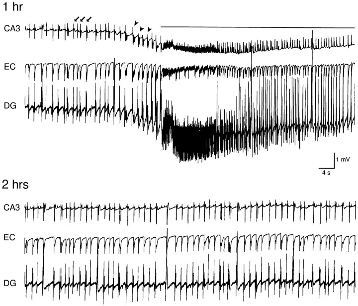
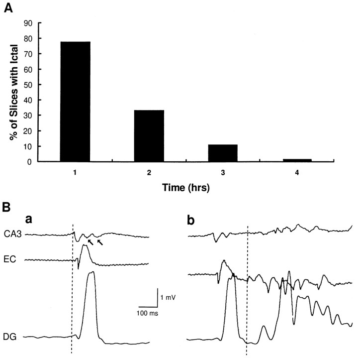
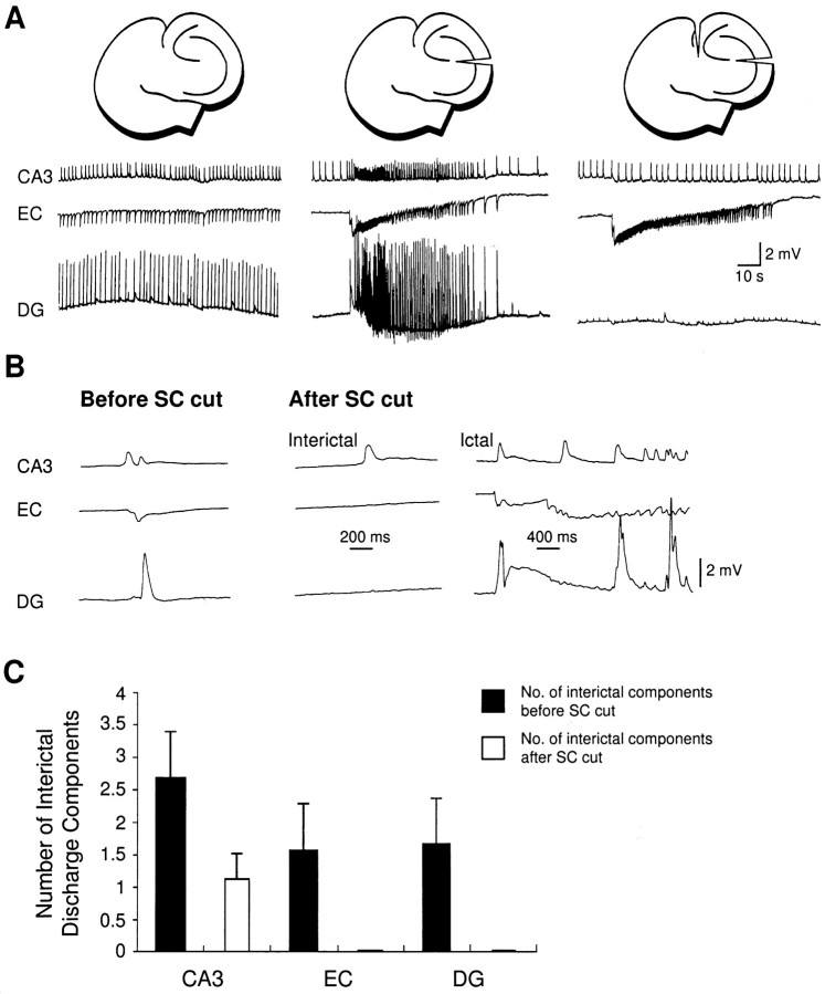
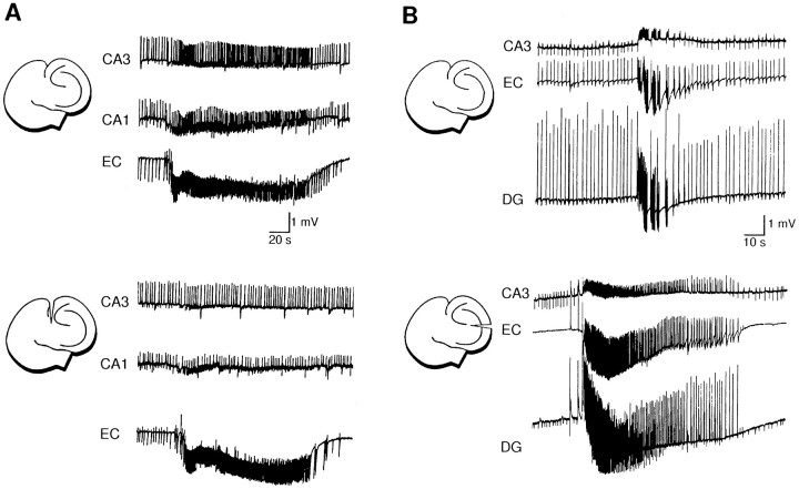
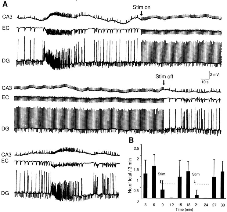
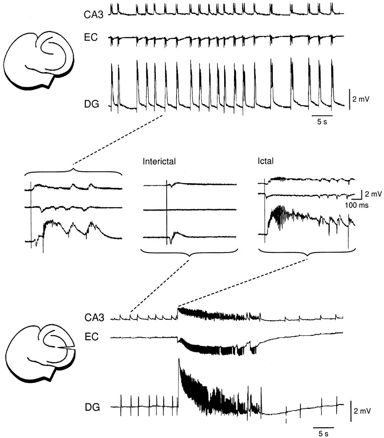
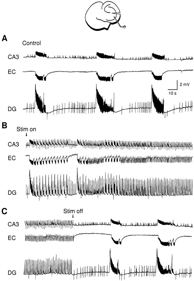
Continuous application of 4-aminopyridine (4-AP, 50 microM) to combined slices of hippocampus-entorhinal cortex obtained from adult mice induces (1) interictal discharges that initiate in the CA3 area and propagate via the hippocampal regions CA1 and subiculum to the entorhinal cortex and return to the hippocampus through the dentate gyrus; and (2) ictal discharges that originate in the entorhinal cortex and propagate via the dentate gyrus to the hippocampus proper. Ictal discharges disappear over time, whereas synchronous interictal discharges continue to occur throughout the experiment. Lesioning the Schaffer collaterals abolishes interictal discharges in CA1, entorhinal cortex, and dentate gyrus and discloses entorhinal ictal discharges that propagate, via the dentate gyrus, to the CA3 subfield. Interictal discharges originating in CA3 also prevent the occurrence of ictal events generated in the entorhinal cortex during application of Mg2+-free medium. In both models, ictal discharge generation recorded in the entorhinal cortex after Schaffer collateral cut is prevented by mimicking CA3 neuronal activity through rhythmic electrical stimulation (0.25-1.5 Hz) of the CA1 hippocampal output region. Our findings demonstrate that interictal discharges of hippocampal origin control the expression of ictal epileptiform activity in the entorhinal cortex. Sectioning the Schaffer collaterals may model the chronic epileptic condition in which cell damage in the CA3 subfield results in loss of CA3 control over the entorhinal cortex. Hence, we propose that the functional integrity of hippocampal output neurons may represent a critical control point in temporal lobe epileptogenesis.


Figures

Fig. 1.
Fig. 1.
Spontaneous epileptiform activity recorded at 1 and 2 hr during continuous bath application of 4-AP. Simultaneous field potential recordings were made in the CA3 stratum radiatum, the deep layers of the entorhinal cortex, and the dentate granule cell layer. Ictal discharge, recorded at 1 hr, is indicated by continuous line, interictal discharges by arrows, and robust interictal discharges with afterdischarge component byarrowheads. At 2 hr during 4-AP application, ictal discharges disappear. In this and the following figures,EC and DG stand for entorhinal cortex and dentate gyrus, respectively.
Fig. 2.
Fig. 2.
A, Percentage histogram of slices generating ictal discharges at different times of 4-AP application. These slices (n = 9) were recorded for >4 hr.B, Expanded traces of interictal (a) and ictal discharges (b) induced by 4-AP (∼1 hr) in an intact entorhinal-hippocampal combined slice. In a, the interictal discharge initiates in the CA3 region and propagates to the entorhinal cortex and the dentate gyrus; arrows point at the late components of the interictal discharge recorded in CA3. Inb, the ictal discharge is preceded by an interictal event with temporal profile similar to that seen in a, whereas the site of origin of the ictal discharge appears to occur in the entorhinal cortex. Dotted lines in aand b were positioned at the time of the earliest visible deflection in the three field potential recordings.
Fig. 4.
Fig. 4.
Effects induced by sectioning the Schaffer collaterals and the perforant pathway on epileptiform discharges recorded ∼2 hr after continuous application of 4-AP.A, Interictal discharges are recorded in CA3, entorhinal cortex, and dentate gyrus in the intact slice (traces, left). Sectioning the Schaffer collaterals abolishes the interictal discharges in entorhinal cortex and dentate gyrus and discloses an ictal discharge that is simultaneously recorded in CA3, entorhinal cortex, and dentate gyrus (traces, middle). Further cutting of the perforant path abolishes the propagation of the ictal discharge to the entorhinal cortex and dentate gyrus (traces, right). B, Expanded traces from the experiment shown in A demonstrate that interictal discharges reenter CA3. They comprise two components in the CA3 of the intact slice (Before SC cut), whereas after sectioning the Schaffer collaterals, a single component is left (After SC cut, Interictal). The onset of an ictal discharge recorded after Schaffer collateral cut is also shown (After SC cut, Ictal).C, Quantitative summary of the effects induced by Schaffer collateral cut on the number of interictal discharge components in CA3, entorhinal cortex (EC) and dentate gyrus (DG) (n = 6;p < 0.05).
Fig. 3.
Fig. 3.
Spontaneous epileptiform activity recorded during application of 4-AP (∼1 hr) in a combined hippocampal–entorhinal cortex slice before and after selective neuronal pathway sectioning. A, Changes in 4-AP-induced activity before and after cut of the perforant path indicate that the ictal discharges originate in the entorhinal cortex. B, Effects induced by Schaffer collateral cut further demonstrate that interictal discharges initiate in the CA3 subfield, because they are abolished in the entorhinal cortex and dentate gyrus. Note also that after Schaffer collateral cut, the ictal discharge duration is increased (n = 6; p < 0.05).
Fig. 7.
Fig. 7.
A, Continuous recordings showing the effect of CA1 stimulation at 1 Hz on the 4-AP-induced epileptiform activity recorded after Schaffer collateral cut. Note the persistence of interictal discharges, presumably of entorhinal origin, before and after the ictal activity. Ictal discharge does not occur during the stimulation period and re-appear on termination of the stimulation.B, Time histogram showing the effect of low-frequency stimulation on ictal discharge occurrence (p< 0.05 for both stimulation protocols). Data were obtained from eight slices for the first stimulation and four slices for the second stimulation protocol.
Fig. 5.
Fig. 5.
Spontaneous epileptiform activity induced by Mg+2-free ACSF before and after Schaffer collateral cut. Before the lesion (top), synchronized interictal discharges are recorded in CA3, entorhinal cortex, and dentate gyrus. Sectioning the Schaffer collaterals (bottom) abolishes interictal discharges in the entorhinal cortex and discloses ictal epileptiform activity that is recorded in the three areas. Expanded traces of the experiment shown in the top andbottom are illustrated in the middle. Note that before the Schaffer collateral cut Mg+2-free-induced interictal discharges consist of multiple components, whereas after the cut (Interictal) they are markedly reduced in duration and number of events. Note also that the ictal discharge (Ictal) is initiated in the entorhinal cortex.
Fig. 6.
Fig. 6.
Effect induced by extracellular stimuli delivered in the CA1 subfield (1 Hz) on the Mg+2-free-induced ictal activity recorded after Schaffer collateral cut.A–C, Continuous recordings demonstrate that low-frequency stimulation prevents the occurrence of ictal discharges that reappear on termination of the stimulation.

Similar articles

Cited by

References

    1. Amaral DG, Witter MP. The three-dimensional organization of the hippocampal formation: a review of anatomical data. Neuroscience. 1989;31:571–591. - PubMed
    1. Avoli M, Barbarosie M, Lücke A, Nagao T, Lopantsev V, Köhling R. Synchronous GABA-mediated potentials and epileptiform discharges in the rat limbic system in vitro. J Neurosci. 1992;16:3912–3924. - PMC - PubMed
    1. Ben-Ari Y. Limbic seizures and brain damage produced by kainic acid: mechanisms and relevance to human temporal lobe epilepsy. Neuroscience. 1985;14:375–403. - PubMed
    1. Bernard C, Wheal HV. A role for synaptic and network plasticity in controlling epileptiform activity in CA1 in the kainic acid-lesioned rat hippocampus in vitro. J Physiol (Lond) 1996;456:127–142. - PMC - PubMed
    1. Bragdon AC, Kojima H, Wilson WA. Suppression of interictal bursting in hippocampus unleashes seizures in the entorhinal cortex: a proepileptic effect of lowering [K+]o and raising [Ca+2]o. Brain Res. 1992;590:128–135. - PubMed

Publication types