Comparative Study
. 1997 Dec 1;17(23):9340-52.
doi: 10.1523/JNEUROSCI.17-23-09340.1997.

Differential brainstem Fos-like immunoreactivity after laryngeal-induced coughing and its reduction by codeine

Affiliations
Comparative Study

Differential brainstem Fos-like immunoreactivity after laryngeal-induced coughing and its reduction by codeine

C Gestreau et al. J Neurosci. .

Abstract

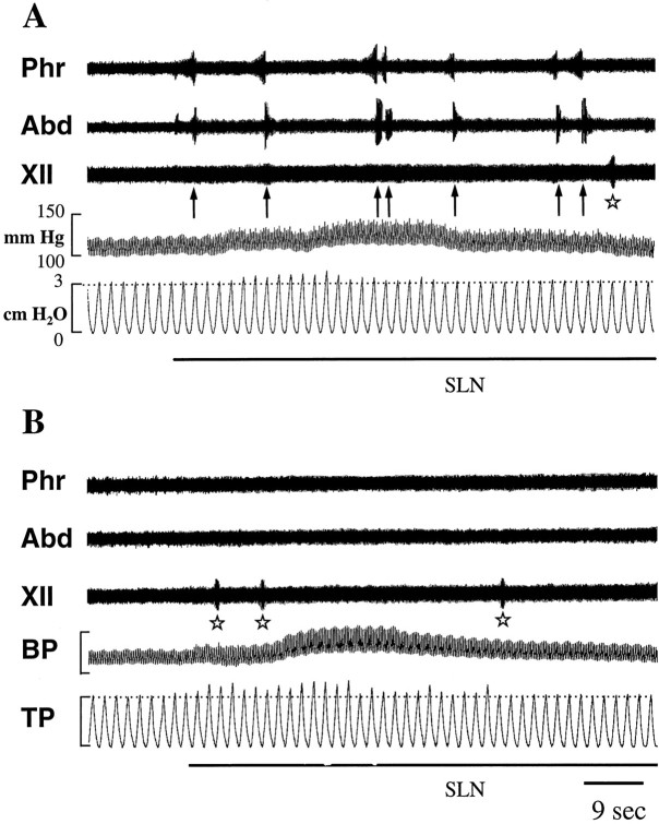
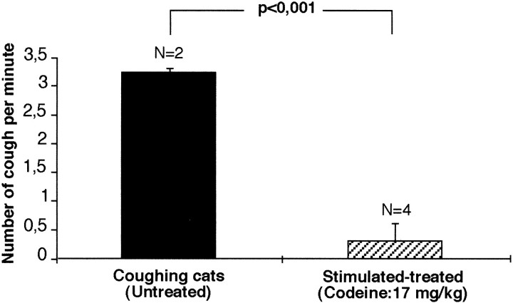
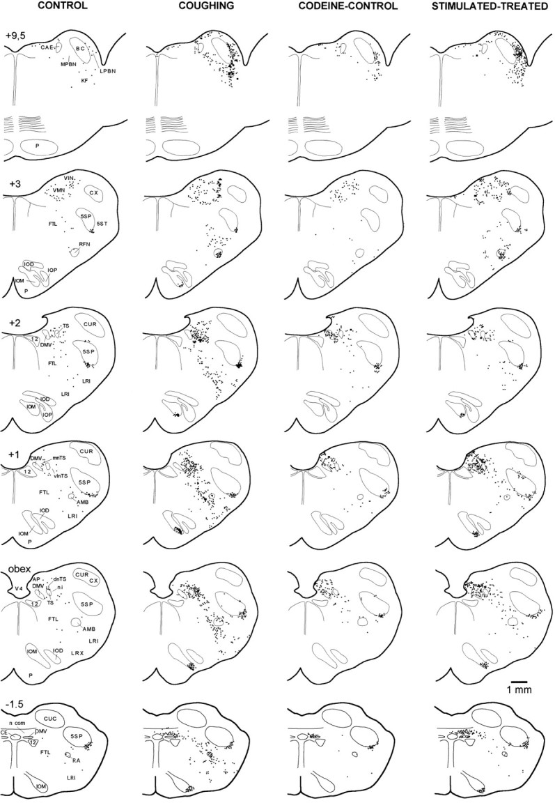
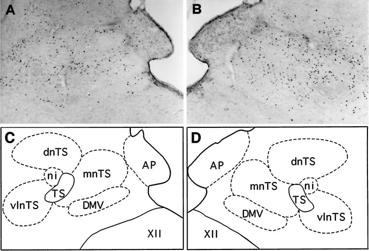
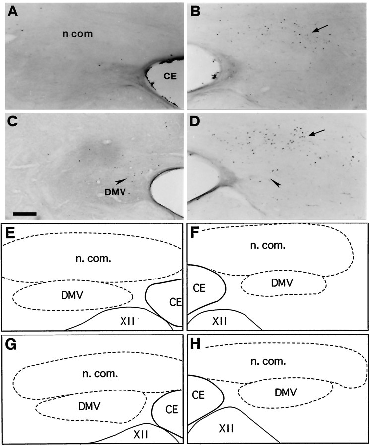
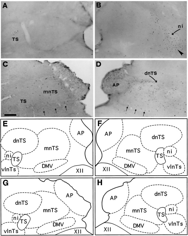
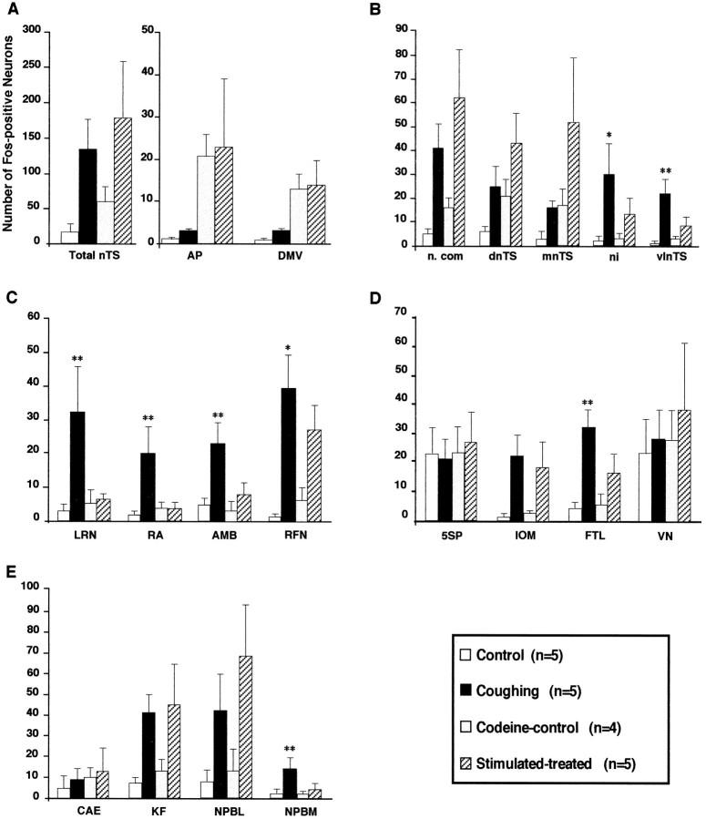
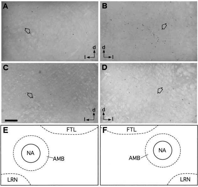
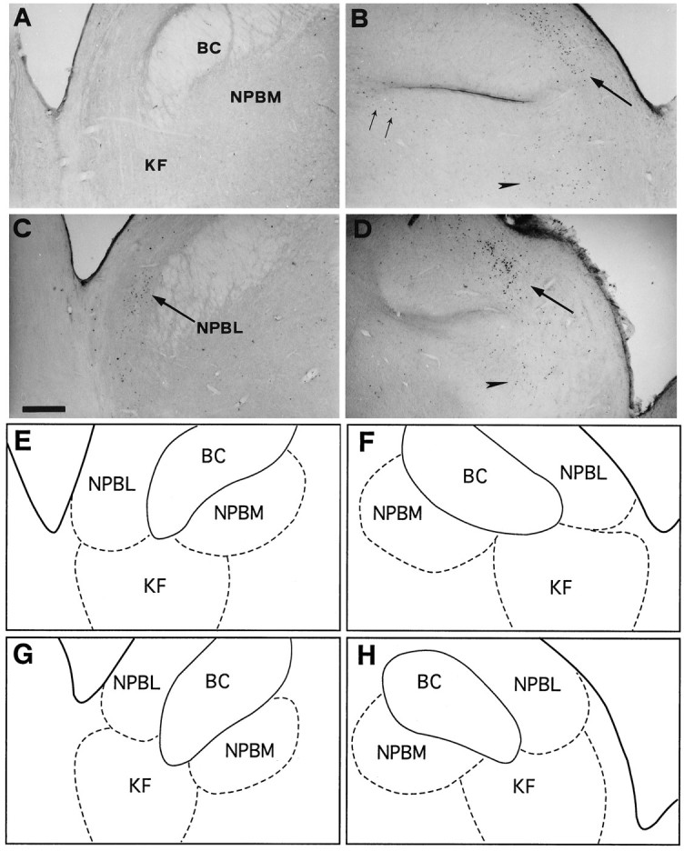
We used the expression of the immediate-early gene c-fos, a marker of neuronal activation, to localize brainstem neuronal populations functionally related to fictive cough (FC). In decerebrate, paralyzed, and ventilated cats, the level of Fos-like immunoreactivity (FLI) was examined in five groups of animals: (1) controls, sham-operated unstimulated animals; (2) coughing cats, including both animals in which FC was elicited by unilateral electrical stimulation of the superior laryngeal nerve (SLN) and (3) those in which FC was elicited by bilateral SLN stimulation; (4) stimulated-treated cats, in which bilateral SLN stimulation was applied after selective blockade of FC by codeine; and (5) codeine controls, sham-operated unstimulated cats subjected to administration of codeine. Fifteen brainstem structures were compared for numbers of labeled cells. Because codeine selectively blocks FC, brainstem nuclei activated specifically during FC were identified as regions showing increased FLI after FC and significant reductions in FLI after FC suppression by codeine in stimulated-treated cats. In coughing animals, we observed a selective immunoreactivity in the interstitial and ventrolateral subdivisions of the nucleus of the tractus solitarius, the medial part of the lateral tegmental field, the internal division of the lateral reticular nucleus, the nucleus retroambiguus, the para-ambigual region, the retrofacial nucleus, and the medial parabrachial nucleus. FLI in all these nuclei was significantly reduced in stimulated-treated cats. Our results are consistent with the involvement of neurons overlapping the main brainstem respiratory-related regions as well as the lateral tegmental field and the lateral reticular nucleus in the neural processing of laryngeal-induced FC.


Figures

Fig. 1.
Fig. 1.
Effects of SLN stimulation (black bars below each panel) (0.2 msec pulses, 5 V, 4 Hz) on phrenic (Phr), iliohypogastric (Abd), and hypoglossal (XII) nerve activities, and on arterial blood (BP) and tracheal (TP) pressures in coughing (A) and stimulated–treated (B) cats. In A, SLN stimulation induced multiple FCs (arrows) and transient hypertension and bronchoconstriction (slight increase in TP peaks abovehorizontal hatched line). In B, after the last intravenous injection of codeine given 15 min before the start of SLN stimulation (see Materials and Methods for details), FCs were abolished, whereas transient changes in BP and TP were not altered. Some buccopharyngeal stages of swallowing (stars) occurred regardless of the antitussive treatment. Stimulus artifacts were erased to improve legibility.
Fig. 2.
Fig. 2.
Antitussive effect of codeine on SLN-induced cough. Codeine significantly reduced the elicitation of FC.
Fig. 3.
Fig. 3.
Distribution of brainstem FLI in transverse standardized hemisections through different rostrocaudal levels of control (unstimulated), coughing (262 SLN-induced coughs), codeine-control, and stimulated–treated (11 SLN-induced coughs) animals. Antitussive effect of codeine was associated with a decrease in FLI in dorsal, medial, and ventrolateral medulla and pons of the stimulated–treated animal. Each square represents one Fos-positive neuron. Rostrocaudal position relative to obex is indicated at top of drawing of each hemisection. 12, Hypoglossal nucleus; 5SP, 5ST, alaminar spinal trigeminal nucleus and tract; AMB, para-ambigual region;BC, brachium conjonctivum; CAE, locus caeruleus; CE, central canal; CUC, CUR, CUX, caudal, rostral, and external cuneate nucleus;DMV, dorsal motor nucleus of the vagus;FTL, lateral tegmental field; IOM, IOD, medial and dorsal accessory inferior olive; IOP, principal nucleus of the inferior olive; KF, Kölliker-Fuse nucleus; LRI, LRX, internal and external divisions of the lateral reticular nucleus; NPBL, NPBM, lateral and medial divisions of the parabrachial nucleus;RA, nucleus retroambiguus; RFN, retrofacial nucleus; TS, tractus solitarius;dnTS, mnTS, n.com, ni, vlnTS, dorsal, medial, commissural, interstitial, and ventrolateral subdivisions of the nucleus tractus solitarius (nTS); VIN, VMN, inferior and medial vestibular nuclei; V4, fourth ventricle.
Fig. 4.
Fig. 4.
Bright-field photomicrographs illustrating patterns of FLI induced in the left (A) and right (B) dorsal vagal complex after FC elicited by stimulation of the right SLN. A and B are from the same transverse section. C and Dare schematic drawings delineating dorsal medullary nuclei shown inA and B, respectively. Note the increase in the number of Fos-positive cells in the ipsilateral nTS (B) as opposed to the contralateral nTS (A). Scale bar, 280 μm. Abbreviations are defined in legend to Figure 3.
Fig. 5.
Fig. 5.
Bright-field photomicrographs of transverse sections illustrating FLI in the left (A, C) and right (B, D) commissural subdivision (n com) of the nTS. Control (A), coughing (B), codeine-control (C), and stimulated–treated (D) cats. FLI was induced in n.com of the two cats subjected to SLN stimulation whether or not they were treated with codeine (arrows in B andD). Codeine induced a few Fos-positive neurons in the dorsal motor nucleus of the vagus nerve (DMV) (arrowheads in C and D).E, F, G, and H are schematic drawings of dorsal medullary nuclei shown in A, B, C, andD, respectively. Outlined structuresindicate selected areas in which labeled cells were counted. Scale bar, 220 μm. CE, Central canal; XII, hypoglossal nucleus.
Fig. 6.
Fig. 6.
Bright-field photomicrographs of transverse sections illustrating FLI in the dorsal vagal complex (nTS, AP, and DMV) of medulla. Control (A), coughing (B), codeine-control (C), and stimulated–treated (D) cats. Note dense labeling in the interstitial (ni, curved arrow) and ventrolateral (arrowhead) subdivisions of the nTS observed only inB. Codeine induced sparse Fos-positive neurons in AP, DMV (three small arrows), and mnTS of codeine-control cat (C). Similar FLI was also observed in AP and DMV of the stimulated–treated (D) cat, associated with an increase in Fos-like expression in the dnTS and mnTS. E, F, G, and H are schematic drawings of dorsal medullary nuclei shown in A, B, C, and D, respectively. Outlined structuresindicate selected areas in which labeled cells were counted. Scale bar, 300 μm. Abbreviations are defined in legend to Figure 3.
Fig. 9.
Fig. 9.
Numbers of Fos-like immunoreactive neurons for the four groups in various brainstem nuclei: A, dorsal vagal complex; B, nTS subnuclei; C, ventral medulla; D, other medullary regions; andE, pons. Histograms represent mean numbers of Fos-positive neurons with superimposed SD. Asterisksindicate significant (*p < 0.05 and **p < 0.01) increases of Fos-positive neurons in coughing cats versus both control and stimulated–treated animals. Abbreviations are defined in legend to Figure 3.
Fig. 7.
Fig. 7.
Bright-field photomicrographs of transverse sections illustrating FLI distribution in the ventral medulla overlapping the AMB and the nucleus ambiguus. Control (A), coughing (B), codeine-control (C), and stimulated–treated (D) cats. Open arrows point to the nucleus ambiguus. Arrowsindicate the dorsal (d) and lateral (l) directions. Note in Bdense labeling ventromedial to the nucleus ambiguus. Eand F are schematic drawings delineating the para-ambigual region (AMB) (hatched line), including the nucleus ambiguus (NA) (continuous line) shown in A andB, respectively. Similarly, E andF also match C and D.Outlined structures indicate selected areas in which labeled cells were counted. Scale bar, 200 μm. Abbreviations are defined in legend to Figure 3.
Fig. 8.
Fig. 8.
Bright-field photomicrographs of transverse sections depicting FLI distribution in the dorsolateral pons. Control (A), coughing (B), codeine-control (C), and stimulated–treated (D) cats. SLN stimulation induced dense FLI in NPBL (large arrows) and KF nucleus (arrowhead) of the two stimulated cats (B, D). FLI evoked in NPBM (two small arrows) of coughing cat (B) decreased after codeine administration (D). E, F, G, andH are schematic drawings of pontine nuclei shown inA, B, C, and D, respectively.Outlined structures indicate selected areas in which labeled cells were counted. Scale bar, 600 μm. Abbreviations are defined in legend to Figure 3.

Similar articles

Cited by

References

    1. Abbadie C, Besson J-M. Effects of morphine and naloxone on basal and evoked Fos-like immunoreactivity in lumbar spinal cord neurons of arthritic rats. Pain. 1993;52:29–39. - PubMed
    1. Bellingham MC, Lipski J. Morphology and electrophysiology of superior laryngeal nerve afferents and post-synaptic neurons in the medulla oblongata of the cat. Neuroscience. 1992;48:205–216. - PubMed
    1. Bereiter DA, Hathaway CB, Benetti AP. Caudal portions of the spinal trigeminal complex are necessary for autonomic responses and display Fos-like immunoreactivity after corneal stimulation in the cat. Brain Res. 1994;657:73–82. - PubMed
    1. Berman AL. The brainstem of the cat: a cytoarchitectonic atlas with stereotaxic coordinates. Milwaukee: University of Wisconsin; 1968.
    1. Bianchi AL, Denavit-Saubié M, Champagnat J. Central control of breathing in mammals: neuronal circuitry, membrane properties, and neurotransmitters. Physiol Rev. 1995;75:1–46. - PubMed

Publication types

MeSH terms