. 1997 Nov 15;11(22):2983-95.
doi: 10.1101/gad.11.22.2983.

The Arabidopsis HY5 gene encodes a bZIP protein that regulates stimulus-induced development of root and hypocotyl

Affiliations

The Arabidopsis HY5 gene encodes a bZIP protein that regulates stimulus-induced development of root and hypocotyl

T Oyama et al. Genes Dev. .

Abstract

Plant developmental processes are controlled by both endogenous programs and environmental stimuli. As a photomorphogenetic mutant, hy5 of Arabidopsis has been isolated and characterized. Our detailed characterization has revealed that the mutant is deficient in a variety of stimulus responses, including gravitropic response and waving growth of roots, as well as light-dependent hypocotyl elongation. In the roots and hypocotyl, the hy5 mutation also affects greening and specific cell proliferation such as lateral root formation and secondary thickening. Those phenotypes indicate that the HY5 gene is responsible for the regulation of fundamental developmental processes of the plant cell: cell elongation, cell proliferation, and chloroplast development. Molecular cloning of the HY5 gene using a T-DNA-tagged mutant has revealed that the gene encodes a protein with a bZIP motif, one of the motifs found in transcriptional regulators. Nuclear localization of the HY5 protein strongly suggests that the HY5 gene modulates the signal transduction pathways under the HY5-related development by controlling expression of genes downstream of these pathways.


Figures
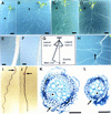
Figure 1
Figure 1
Phenotypes of hy5 mutants. (AD) Plants at 20 DAG grown on vertically positioned agar plates supplemented with sucrose. Wild-type of Landsberg erecta (Ler) ecotype (A), hy5-1 (B), hy5–Ks50 (C), and hy5-215 (D) are shown. The arrowhead in each panel indicates a lateral root. Scale bars, 10 mm. (E,F) Root hairs of wild-type Ler ecotype (E) and hy5-1 (F). The seedlings were grown on agar plates set in a vertical position. Scale bars, 100 μm. (G) A diagram, indicating how to measure angles formed by roots and gravity orientation. Roots of seedlings elongate on an agar plate set in a vertical position. Plus (+) and minus (−) degrees represent a clockwise angle from the gravity orientation and a counterclockwise angle, respectively (see Table 3). (H) A magnified view of Fig. 2B, showing a secondary lateral root of hy5-1 (arrow). (I,J) Wavy pattern of wild-type of Ler ecotype (I) and hy5-1 (J). Seedlings were grown on an agar plate for 3 days set in a vertical position. Then the plate was tilted to 45° (arrows indicate the positions of root tips at the time of the position change), and the seedlings were grown for 3 days. (K,L) Cellular organization in the secondarily thickened roots of wild-type Ler ecotype (K) and hy5-1 (L). Transverse sections were made at the same position of main roots of seedlings at 20 DAG grown on agar plates supplemented with sucrose and stained with toluidine blue (see Materials and Methods). A well-lignified xylem vessel, a peridremal cell, and a fiber element are indicated by a star, a large arrow, and a small arrow, respectively. Scale bars, 50 μm.
Figure 2
Figure 2
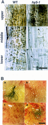
Greening in the hypocotyl and roots. (A) Chloroplasts in the hypocotyl of wild-type Ler ecotype (left) and hy5-1 (right) plants at 20 DAG grown on agar plates supplemented with sucrose. Upper, middle, and lower parts of the hypocotyl are shown in respectively labeled panels. Scale bar, 25 μm. (B) Roots of plants at 30 DAG grown in a liquid medium. Leaves and the hypocotyl are removed from the plants. Wild-type of Ler ecotype (a), hy5-1 (b), hy5–Ks50 (c), and cop1-6 (d) are shown. Scale bar, 10 mm.
Figure 3
Figure 3
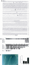
Sequence analysis of the HY5 gene. (A) Genomic DNA sequence and deduced amino acid sequence of HY5. The genomic sequence from SacI to PstI in the HY5 locus of the Ws wild type is shown. The first G in the SacI site is designated as number 1. Numbers in italics indicate the amino acid sequence of the HY5 protein. Exons are shown in uppercase letters. The bZIP domain is underlined. In hy5–Ks50, sequences between t-147 and t-935 nucleotides (in boldface at arrowheads) are deleted and a T-DNA concatemer is inserted. In hy5-1, the nucleotide C-811 (white letter) is replaced by T, which results in a stop of translation. In hy5-215, the nucleotide g-1117 (white letter), which is the last nucleotide in the first intron, is replaced by an a. The HY5 cDNA sequence has been deposited in DDBJ/GenBank/EMBL databases (accession no. AB005295). (B) Amino acid comparison of plant bZIP proteins. The deduced HY5 amino acid sequence is compared with the sequences of eight related proteins in plants: STF1A from soybean (GenBank accession no. L28003, J.-C. Hong, pers. comm.), TGA-1b and TGA-1a from tobacco (Katagiri et al. 1989), EmBP-1 from wheat (Guiltinan et al. 1990), HBP-1a and HBP-1b from wheat (Tabata et al. 1989, 1991), GBF-1 from Arabidopsis (Schindler et al. 1992a), and OPAQUE2 from maize (Hartings et al. 1989; Schmidt et al. 1990). White letters represent residues identical to the HY5 sequence. A serine residue that is predicted to be phosphorylated by CKII is indicated (○). The basic region (from K-90 to K-109) is overlined. (•) The heptad repeat of leucines in the leucine zipper region. Asterisks (*) indicate the carboxyl-terminus of a given protein. (C) Complementation of hy5–Ks50 with a genomic sequence including the HY5 gene. (Left) Plants at 20 DAG grown in the light on a vertically positioned agar plate supplemented with sucrose. The plants are wild-type of Ws ecotype, hy50–Ks50 transformed with the genomic sequence, and hy5–Ks50 (from left to right). Leaves were removed from the plants. Scale bar, 10 mm. (Right) Bar graph of hypocotyl lengths of seedlings at 7 DAG grown in the light. The bars represent wild-type of Ws ecotype, hy5–Ks50 transformed with the genomic sequence, and hy5-Ks50, respectively. Numbers of the measured samples are 18 (wild-type) or 40 (the transformant and hy5–Ks50).
Figure 4
Figure 4
Northern blot analysis of HY5 mRNA. An aliquot of the total RNA (10 μg/lane) was subjected to agarose gel electrophoresis, transferred to nylon membranes, and hybridized with the probe of HY5 cDNA, as described in Materials and Methods. The same membranes were subsequently hybridized with the probe of the αTUBULIN (αTUB) coding region of Arabidopsis (Kopczak et al. 1992). rRNA was detected by staining gels with ethidium bromide. (A) Total RNA was extracted from roots of plants at 30 DAG grown in a liquid medium. (B) The total RNA was extracted from root, hypocotyl, and cotyledon tissues isolated from light-grown seedlings of wild-type (Ler) at 3 DAG on agar plates supplemented with sucrose. Leaf, stem, and floral organs were isolated from mature wild-type plants grown in soil. Samples of the floral organs contained different tissues of the inflorescent meristem, floral buds, and mature flowers. (C) Total RNA was extracted from light- and dark-grown seedlings of wild-type Ler ecotype and the cop1-6 mutant or from light-grown seedlings of the hy2 and the hy4 mutants. All seedlings were grown on agar plates supplemented with sucrose for 3 days. (D) Total RNA was extracted from roots of plants at 30 DAG of each genotype grown in a liquid medium.
Figure 5
Figure 5
Subcellular localization of the HY5 protein. Protoplasts were prepared from roots of seedlings at 7 DAG grown on agar plates supplemented with sucrose in the light. The top and bottom panels show the wild-type Ws ecotype and hy5-Ks50, respectively. The left and right panels show DAPI staining of the nuclei and Texas Red imaging of the same cells detected with anti-HY5 antiserum, respectively. Scale bar, 10 μm.

Similar articles

Cited by

References

    1. Aeschbacher RA, Schiefelbein JW, Benfey PN. The genetic and molecular basis of root development. Annu Rev Plant Physiol Plant Mol Biol. 1994;45:25–45.
    1. Ahmad M, Cashmore AR. HY4 gene of A. thaliana encodes a protein with characteristics of a blue-light photoreceptor. Nature. 1993;366:162–166. - PubMed
    1. Ang L-H, Deng X-W. Regulatory hierarchy of photomorphogenic loci: allele-specific and light-dependent interaction between the HY5 and COP1 loci. Plant Cell. 1994;6:613–628. - PMC - PubMed
    1. Arnon DI. Copper enzymes in isolated chloroplasts. Polyphenoloxidase in Beta vulgaris. Plant Physiol. 1949;24:1–15. - PMC - PubMed
    1. Ausubel FM, Brent R, Kingston RE, Moore DD, Seidman JG, Smith JA, Struhl K. Current protocols in molecular biology. New York, NY: Greene/Wiley; 1987.

Publication types

MeSH terms