. 1997 Nov 15;11(22):2996-3006.
doi: 10.1101/gad.11.22.2996.

The LKLF transcription factor is required for normal tunica media formation and blood vessel stabilization during murine embryogenesis

Affiliations

The LKLF transcription factor is required for normal tunica media formation and blood vessel stabilization during murine embryogenesis

C T Kuo et al. Genes Dev. .

Abstract

The transcriptional programs that regulate blood vessel formation are largely unknown. In this paper, we examine the role of the zinc finger transcription factor LKLF in murine blood vessel morphogenesis and homeostasis. By in situ hybridization and immunohistochemistry, we show that LKLF is expressed as early as embryonic day 9.5 (E9.5) in vascular endothelial cells throughout the developing mouse embryo. To better understand the function of LKLF, we used homologous recombination in embryonic stem (ES) cells to generate LKLF-deficient (LKLF-/-) mice. Both angiogenesis and vasculogenesis were normal in the LKLF-/- mice. However, LKLF-/- embryos died between E12.5 and E14.5 from severe intra-embryonic and intra-amniotic hemorrhaging. This bleeding disorder was associated with specific defects in blood vessel morphology. Umbilical veins and arteries in the LKLF-/- embryos displayed an abnormally thin tunica media and aneurysmal dilatation before rupturing into the amniotic cavity. Similarly, vascular smooth muscle cells in the aortae from the LKLF-/- animals displayed a cuboidal morphology and failed to organize into a compact tunica media. Consistent with these findings, electron microscopic analyses demonstrated endothelial cell necrosis, significant reductions in the number of vessel-wall pericytes and differentiating smooth muscle cells, and decreased deposition of extracellular matrix in the LKLF-/- vessels. Despite these defects, in situ hybridization demonstrated normal expression of platelet-derived growth factor B, Tie1, Tie2, transforming growth factor beta, and heparin-binding epidermal growth factor in the vasculature of the LKLF-/- embryos. Therefore, LKLF defines a novel transcriptional pathway in which endothelial cells regulate the assembly of the vascular tunica media and concomitant vessel wall stabilization during mammalian embryogenesis.


Figures

Figure 1
Figure 1
Embryonic expression of LKLF. In situ hybridization analyses of E9.5 (A,B) and E12.5 (C,D) wild-type mouse embryos using radio-labeled sense and antisense LKLF cRNA probes. Note LKLF expression in the developing vasculature including the dorsal aorta (da) of the E9.5 embryo. The areas of highest LKLF expression in the E12.5 embryo are in the umbilical vessels (uv), the intervertebral arteries (iva), the vertebrae (vert), and the smaller vessel structures in the head and tail regions. Bar, 1 mm.
Figure 2
Figure 2
LKLF expression in embryonic vascular endothelial cells. Histological (A–C), in situ hybridization (D–I), and immunohistochemical (J–L) analyses of developing vessels in E9.5 wild-type mouse embryos (A,D,G,J), E12.5 embryonic brain (B,E,H,K), and E12.5 umbilical vessels (C,F,I,L). (A–C) H + E staining. (D–I) In situ hybridizations using radio-labeled sense and antisense LKLF cRNA probes. Note the intracerebral vessels (v) and capillaries (c), the umbilical arteries (ua), and veins (uv). The fluorescent signal in the lumens of the umbilical vessels seen with both sense and antisense probes reflects RBC autoflourecence, not hybridization. (J–L) Immunohistochemical analyses using an anti-CD34 monoclonal antibody. Note CD34+ endothelial cells lining the endoluminal surface of the intracerebral vessels as well as the umbilical veins and arteries.
Figure 3
Figure 3
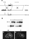
Targeted disruption of the LKLF gene. (A) Schematic representation of the LKLF targeting strategy. (Top) Partial restriction endonuclease map of the murine LKLF locus. Exons (E1-E3) are shown as boxes, the coding region is solid, the regions that encode the zinc fingers are hatched, the 5′ and 3′ UTRs are open. (E) EcoRI. (Middle) Structure of the LKLF-targeting construct containing the HSV-tk (tk) and neomycin (neoR) resistance genes under the control of the mouse phosphoglycerate kinase (PGK) promoter. (Bottom) Structure of the targeted LKLF allele containing a deletion of the entire LKLF gene. The probe used in Southern blot analyses is shown (probe). This targeting construct has been described previously (Kuo et al. 1997). (B) Southern blot analysis of offspring from an LKLF+/− × LKLF+/− mating. EcoRI digestion of E11.5 embryonic yolk-sac DNA hybridized to the genomic probe shown in A. The wild-type locus (wt) is 3.5 kb and the targeted locus (t) is 2.1 kb. (C,D) In situ hybridization of E11.5 wild-type (C) and LKLF−/− (D) embryo sections with an antisense LKLF cRNA probe derived from the full-length LKLF cDNA. Note LKLF expression in vascular endothelial cells throughout the wild-type embryo and the absence of LKLF expression in the LKLF−/− embryo.
Figure 4
Figure 4
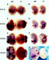
Intra-embryonic and intra-amniotic hemorrhages in LKLF−/− embryos. Photomicrographs of E11.5–E14.5 LKLF−/− embryos with the amnion, yolk sac, and placenta intact (A,C,E,G) or with extraembryonic membranes removed (B,D,F,H). Note the lack of detectable morphological differences between E11.5 wild-type and LKLF−/− embryos. E12.5 LKLF−/− embryos display intra-embryonic hemorrhages around the cardiac out-flow tract and in the abdomen (arrows). The yolk sac vessels from these embryos lack blood. E13.5 LKLF−/− embryos demonstrate severe intra-amniotic and intra-embryonic (arrows) hemorrhages. E14.5 LKLF−/− embryos have exsanguinated into the amniotic cavity.
Figure 5
Figure 5
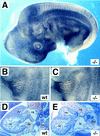
Normal vasculogenesis and angiogenesis in the LKLF−/− embryos. (A–C) Whole-mount immunohistochemical staining with an anti-PECAM-1 monoclonal antibody. Wild-type (wt) and LKLF−/− (−/−) embryos were stained with an anti-PECAM-1 antibody and visualized by low power (A) or high power (B,C) photomicroscopy. Note the normal vascular patterning, capillary plexes, and capillary sprouting in the LKLF−/− embryo. (D,E) H + E-stained embryo sections from E12.5 wild-type (wt) and LKLF−/− (−/−) embryos. Note the normal cardiac morphology and the normal size of the dorsal aorta (da) in the LKLF−/− embryo. (ra) Right atrium; (rv) right ventricle; (la) left atrium; (lv) left ventricle.
Figure 6
Figure 6
Umbilical vessel defects in the LKLF−/− embryos. (A,B) H + E staining of umbilical artery sections from E12.5 wild-type (wt) and LKLF−/− (−/−) embryos. Note aneurysm formation in the umbilical artery of the LKLF−/− embryo. (C,D) H + E staining of umbilical vein sections from E12.5 wild-type and LKLF−/− embryos. Note the marked reduction in the number of pericytes and smooth muscle cells surrounding the LKLF−/− umbilical vein. (E–H) Immunohistochemical analyses of umbilical vessel sections using an anti-CD34 (α-CD34) monoclonal antibody. Note the presence of vascular endothelial cells in the umbilical arteries and veins of both the wild-type and LKLF−/− embryos. Both the umbilical artery and vein in this section show frank rupture (arrows). (I–L) Immunohistochemical analyses of umbilical vessel sections using an anti-smooth muscle α-actin (α-SMαA) antibody. Note the significant reduction in the number of differentiating smooth muscle cells surrounding the arterial aneurysm and the umbilical vein in the LKLF−/− embryo. Bar, 25 μm.
Figure 7
Figure 7
Reduced pericytes, VSMCs, and extracellular matrix deposition in the LKLF−/− vessels. Electron microscopic analyses of umbilical veins from E12.5 wild-type (wt) and LKLF−/− (−/−) embryos. Note the reduction in the number of pericytes (p) and developing smooth muscle cells surrounding the endothelial cells (e) in the LKLF−/− umbilical vein. In addition, LKLF−/− umbilical veins contained less extracellular matrix (m) as compared with the wild-type vein. Necrotic LKLF−/− endothelial cells (e*) were seen consistently in the LKLF−/− veins. (lum) Vessel lumen.
Figure 8
Figure 8
Aortic defects in the LKLF−/− embryos. (A,B) H + E staining of aortic sections from wild-type (wt) and LKLF−/− (−/−) embryos. Note the disorganized tunica media composed of cuboidal VSMCs with enlarged nuclei in the LKLF−/− aorta. (C,D) Immunohistological analyses of aortic sections stained with an anti-CD34 (α-CD34) monoclonal antibody. Note the presence of endothelial cells in both the wild-type and LKLF−/− aortae. (E,F) Immunohistological analyses of aortic sections stained with an anti-smooth muscle α-actin antibody (α-SMαA). Bar, 25 μm.
Figure 9
Figure 9
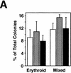
Normal myelomonocytic, platelet, and erythroid development in the LKLF−/− embryos. (A) Quantitative analysis of hematopoietic colonies differentiated in vitro from E11.5 LKLF−/− fetal liver cells. (Open bar) Wild type; (hatched bar) +/−; (solid bar) −/−. (B) Wright-Giemsa-stained peripheral blood smear from an E12.5 LKLF−/− embryo. (e) Enucleated erythrocyte; (ne) nucleated erythrocyte; (p) platelet.
Figure 9
Figure 9
Normal myelomonocytic, platelet, and erythroid development in the LKLF−/− embryos. (A) Quantitative analysis of hematopoietic colonies differentiated in vitro from E11.5 LKLF−/− fetal liver cells. (Open bar) Wild type; (hatched bar) +/−; (solid bar) −/−. (B) Wright-Giemsa-stained peripheral blood smear from an E12.5 LKLF−/− embryo. (e) Enucleated erythrocyte; (ne) nucleated erythrocyte; (p) platelet.

References

    1. Anderson KP, Kern CB, Crable SC, Lingrel JB. Isolation of a gene encoding a functional zinc finger protein homologous to erythroid Kruppel-like factor: Identification of a new multigene family. Mol Cell Biol. 1995;15:5957–5965. - PMC - PubMed
    1. Bieker JJ, Southwood CM. The erythroid Kruppel-like factor transactivation domain is a critical component for cell-specific inducibility of a β-globin promoter. Mol Cell Biol. 1995;15:852–860. - PMC - PubMed
    1. Bradley A. Production and analysis of chimeric mice. In: Robertson EJ, editor. Tetratocarcinomas and embryonic stem cells, a practical approach. Oxford, UK: IRL Press Limited; 1987. pp. 113–151.
    1. Carmeliet P, Ferreira V, Breier G, Pollefeyt S, Kieckens L, Gertsenstein M, Fahrig M, Vandenhoeck A, Harpal K, Eberhardt C, Declercq C, Pawling J, Moons L, Collen D, Risau W, Nagy A. Abnormal blood vessel development and lethality in embryos lacking a single VEGF allele. Nature. 1996;380:435–439. - PubMed
    1. Crossley M, Whitelaw W, Perkins A, Williams G, Fujiwara Y, Orkin SH. Isolation and characterization of the cDNA encoding BKLF/TEF-2, a major CACCC-box-binding protein in erythroid cells and selected other cells. Mol Cell Biol. 1996;16:1695–1705. - PMC - PubMed

Publication types

LinkOut - more resources