. 1997 Nov 15;11(22):3046-60.
doi: 10.1101/gad.11.22.3046.

Phosphorylation- and ubiquitin-dependent degradation of the cyclin-dependent kinase inhibitor Far1p in budding yeast

Affiliations

Phosphorylation- and ubiquitin-dependent degradation of the cyclin-dependent kinase inhibitor Far1p in budding yeast

S Henchoz et al. Genes Dev. .

Abstract

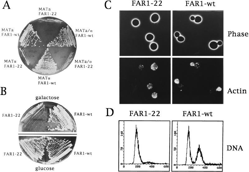
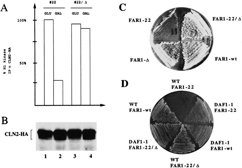
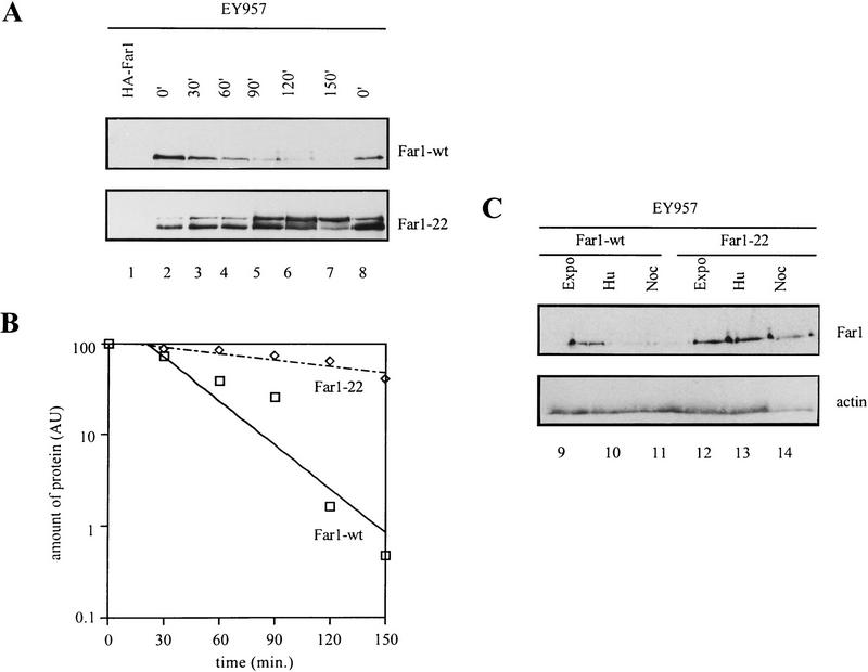
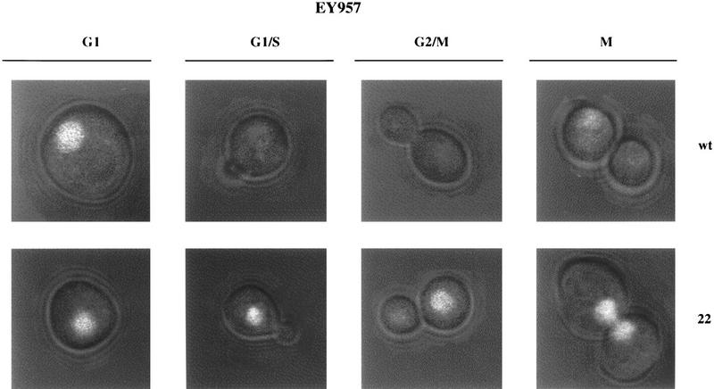
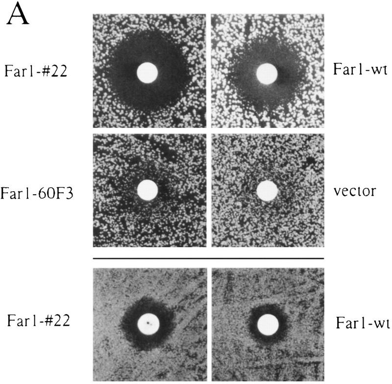
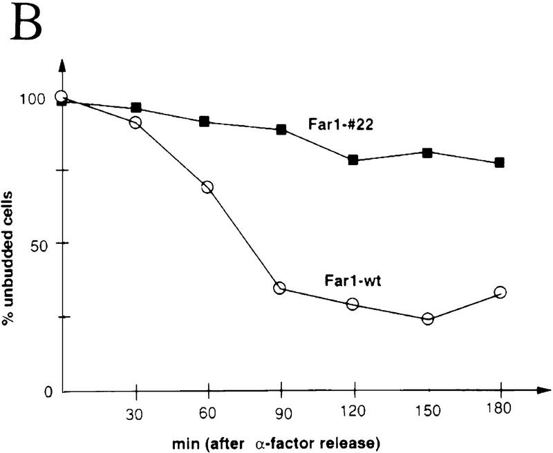
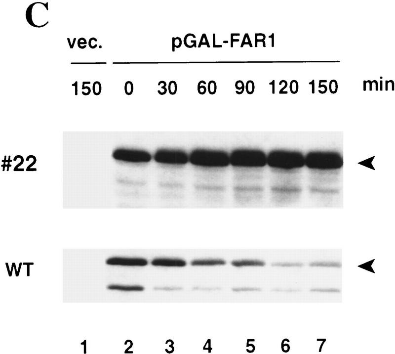
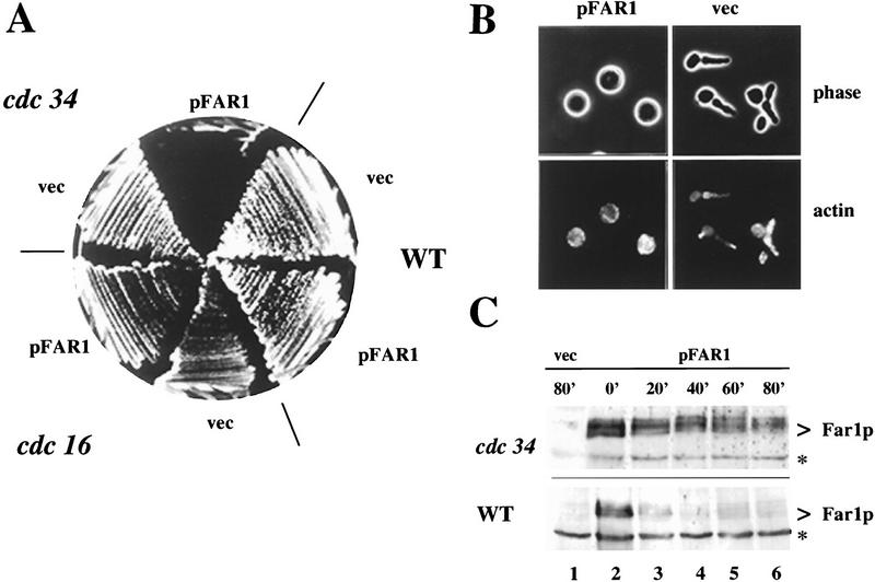
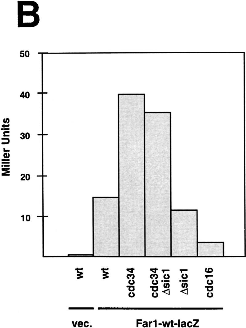
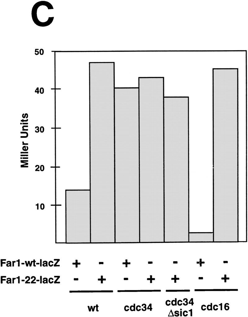
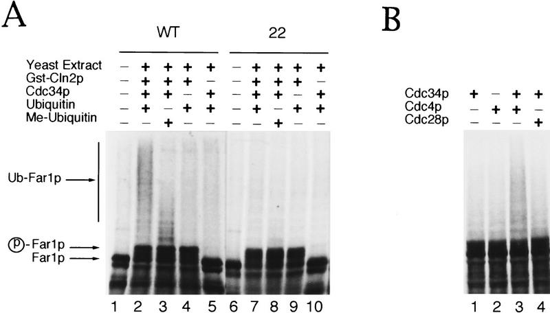
Cyclin-dependent kinase inhibitors (CKIs) play key roles in controlling the eukaryotic cell cycle by coordinating cell proliferation and differentiation. Understanding the roles of CKIs requires knowledge of how they are regulated both through the cell cycle and in response to extracellular signals. Here we show that the yeast CKI, Far1p, is controlled by ubiquitin-dependent proteolysis. Wild-type Far1p was stable only in the G1 phase of the cell cycle. Biochemical and genetic evidence indicate that its degradation required the components of the G1-S ubiquitination system, Cdc34p, Cdc4p, Cdc53p, and Skp1p. We isolated a mutant form of Far1p (Far1p-22) that was able to induce cell cycle arrest in the absence of alpha-factor. Cells that overexpress Far1-22p arrested in G1 as large unbudded cells with low Cdc28p-Clnp kinase activity. Wild-type Far1p, but not Far1-22p, was readily ubiquitinated in vitro in a CDC34- and CDC4-dependent manner. Far1-22p harbors a single amino acid change, from serine to proline at residue 87, which alters phosphorylation by Cdc28p-Cln2p in vitro. Our results show that Far1p is regulated by ubiquitin-mediated proteolysis and suggest that phosphorylation of Far1p by the Cdc28p-Clnp kinase is part of the recognition signal for ubiquitination.


Figures

Figure 1
Figure 1
Cells producing Far1-22p arrest their cell cycle in G1 independently of the mating pathway. (A) Haploid a (K699), α (K700), or a/α diploid cells (YMP562) were transformed with a plasmid expressing either wild-type Far1p or Far1-22p from the inducible GAL promoter and grown on medium containing galactose. Note that cells expressing Far1-22p were unable to form colonies. (B) fus3Δ cells (K2297) that carry a plasmid coding for either wild-type Far1p or Far1-22p from the inducible GAL promoter were grown on media containing galactose (top, GAL promoter on) or glucose (bottom; GAL promoter off). (C,D) Cells expressing wild-type Far1p (YMP128, right panels) or Far1-22p (YMP126, left panels) from the inducible GAL promoter were analyzed 6 hr after addition of galactose. (C) (Top) Phase-contrast photographs; (bottom) actin visualized after staining with rhodamine phalloidin. (D) Flow cytometric DNA quantification.
Figure 2
Figure 2
Cells producing Far1-22p arrest in G1 by inhibition of the G1 kinase, Cdc28p–Clnp. (A,B) Cln2p tagged at its carboxyl terminus with three copies of the HA epitope (Cln2–HA) was immunoprecipitated from extracts prepared from cells (YMT263) expressing either Far1-22p or for control, an inactive mutant form of Far1p, Far1-22/Δ285–350, from the inducible GAL promoter. Cells were grown in media containing galactose (GAL, GAL promoter on) or glucose (GLU, GAL promoter off). Cln2–HAp-associated kinase activity was measured with histone H1 as a substrate and quantified (A). The kinase activity associated with Cln2–HAp from cells grown in glucose was normalized to 100%. Similar amounts of Cln2–HAp were immunoprecipitated in each assay as shown by immunoblotting (B). (C) Growth inhibition caused by production of Far1-22p was dependent on the ability of Far1p to bind to the Cdc28p–Clnp kinase. The following Far1p proteins were analyzed: wild-type Far1p, Far1-22p, Far1-Δ285–350, which is unable to bind to Cdc28p–Clnp (Peter et al. 1993), and Far1-22/Δ285–350. (D) Growth inhibition caused by expression of Far1-22p was rescued by co-overexpression of a stable G1-cyclin, Cln3p (DAF1). DAF1-1 cells (IH2517), which express a stable form of the G1 cyclin Cln3p or isogenic wild-type cells (IH2518), were transformed with plasmids expressing either wild-type Far1p, Far1-22p, or Far1-22/Δ285–350p from the inducible GAL promoter.
Figure 3
Figure 3
Far1-22p has an increased half-life and is present throughout the cell cycle. (A) Cells (K699) that carry a plasmid coding for either wild-type Far1–GFPp or Far1-22p–GFP from the inducible GAL promoter were grown in raffinose and expression was induced by addition of galactose for 5 hr. Glucose was then added to shut off the GAL promoter, and samples were taken every 30 min as indicated and immunoblotted for the presence of Far1–GFP fusion protein (lanes 1–8). The specificity of the GFP antibodies was confirmed by including cells expressing an HA-tagged version of Far1p (lane 1). (B) Quantitation of the GAL shutoff experiments. The degradation of wild-type Far1p-GFP and Far1-22-GFP proteins was quantified by PhosphorImager and plotted against the time after repressing the GAL promoter. (C) Far1–22p, but not wild-type Far1p, is present throughout the cell cycle. EY957 cells producing wild-type Far1p–GFP (lanes 9–11) or Far1–22p-GFP (lanes 12–14) from the inducible GAL promoter were grown in medium containing raffinose (GAL promoter off), arrested in S phase with hydroxyurea (HU) or in mitosis with nocodazole (Noc), and then expression was induced by addition of galactose. Control cells were treated identically except that they were not exposed to the drugs (expo). After 5 hr, cells were analyzed for the presence of Far1p–GFP by immunoblotting with antibodies specific for GFP (top panel). Equal loading was confirmed by immunoblotting the samples with antibodies against actin (bottom panel).
Figure 4
Figure 4
Wild-type Far1p, but not Far1–22p, is expressed in a cell cycle-dependent manner. Wild-type Far1p or Far1-22p was fused at its carboxyl terminus to GFP and produced in yeast (EY957) from the inducible GAL promoter. Note that wild-type Far1p–GFP (top row) was found in the nucleus of unbudded cells (G1), but staining disappeared in small budded cells (G1/S), large budded cells (G2/M), and cells in mitosis (M). In contrast, Far1-22p–GFP (bottom row) could be detected in the nucleus of cells at all stages of the cell cycle.
Figure 5
Figure 5
Degradation of Far1p is required for cells to recover efficiently from α-factor induced cell cycle arrest. (A) cell cycle arrest was assayed by halo formation of far1Δ cells transformed with single-copy plasmids carrying wild-type FAR1, FAR1-22, FAR1-60F3 (Peter et al. 1993), or no insert. (Top two rows) bar1Δ cells (K2180), (bottom row) BAR1+ cells (YMP1054). Filter disks contain 0.1 μg (top two rows) and 10 μg of α-factor (bottom row). Note that cells producing Far1-22p are hypersensitive to pheromone and that halos fill in inefficiently. (B,C) Cells producing Far1-22p are unable to re-enter the cell cycle. Cells deleted for FAR1 (K2180) producing wild-type Far1p or Far1-22p from the GAL promoter were grown in raffinose medium to an OD600 of 0.4, at which time cells were arrested by the simultaneous addition of α-factor and galactose. After 3 hr, cells were washed and reinoculated in fresh medium, without α-factor, but containing galactose. Every 30 min, samples were harvested. Re-entry into the cell cycle was monitored microscopically by analyzing the percentage of unbudded cells (B) Levels of wild-type Far1p (bottom panel) and Far1-22p (top panel) were determined after immunoblotting extracts with Far1p antibodies (C, lanes 1–7). Note that in contrast to cells producing wild-type Far1p, Far1-22p is accumulating in G1 and cells are unable to form a bud.
Figure 5
Figure 5
Degradation of Far1p is required for cells to recover efficiently from α-factor induced cell cycle arrest. (A) cell cycle arrest was assayed by halo formation of far1Δ cells transformed with single-copy plasmids carrying wild-type FAR1, FAR1-22, FAR1-60F3 (Peter et al. 1993), or no insert. (Top two rows) bar1Δ cells (K2180), (bottom row) BAR1+ cells (YMP1054). Filter disks contain 0.1 μg (top two rows) and 10 μg of α-factor (bottom row). Note that cells producing Far1-22p are hypersensitive to pheromone and that halos fill in inefficiently. (B,C) Cells producing Far1-22p are unable to re-enter the cell cycle. Cells deleted for FAR1 (K2180) producing wild-type Far1p or Far1-22p from the GAL promoter were grown in raffinose medium to an OD600 of 0.4, at which time cells were arrested by the simultaneous addition of α-factor and galactose. After 3 hr, cells were washed and reinoculated in fresh medium, without α-factor, but containing galactose. Every 30 min, samples were harvested. Re-entry into the cell cycle was monitored microscopically by analyzing the percentage of unbudded cells (B) Levels of wild-type Far1p (bottom panel) and Far1-22p (top panel) were determined after immunoblotting extracts with Far1p antibodies (C, lanes 1–7). Note that in contrast to cells producing wild-type Far1p, Far1-22p is accumulating in G1 and cells are unable to form a bud.
Figure 6
Figure 6
Overexpression of wild-type Far1p is lethal in cdc34 cells at the semipermissive temperature because of stabilization of the Far1 protein. (A) Temperature-sensitive cdc16 (K4102) and cdc34 (YMT670) mutants, as well as isogenic wild-type cells (K699), were transformed with a plasmid carrying wild-type FAR1 under the control of the inducible GAL promoter or an empty vector (vec) for control. Transformants were plated on media containing galactose and incubated at 30°C. Wild-type and cdc16 mutant strains tolerate overexpression of Far1p, whereas cdc34 mutants do not. All strains grew normally when plated on medium containing glucose (data not shown). (B) cdc34 cells (YMT670) carrying a plasmid expressing Far1p from the inducible GAL promoter (pFar1; left panels) or control plasmid with no insert (vec; right panels) were grown in raffinose at 30°C, at which time expression of Far1p was induced by addition of galactose. After 5 hr, cells were fixed and analyzed by phase contrast microscopy (Phase, top panels) or after actin staining with rhodamine phalloidin (Actin, bottom panels). cdc34 cells producing Far1p arrest with a morphology indistinguishable from that of wild-type cells producing Far1-22p (Fig. 1C). (C) The half-life of wild-type Far1p was determined in cdc34 far1Δ (top panel; YMP1056) and far1Δ cells (bottom panel; YMP1054), which express Far1p from the GAL promoter. After repression of the GAL promoter at the indicated times (minutes), aliquots were removed and examined by immunoblotting as described. The specificity of the Far1p antibodies was confirmed by analysis of cells carrying an empty vector (vec; lane 1). The position of Far1p is marked by an arrowhead; the asterisks mark an unspecific protein recognized by the Far1p antiserum.
Figure 7
Figure 7
Overexpression of Far1p is lethal in skp1 mutants in an allele-specific fashion and Far1p accumulates in cdc34 mutants independent of the cell cycle stage. (A) The effect of overexpression of Far1p was tested in specific temperature-sensitive alleles of SKP1 that either cause arrest in G1 (skp1-11, Y552) or in G2 (skp1-12, Y554) as described in Fig. 6. Only cells harboring the G1-specific allele of SKP1 were arrested by overexpression of wild-type Far1p. (B) Far1p accumulates in cdc34 (YMT670) and cdc34 Δsic1 cells (ES464). The indicated strains were arrested at 37°C and the accumulation of a Far1–LacZ fusion protein was quantified and plotted as Miller Units deduced from three independent experiments. Note that Far1–LacZp accumulates in cdc34 mutants independently of the cell cycle arrest point; no accumulation was observed in cdc16 cells or cells deleted for SIC1. (C) Far1-22–LacZp accumulates in wild-type (K699) and cdc16 (K4102) mutant cells but is produced at levels similar to wild-type Far1–LacZp in cdc34 (YMT670) and cdc34 Δsic1 (ES464) cells.
Figure 7
Figure 7
Overexpression of Far1p is lethal in skp1 mutants in an allele-specific fashion and Far1p accumulates in cdc34 mutants independent of the cell cycle stage. (A) The effect of overexpression of Far1p was tested in specific temperature-sensitive alleles of SKP1 that either cause arrest in G1 (skp1-11, Y552) or in G2 (skp1-12, Y554) as described in Fig. 6. Only cells harboring the G1-specific allele of SKP1 were arrested by overexpression of wild-type Far1p. (B) Far1p accumulates in cdc34 (YMT670) and cdc34 Δsic1 cells (ES464). The indicated strains were arrested at 37°C and the accumulation of a Far1–LacZ fusion protein was quantified and plotted as Miller Units deduced from three independent experiments. Note that Far1–LacZp accumulates in cdc34 mutants independently of the cell cycle arrest point; no accumulation was observed in cdc16 cells or cells deleted for SIC1. (C) Far1-22–LacZp accumulates in wild-type (K699) and cdc16 (K4102) mutant cells but is produced at levels similar to wild-type Far1–LacZp in cdc34 (YMT670) and cdc34 Δsic1 (ES464) cells.
Figure 8
Figure 8
Wild-type Far1p but not Far1-22p is ubiquitinated in vitro. Wild-type Far1p (A, lanes 1–5; B, lanes 1–4) or Far1-22p (A, lanes 6–10) synthesized in vitro in the presence of [35S]methionine was incubated in fractionated extracts prepared from cells lacking CLN1,2, and 3 (A) or cells deleted for CLN1,2, and 3, which are also temperature-sensitive for cdc4 (B). These extracts are devoid of Cdc34p and contain low levels of ubiquitin (Verma et al. 1997a). (A) Wild-type Far1p was ubiquitinated if the extract was supplemented with the addition of Cdc34p, GST–Cln2p, and ubiquitin (lane 2). Reduced accumulation of high molecular weight ubiquitin conjugates was observed if methyl-ubiquitin was added to block chain extension (lane 3). No ubiquitination was observed with Far1-22p (lanes 6–10). (B) Ubiquitination of wild-type Far1p was dependent on Cdc4p (lanes 1 and 4). Ubiquitination in cdc4-extracts was restored only by the simultaneous addition of purified Cdc34p and baculo-infected insect lysate containing Cdc4p (lane 3). The addition of insect lysate containing Cdc28p serves as a specificity control (lane 4).
Figure 9
Figure 9
Far1p is phosphorylated by Cdc28p–Cln2p kinase on Ser-87 in vitro. (A) Full-length wild-type Far1p was produced in E. coli, purified as a GST fusion protein and phosphorylated in vitro with Cdc28p–Cln2p kinase immunoprecipitated from yeast extracts (lane 3). As a control, the kinase assays were performed with GST alone (lane 2) or without substrate (lane 1). Phosphorylated proteins were analyzed by autoradiography (top left panel) and immunoblotting with specific antibodies against Far1p (bottom left panel). The arrowhead marks the position of GST-Far1 protein. A fragment containing amino acids 50–340 of either wild-type Far1p (lane 4) or Far1-22p (lane 5) was expressed as a GST fusion protein in E. coli and phosphorylated in vitro by immunoprecipitated Cln2–HAp-associated kinase. Kinase reactions were also carried out with histone H1 (lane 7) or in the absence of substrate (lane 6). The specificity of the immunoprecipitated Cln2p-associated kinase was confirmed by including an untagged Cln2p (lane 8). The addition of equal amounts of GST–Far1p in the kinase reactions was verified by immunoblotting with antibodies against Far1p (bottom right panel). The arrowhead points to the position of the GST-Far150-340 protein; the bracket marks the position of phosphorylated histone H1. (B) Sequencing of the FAR1-22 allele uncovered a single mutation that changes Ser-87 to proline. Note that Ser-87 is followed by a proline residue and is, therefore, a potential CDK phosphorylation site (Nigg 1995). (C) Ser-87 is phosphorylated by Cdc28p–Cln2p kinase in vitro. A fragment containing amino acids 50–340 of either wild-type Far1p (top panel) or Far1p-S87T, in which Ser-87 was replaced by a threonine residue (bottom panel), was phosphorylated in vitro by immunoprecipitated Cln2–HAp-associated kinase and phosphoamino acid analysis was performed. (S) The position of phosphoserine; (T) The position of phosphothreonine.
Figure 10
Figure 10
A model for the degradation of Far1p. Far1p binds to the Cdc28p–Clnp kinase, which in turn phosphorylates Far1p on Ser-87. Phosphorylated Far1p is recognized by the ubiquitination machinery composed of Cdc34p, Cdc53p, Cdc4p, and Skp1p, which ubiquitinates Far1p. Ubiquitinated Far1p is then degraded by the proteasome.

Similar articles

Cited by

References

    1. Ausubel FM, Brent R, Kingston RE, Moore DD, Seidman JG, Smith JA, Struhl K. Current protocols in molecular biology. New York, NY: Greene Publishing Associates and Wiley-Interscience; 1991.
    1. Bai C, Sen P, Hofmann K, Ma L, Goebl M, Harper JW, Elledge SJ. SKP1 connects cell cycle regulators to the ubiquitin proteolysis machinery through a novel motif, the F-box. Cell. 1996;86:263–274. - PubMed
    1. Banergee A, Gregori L, Xu Y, Chau V. The bacterially expressed yeast Cdc34 gene product can undergo autoubiquitination to form a multiubiquitin chain-linked protein. J Biol Chem. 1993;268:5668–5675. - PubMed
    1. Barral Y, Jentsch S, Mann C. G1 cyclin turnover and nutrient uptake are controlled by a common pathway in yeast. Genes & Dev. 1995;9:399–409. - PubMed
    1. Brandeis M, Hunt T. The proteolysis of mitotic cyclins in mammalian cells persists from the end of mitosis until the onset of S phase. EMBO J. 1996;15:5280–5289. - PMC - PubMed

Publication types

MeSH terms

Substances