. 1997 Nov 15;11(22):3096-108.
doi: 10.1101/gad.11.22.3096.

Transcriptional activity of pannier is regulated negatively by heterodimerization of the GATA DNA-binding domain with a cofactor encoded by the u-shaped gene of Drosophila

Affiliations

Transcriptional activity of pannier is regulated negatively by heterodimerization of the GATA DNA-binding domain with a cofactor encoded by the u-shaped gene of Drosophila

M Haenlin et al. Genes Dev. .

Abstract

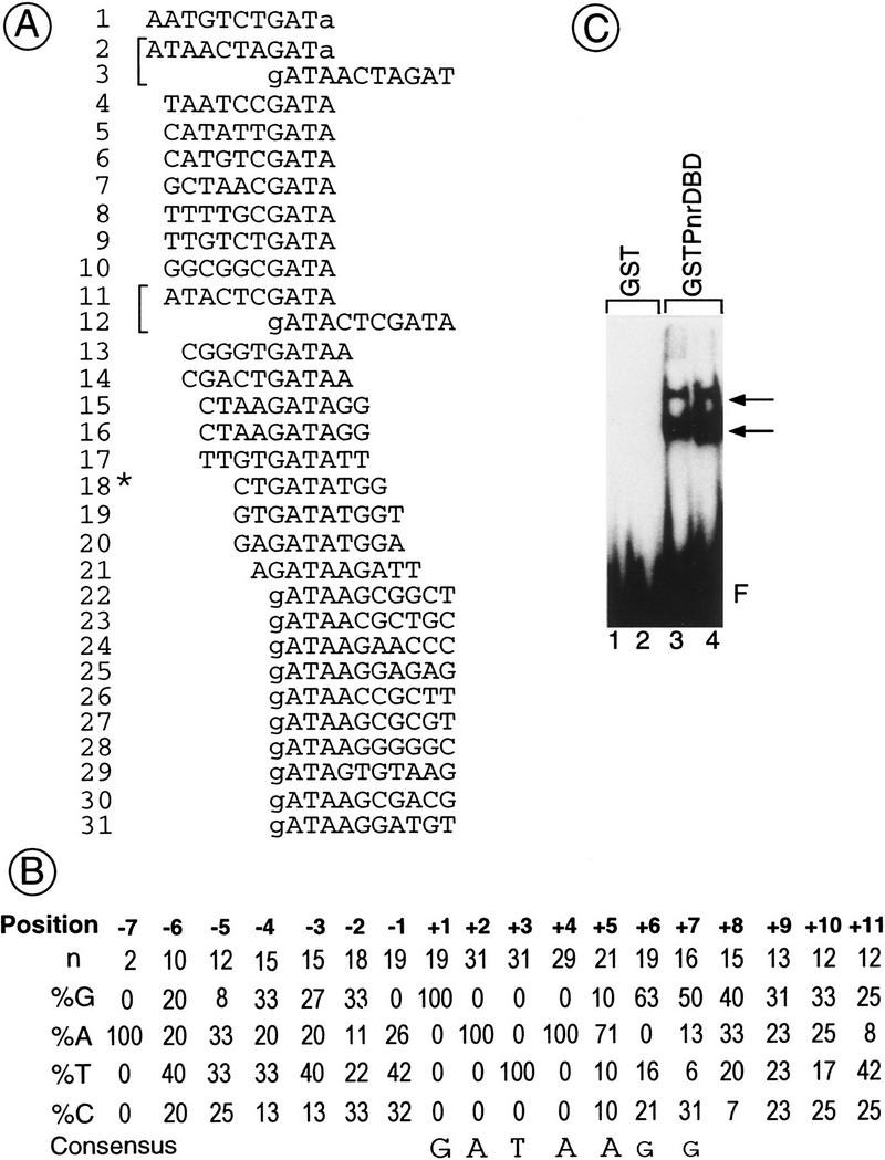
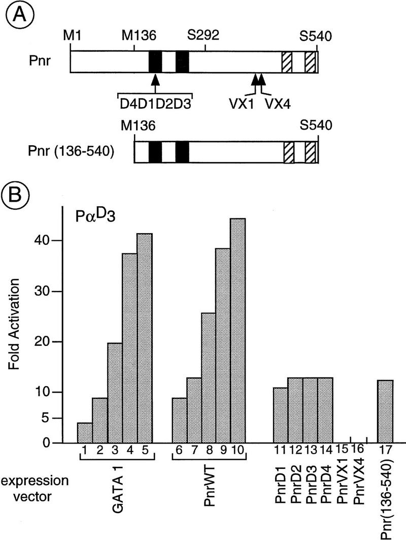
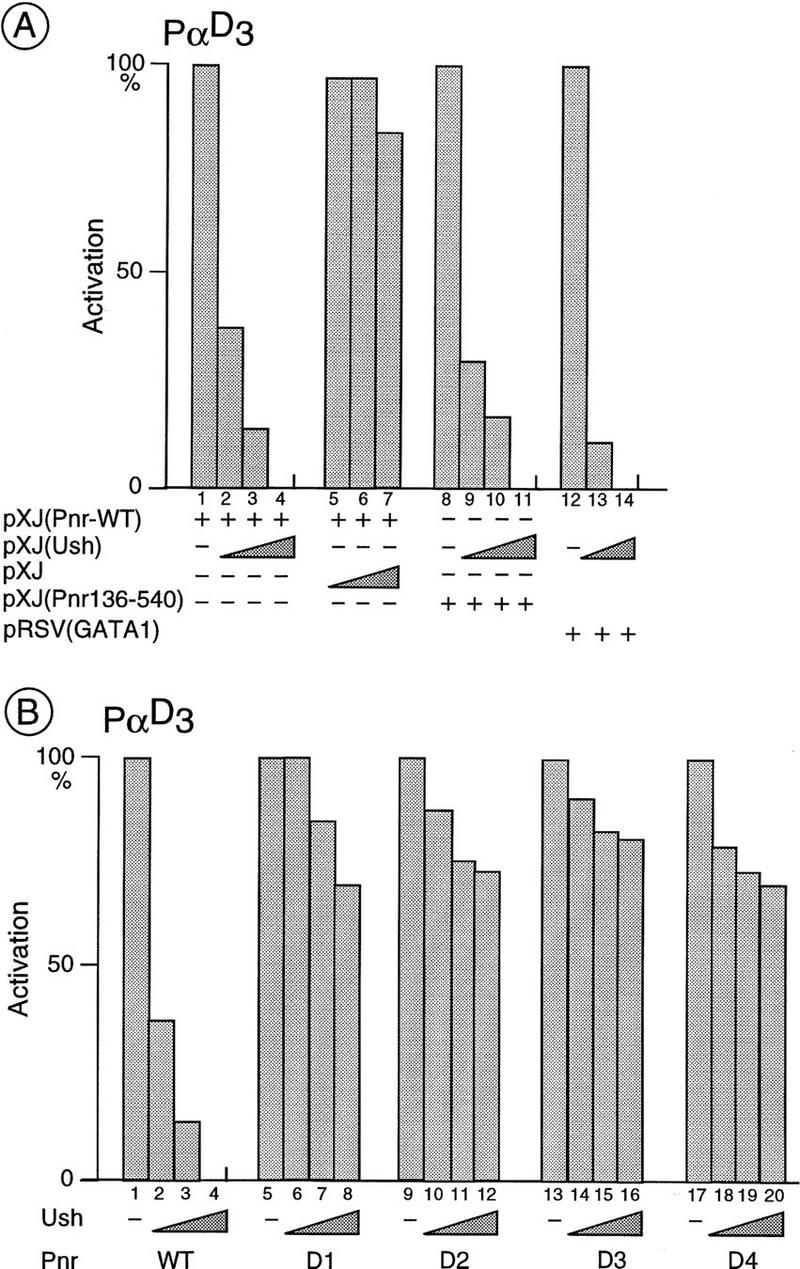
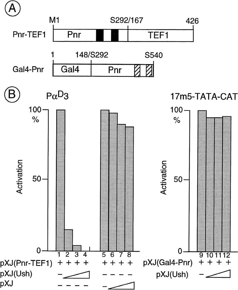
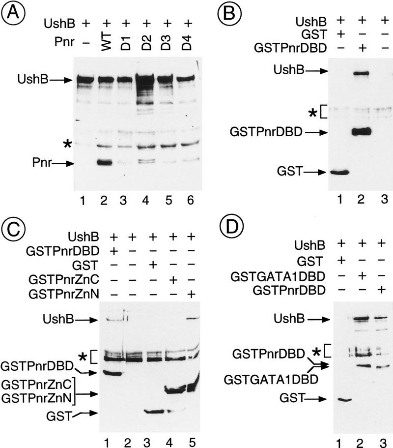
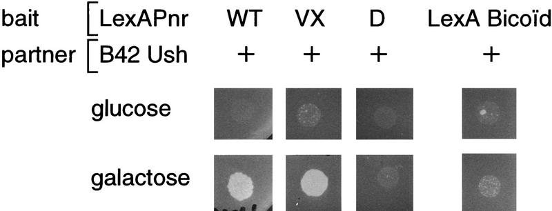
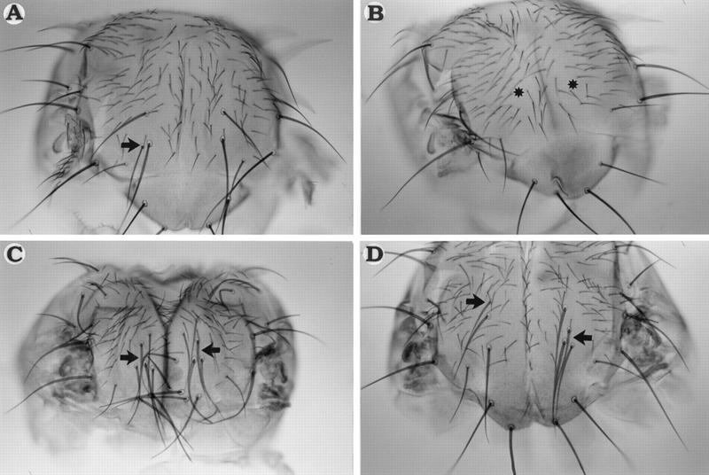
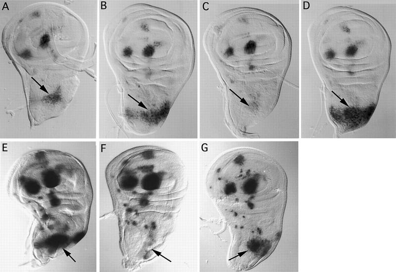
The genes pannier (pnr) and u-shaped (ush) are required for the regulation of achaete-scute during establishment of the bristle pattern in Drosophila. pnr encodes a protein belonging to the GATA family of transcription factors, whereas ush encodes a novel zinc finger protein. Genetic interactions between dominant pnr mutants bearing lesions situated in the amino-terminal zinc finger of the GATA domain and ush mutants have been described. We show here that both wild-type Pannier and the dominant mutant form activate transcription from the heterologous alpha globin promoter when transfected into chicken embryonic fibroblasts. Furthermore, Pnr and Ush are found to heterodimerize through the amino-terminal zinc finger of Pnr and when associated with Ush, the transcriptional activity of Pnr is lost. In contrast, the mutant pnr protein with lesions in this finger associates only poorly with Ush and activates transcription even when cotransfected with Ush. These interactions have been investigated in vivo by overexpression of the mutant and wild-type proteins. The results suggest an antagonistic effect of Ush on Pnr function and reveal a new mode of regulation of GATA factors during development.


Figures

Figure 1
Figure 1
Definition of a consensus-binding site for Pnr. Degenerated oligonucleotides were loaded on a GST column where a fusion protein between the GST and the Pnr–DBD is bound (GST–Pnr–DBD). After elution, the selected oligonucleotides were PCR amplified and loaded again on the column (see Materials and Methods for details). After four cycles of selection and PCR amplification, the oligonucleotides were both subcloned for sequence analysis and used as a template in an electrophoretic mobility-shift assay (EMSA). (A) Alignment of 29 Pnr binding sequences recovered after four rounds of selection. The nucleotides included in the random region are shown in uppercase letters, whereas the bordering G and A nucleotides are in lowercase letters. The sequences are aligned with respect to the GATA motif. Brackets indicate oligonucleotides containing a repeat of the GATA sequence. The asterisk (*) denotes a template containing only nine nucleotides in the random region, which may correspond to either incomplete oligonucleotide synthesis or to PCR artifacts. (B) Consensus-binding site derived from the frequencies of the bases at the sites selected. (C) Autoradiography of an EMSA using as a template the pool of oligonucleotides selected and as a protein extract either purified GST (lanes 1,2: 20 and 200 ng, respectively) or GST–Pnr–DBD (lanes 3,4: 20 and 200 ng, respectively). (F) Unbound oligonucleotides. The arrows indicate two complexes of which the one migrating more slowly may correspond either to a template containing two GATA sequences (A) with one molecule bound to each motif or to a dimer bound to a template containing a single GATA sequence.
Figure 2
Figure 2
Transactivation of the α-globin promoter sequences by different forms of Pnr in chicken embryonic fibroblasts (CEF). (A) Structural features of the different pnr proteins used in the present study indicating the two zinc fingers (solid boxes) and two sequences organized as putative amphipathic α helices (hatched boxes). The point mutations associated with the pnrD alleles that result in a single amino acid exchange are located in the amino zinc finger, whereas the lesions associated with the pnrVX1/4 alleles and corresponding to a frameshift mutation in the open reading frame are localized in the amino-terminal of the two amphipathic helices. The amino acids are indicated with the single letter code and numbering refers to that given in Ramain et al. (1993). The methionine-136 (M136) is used as an internal start codon in the truncated protein Pnr (136–540) encoded by the pXJ(Sph–Not) expression vector (see Materials and Methods). In contrast, the chimeric proteins (see Fig. 4) contain either the amino acids methionine-1 to serine-292 (chimera Pnr–TEF-1) or the amino acids serine-292 to serine-540 (chimera Gal4–Pnr). (B) Transactivation of the α-globin promoter by the different forms of Pnr. The CEF cells were cotransfected with the α-globin reporter (6 μg of PαD3) and the expression vector for cGATA-1 (lanes 1–5; 25, 50, 100, 250, and 500 ng), wild-type Pnr (PnrWT; lanes 6–10; 25, 50, 100, 250, and 500 ng) and PnrD1, PnrD2, PnrD3, PnrD4, PnrVX1, PnrVX4, and Pnr (136–540), (lanes 11–17: 50 ng each). The level of activation is expressed relative to the reporter alone and as the average (s.d. ± 20%) of three independent experiments performed with two independent DNA preparations.
Figure 3
Figure 3
In vivo interactions between Ush and Pnr or cGATA-1 in CEFs. (A) Activation of the α-globin promoter sequences by cGATA-1 and wild-type Pnr is reduced strongly in the presence of Ush. CEF cells were cotransfected with the α-globin reporter (6 μg of PαD3) and an expression vector either for the full-length Pnr (lanes 1–7, 40 ng), or the truncated Pnr (136–540) (lanes 8–11, 40 ng), or cGATA-1 (lanes 12–14, 40 ng) and increasing amounts of an expression vector for Ush (lanes 2, 9, 13: 20 ng; lanes 3, 10, 14: 40 ng; lanes 4, 11: 80 ng). Cotransfection with the empty pXJ expression vector (lanes 5–7: 20, 40, and 80 ng, respectively) is used as a control. The CAT activities are expressed relative to the Pnr and cGATA-1 expression vectors alone, where the CAT activity is fixed arbitrarily at 100%. In each case, they represent the average (s.d. ± 20%) of three independent experiments performed with two independent DNA preparations. (B) Activation of the α-globin promoter sequences by the mutant protein PnrD is affected poorly by Ush. CEF cells are cotransfected with the α-globin reporter (6 μg of PαD3), an expression vector for either the wild-type Pnr or one of the mutated PnrD proteins (40 ng in each case; lanes 1–4: wild-type Pnr; lanes 5–8: PnrD1; lanes 9–12: PnrD2; lanes 13–16: PnrD3; lanes 17–20: PnrD4) and increasing amounts of a Ush expression vector (lanes 2,6,10,14,18: 20 ng; lanes 3,7,11,15,19: 40 ng; lanes 4,8,12,16,20: 80 ng). The activation represents the average (s.d. ± 20%) of three independent experiments performed with two independent DNA preparations and is expressed as a percentage of the full activation (100%) seen with Pnr alone.
Figure 4
Figure 4
In vivo interactions between Ush and Pnr–DBD containing chimeric proteins. (A) Structural features of the chimeras used in the present study. The Pnr–TEF-1 chimera results from a fusion of the amino terminus of Pnr (M1–S292) including the DNA-binding zinc fingers (solid boxes), and the amino acids 167–426 of the SV40-enhancer binding protein TEF-1 that bears an activation function. The Gal4–Pnr chimera results from a fusion of the Gal4–DNA-binding domain (amino acids 1–148) and the carboxyl terminus of Pnr (S292–S540) containing the two α helices (hatched boxes). (B) Specific inhibition by Ush of the transcriptional activation induced by the Pnr–DBD-containing chimera. CEFs were cotransfected with either the α-globin reporter (6 μg of PαD3), or the upstream activating sequence (UAS) reporter (1 μg of 17m5–TATA–CAT), an expression vector encoding either the Pnr–TEF-1 (lanes 1–8: 40ng), or the Gal4–Pnr chimera (lanes 9–12: 40ng), and increasing amounts of a Ush expression vector (lanes 2,10: 20ng; lanes 3,11: 40ng; lanes 4,12: 80ng). Cotransfection of the empty pXJ expression vector (lanes 6–8: 20, 40, and 80 ng, respectively) is used as a control. The activation represents the average (s.d. ± 20%) of three independent experiments performed with two independent DNA preparations and is expressed as a percentage of the full activation (100%) seen with the chimera alone.
Figure 5
Figure 5
Direct in vitro protein–protein interactions between Ush and Pnr. Cos cells were cotransfected with 5 μg of each expression vector, as denoted above each panel, and the corresponding protein extracts were either immunoprecipitated (A) with the B10 monoclonal antibody raised against the B epitope of the estrogen receptor inserted into the Ush open reading frame or incubated with glutathione–agarose (B–D). Selected proteins were analyzed by Western blot. Ush–B is detected with the B10 antibody, Pnr and PnrD with the specific monoclonal antibody 2B8, and the GST fusion proteins with the GST-specific monoclonal antibody 1D10. (A) In vitro interactions between Ush and wild-type Pnr as well as PnrD. Arrows indicate the Ush–B (lanes 1–6) and Pnr (lanes 2–6: wild-type Pnr, PnrD1, PnrD2, PnrD3 and PnrD4, respectively). The asterisk (*) denotes the location of the immunoglobulin heavy chain. (B) In vitro interactions between Ush and the isolated Pnr–DBD. Arrows indicate Ush–B (lane 2), GST (lane 1), and the fusion GST–Pnr–DBD (lane 2). The asterisk denotes artifactual bands. (C) In vitro interactions between Ush and Pnr isolated zinc fingers. Arrows indicate Ush–B (lanes 1,5), GST alone (lane 3), the fusion GST–Pnr–DBD (lane 1), and the fusion proteins GST–Pnr–ZnC (lane 4) and GST–Pnr–ZnN (lane 5). The asterisk denotes artifactual bands. (D) In vitro interactions between Ush and the cGATA–1–DBD. Arrows indicate Ush–B (lanes 2,3), GST alone (lane 1), and the fusion proteins GST–Pnr–DBD (lane 2) and GST-GATA-1–DBD (lane 3). The asterisk denotes artifactual bands.
Figure 6
Figure 6
In vivo interactions between Ush and Pnr in yeast. Yeast transformed by an expression vector encoding the bait LexA–Pnr (the different forms of the protein were wild-type Pnr, PnrVX1, or PnrD1) and an expression vector encoding the partner B42–Ush were plated on a medium lacking histidine, trytophane, and leucine, with either glucose, where the expression of B42–Ush is repressed, or with galactose, where expression of the partner is induced. The results were the same for LexA–PnrVX4, LexA–PnrD2, LexA–PnrD3, and LexA–PnrD4 (data not shown). The specificity of the interaction is further demonstrated by the use of the LexA–Bicoid unrelated fusion protein. Levels of LexA–Pnr fusion proteins in the different strains were monitored by Western blot analysis using the Pnr-specific monoclonal antibody 2B8 (data not shown).
Figure 7
Figure 7
Effect of overexpression of the different pnr proteins on development of the dorsocentral bristles. Homozygous transgenic strains carrying a UAS–Pnr transposon were crossed with pnrMD237/TM6b; Tb flies and the Tb+ flies carrying the pnrMD237 driver were analyzed. Arrows denote additional dorsocentral bristles in A, C, and D, whereas asterisks (*) indicate the positions of the missing bristles in B. (A) Strain UAS–Pnr+. (B) Strain UAS–PnrVX4. (C) The transgenic strain UAS–PnrD4. (D) Strain UAS–Pnr+ (same as in (A) in the presence of a reduced amount of Ush [Df(3R)ushRev18, ush−/+].
Figure 8
Figure 8
The effects of overexpression of the different forms of pnr proteins and ush on the lacZ reporter whose expression is driven by the dorsocentral-specific enhancer sequences (Gomez-Skarmeta et al. 1995). Arrows denote lacZ expression in the dorsocentral region of the thoracic disc. (A–D) Homozygous transgenic flies carrying a UAS–Pnr transposon were crossed with DC–enhancer–sc–lacZ; pnrMD237/TM6B Tb flies, and the resulting larvae of the appropriate genotype (see below) were dissected and the imaginal discs stained for β-galactosidase activity. In each case, the reaction was left for 3 hr at 22°C; longer reaction times led to background staining, which prevents comparison between the different pnr proteins. (E–G) Overexpression of ush leads to the reduction of ac–sc expression in pnrMD237/+ (F) but not pnrD/pnrMD237 mutants (G). In E–G the reaction was left for 6 hr at 37°C to emphasize the fact that overexpression of Ush leads to complete repression of the lacZ reporter.(A) Genotype (UAS-PnrWT/lacZ; TM6B, Tb/+); (B) genotype (UAS–PnrWT/lacZ; pnrMD237/+); (C) genotype (UAS–PnrVX4/lacZ; pnrMD237/+); (D) genotype (UAS–PnrD4/lacZ; pnrMD237/+); (E) genotype (UAS–Ush/lacZ; TM6B, Tb/+); (F) genotype (UAS–Ush/lacZ; pnrMD237/TM6B, Tb); (G) genotype (UAS–Ush/lacZ; pnrMD237/pnrD1).

References

    1. Abel T, Michelson AM, Maniatis T. A Drosophila GATA family member that binds to Adh regulatory sequences is expressed in the developing fat body. Development. 1993;119:623–633. - PubMed
    1. Andrews NC, Orkin SH. Transcriptional control of erythropoiesis. Curr Opin Hematol. 1994;1:119–124. - PubMed
    1. Brand AH, Perrimon N. Targeted gene expression as a means of altering cell fates and generating dominant phenotypes. Development. 1993;118:401–415. - PubMed
    1. Calleja M, Moreno E, Pelaz S, Morata G. Visualization of gene expression in living adult Drosophila. Science. 1996;274:252–255. - PubMed
    1. Campuzano S, Modolell J. Patterning of the Drosophila nervous system: The achaete-scute gene complex. Trends Genet. 1992;8:202–208. - PubMed

Publication types

MeSH terms