. 1997;6(5):315-32.

Identification of transcription factories in nuclei of HeLa cells transiently expressing the Us11 gene of herpes simplex virus type 1

Affiliations

Identification of transcription factories in nuclei of HeLa cells transiently expressing the Us11 gene of herpes simplex virus type 1

F Puvion-Dutilleul et al. Gene Expr. 1997.

Abstract

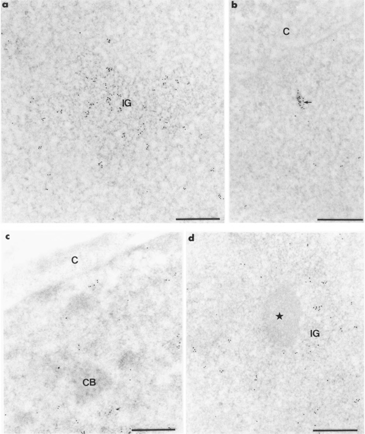
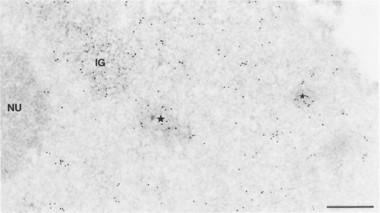
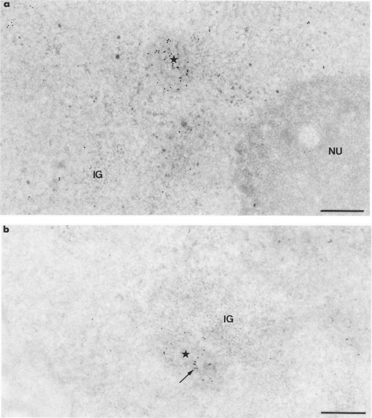
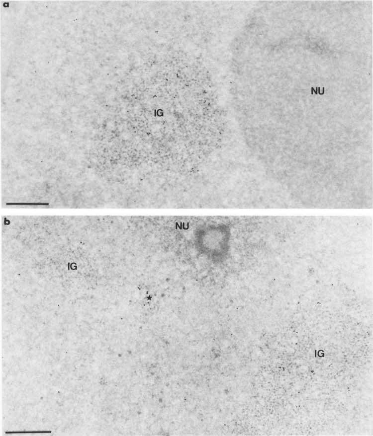
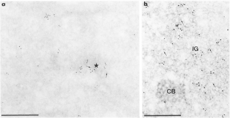
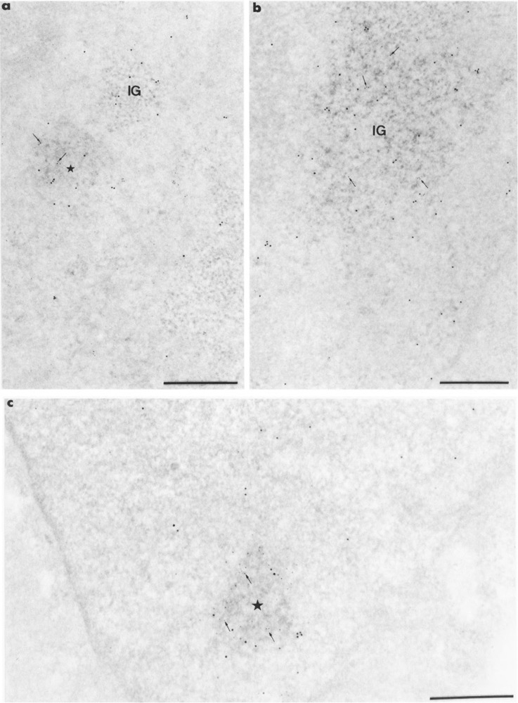
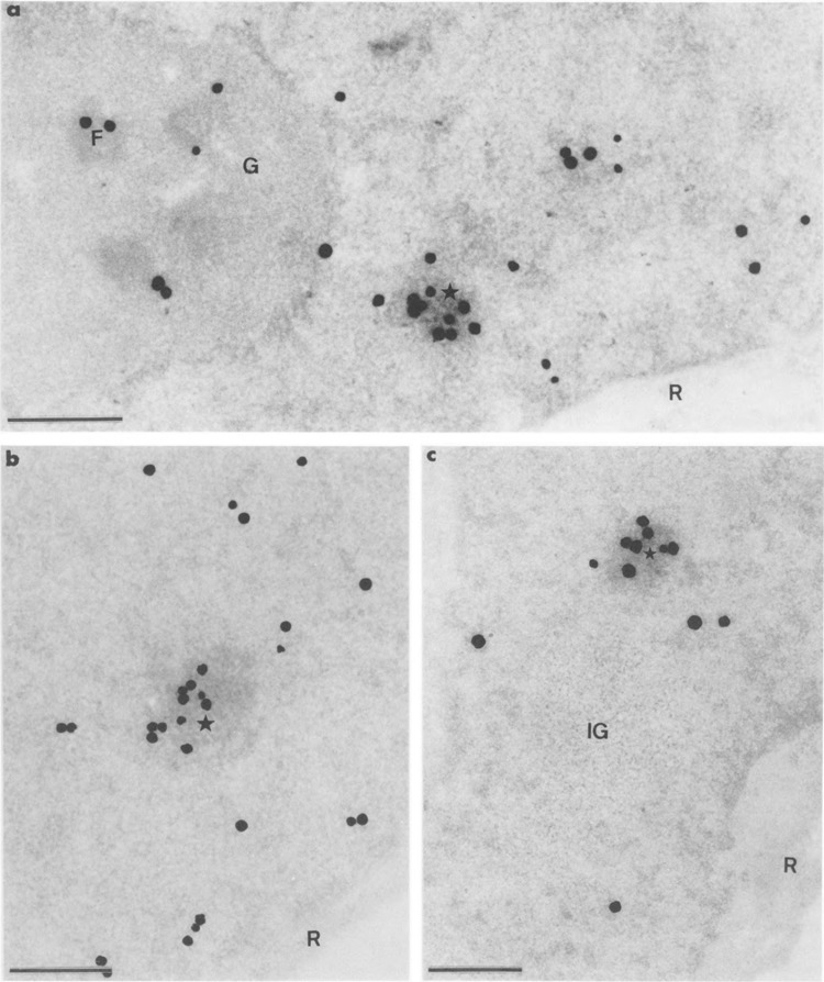
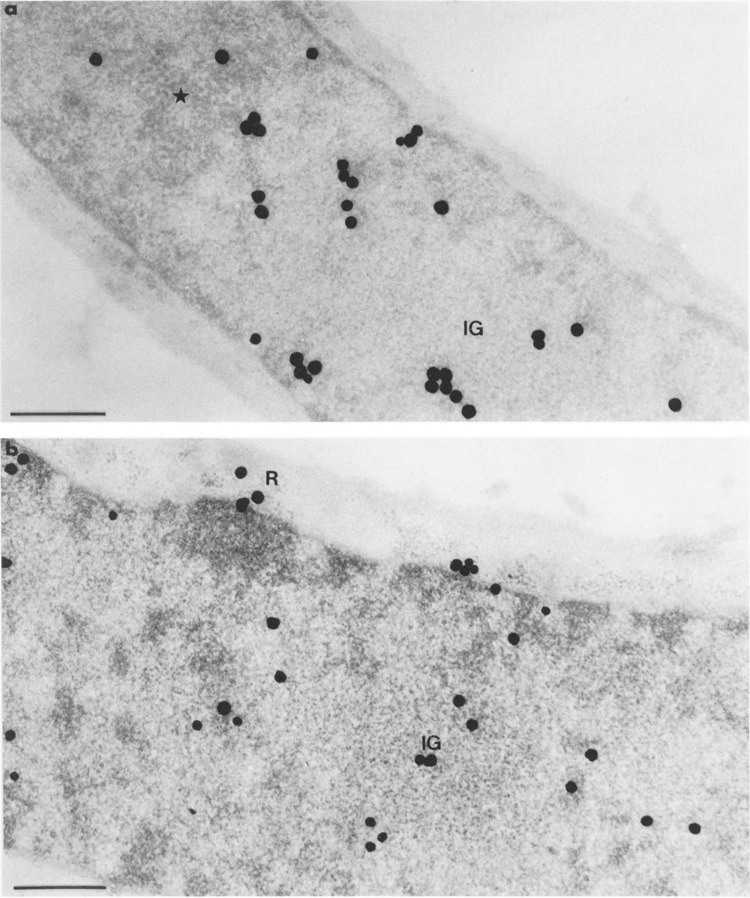
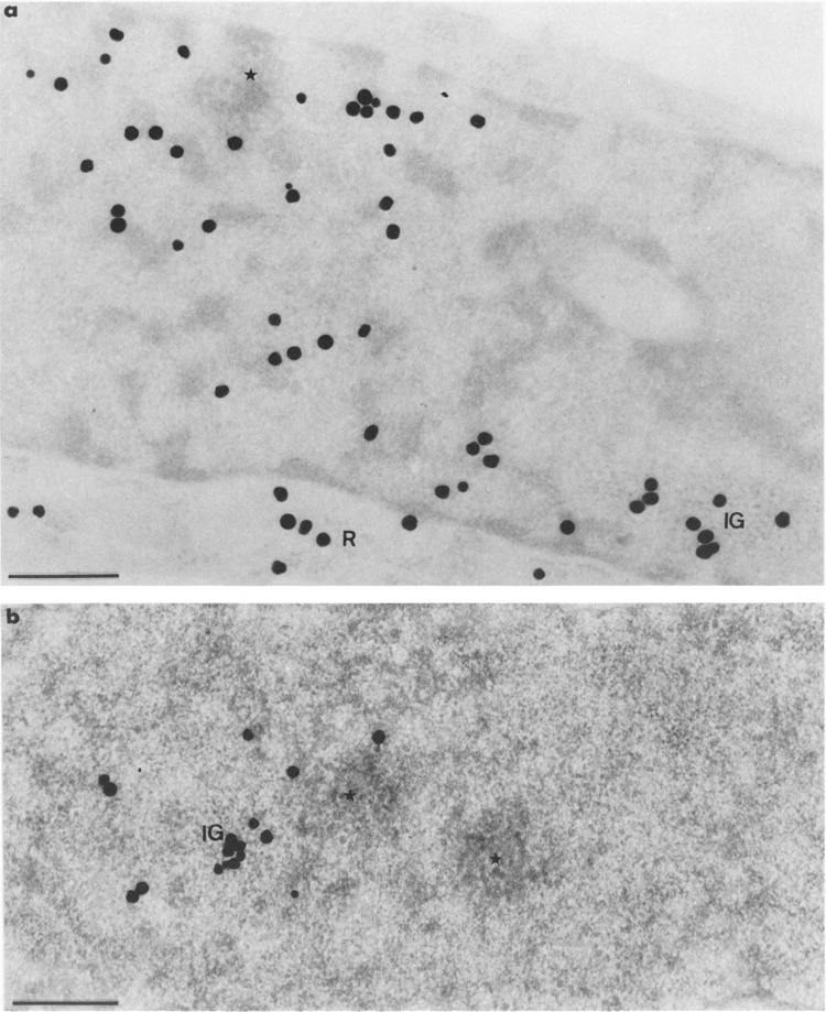
Nuclear distribution and migration of herpes simplex virus type 1 Us11 transcripts were studied in transient expression at the ultrastructural level and compared to that of RNA polymerase II protein. Transcription was monitored by autoradiography following a short pulse with tritiated uridine. Us11 transcripts accumulated mainly over the foci of intermingled RNP fibrils as demonstrated by the presence of silver grains localizing incorporated radioactive uridine superimposed to these structures in which the presence of Us11 RNA and poly(A) tails was previously demonstrated. Silver grains were also scattered over the remaining nucleoplasm but not in the clusters of interchromatin granules, and over the dense fibrillar component of the nucleolus as in control, nontransfected HeLa cells. Pulse-chase experiments revealed the transient presence of migrating RNA in the clusters of interchromatin granules. RNA polymerase II was revealed by immunogold labeling following the use of two monoclonal antibodies: mAb H5, which recognizes the hyperphosphorylated form of the carboxy-terminal domain (CTD) of the molecule, and mAb 7C2, which recognizes both its hyperphosphorylated and unphosphorylated forms. The two mAbs bind to the newly formed Us11 transcription factories and the clusters of interchromatin granules of transfected cells. In control cells, however, clusters of interchromatin granules were labeled with mAb H5 but not with mAB 7C2. Taken together, our data demonstrate the involvement of the clusters of interchromatin granules in the intranuclear migration of Us11 RNA in transient expression. They also suggest the occurrence of changes in the accessibility of the RNA polymerase II CTD upon expression of the Us11 gene after transfection by exposing some epitopes, otherwise masked in nontransfected cells.


Figures

FIG. 1
FIG. 1
Specificity of mAb 7C2. About 1 μg of DE1.0, a 1 M KCl eluate from a DEAE-5PW chromatography corresponding to a partially purified RNA polymerase II fraction from HeLa cells [see (25)], was loaded on a 5% polyacrylamide SDS gel (29). After electrophoresis, the proteins were transferred to a nitrocellulose membrane and incubated with the mAb 7C2. Specific immune complexes were revealed by the ECL Western blotting detection system (Amersham International pic, Buckinghamshire, UK), as previously described (8). The positions of the phosphorylated (Ho) and unphosphorylated (11a) largest subunit of RNA polymerase II are indicated.
FIG. 2
FIG. 2
Intranuclear distribution of RNA polymerase II molecules in HeLa cells following the use of mAb H5. Formaldehyde fixation, Lowicryl embedding, and uranyl acetate staining. Bars represent 0.5 μm. (a, b) Gold particles are numerous over a cluster of interchromatin granules (IG) in (a) and over a small cluster of RNP fibrils (arrow) in (b). They are scattered over the surrounding nucleoplasm, (c, d) The coiled body (CB) in (c) and the interchromatin granule-associated zone (star) in (d) are devoid of gold particles. C: cytoplasm; IG: cluster of interchromatin granules.
FIG. 3
FIG. 3
Intranuclear distribution of RNA polymerase II molecules in a cell transiently expressing the Us11 gene, following the use of mAb H5. Formaldehyde fixation, Lowicryl embedding, and uranyl acetate staining. Bars represent 0.5 μm. Gold particles are mainly observed over the cluster of interchromatin granules (IG) and the clumps of intermingled RNP fibrils induced by transfection (stars). They are scattered over the surrounding nucleoplasm and absent over the nucleolus (NU).
FIG. 4
FIG. 4
Intranuclear distribution of RNA polymerase II molecules in cells transiently expressing the Us11 gene, following the use of mAb 7C2. Formaldehyde fixation, Lowicryl embedding, and uranyl acetate staining. Bars represent 0.5 μm. (a) Gold particles accumulate over a roundish focus of intermingled RNP fibrils (star) whereas they are scattered over the surrounding nucleoplasm. IG: cluster of interchromatin granules; NU: nucleolus, (b) The focus of intermingled viral RNP fibrils (star) is located near a cluster of interchromatin granules (IG). Labeling over the focus of RNP fibrils is exactly superimposed to its individual fibrils (arrow).
FIG. 5
FIG. 5
Intranuclear distribution of RNA polymerase II molecules in cells transiently expressing the Us11 gene, following the use of mAb 7C2. Formaldehyde fixation, Lowicryl embedding, and uranyl acetate staining. Bars represent 0.5 μm. (a) Gold particles are numerous over the large cluster of interchromatin granules (IG) and are markedly rare in the surrounding nucleoplasm. The nucleolus (NU) is entirely devoid of labeling, (b) Two clusters of interchromatin granules (IG) are present, only one being decorated with gold particles (IG on the right). Between these two clusters is a small focus of intermingled RNP fibrils (star) that is labeled as in Fig. 4a, b. Once again the nucleolus (NU) is unlabeled.
FIG. 6
FIG. 6
Concomitant detection of mAb H5 and mAb 7C2 antigens of RNA polymerase II molecules with 10- and 5-nm gold particles, respectively, in cells transiently expressing the Us11 gene. Formaldehyde fixation, Lowicryl embedding, and uranyl acetate staining. Bars represent 0.5 μm. (a, b) Mixed 5-nm (arrows) and 10-nm gold particles are present over the clump of intermingled viral RNP fibrils (star) in (a) and the cluster of interchromatin granules (IG) in (b). Individual 5- and 10-nm gold particles are present in the surrounding nucleoplasm. CB: coiled body.
FIG. 7
FIG. 7
Colocalization of RNA polymerase II molecules and Us11 RNA on Lowicryl sections of cells transiently expressing Us11 gene. Formaldehyde in (a, b) and glutaraldehyde in (c) fixation. Uranyl acetate staining. Bars represent 0.5 μm. (a, b) mAb 7C2 and biotinylated Us11 DNA probe combination. Gold particles of 5-nm diameter label the protein whereas the 10-nm gold particles label the viral RNA. (a) Mixed 5-nm (arrows) and 10-nm gold particles are present over the focus of intermingled viral RNP fibrils (star) whereas 10-nm gold particles are present only over the small cluster of interchromatin granules (IG). Individual 5- and 10-nm gold particles are present in the surrounding nucleoplasm, (b) Mixed 5-nm (arrows) and 10-nm gold particles are present over the large cluster of interchromatin granules (IG). Individual 5- and 10-nm gold particles are present in the surrounding nucleoplasm, (c) mAb H5 and biotinylated Us11 DNA probe combination. Gold particles of 10-nm diameter label the protein, whereas the 5-nm gold particles label the viral RNA. Mixed 5-nm (arrows) and 10-nm gold particles are present over the focus of intermingled viral RNP fibrils (star).
FIG. 8
FIG. 8
Autoradiograms of cells transiently expressing Us11 gene and pulse-labeled during 6 min with tritiated uridine just before fixation. Epon embedding and uranyl acetate staining. Bars represent 0.5 μm. (a) Silver grains that localize the sites of transcription of both Us11 genes and cellular genes are numerous over the focus of intermingled RNP fibrils (star) and are also present over areas of the surrounding nucleoplasm and over the dense fibrillar component (F) of the nucleolus. This indicates the persistent transcriptional activity of the cellular genes, including the ribosomal RNA genes. The granular component (G) of the nucleolus and the cytoplasmic ribosomes (R) are devoid of labeling, (b) Part of nucleoplasm. The focus of intermingled RNP fibrils (star) is intensely labeled. The concomitant presence of silver grains over the surrounding nucleoplasm demonstrates once again that transcription of cellular genes is still occurring. Cytoplasmic ribosomes (R) are devoid of silver grains, (c) Part of nucleoplasm in which the intensely labeled focus of intermingled RNP fibrils (star) is located near an unlabeled large cluster of interchromatin granules (IG). Once again the cytoplasmic ribosomes (R) are devoid of silver grains.
FIG. 9
FIG. 9
Autoradiograms of cells transiently expressing Us11 gene and pulse-labeled with tritiated uridine as in Fig. 8, then submitted to a chase of 30 min before fixation. Epon embedding. Bars represent 0.5 μm. (a) Uranyl acetate staining. Silver grains are present at the border of the focus of intermingled viral RNP fibrils (star), but not inside. Silver grains are also present in the large cluster of interchromatin granules (IG) as well as in the remaining nucleoplasm, (b) Uranyl acetate and lead citrate staining. The cluster of interchromatin granules (IG) is clearly labeled. Silver grains are also scattered over the nucleoplasm and some grains are present over the cytoplasmic ribosomes (R).
FIG. 10
FIG. 10
Autoradiograms of cells transiently expressing Us11 gene and pulse-labeled with tritiated uridine as in Fig. 8, then submitted to a chase of 60 min before fixation. Epon embedding. Bars represent 0.5 μm. (a) Uranyl acetate staining. The cluster of interchromatin granules (IG) is labeled whereas there is no labeling over the small focus of intermingled viral RNP fibrils (star). Silver grains are scattered over the remaining nucleoplasm and over the cytoplasmic ribosomes (R). (b) EDTA regressive staining. Part of nucleoplasm showing a labeled cluster of interchromatin granules (IG) and two unlabeled foci of intermingled RNP fibrils (stars), one of which being contiguous to the labeled cluster of interchromatin granules.

Similar articles

Cited by

References

    1. Bachellerie J. P.; Puvion E.; Zalta J. P. Ultra-structural organization and biochemical characterization of chromatin-RNA-protein complexes isolated from mammalian cell nuclei. Eur. J. Biochem. 58:327–337; 1975. - PubMed
    1. Bernhard W. A new staining procedure for electron microscopical cytology. J. Ultrastruct. Res. 27:250–265; 1969. - PubMed
    1. Besse S.; Diaz J. J.; Pichard E.; Kindbeiter K.; Madjar J. J.; Puvion-Dutilleul F. In situ hybridization and immuno-electron microscope analyses of herpes simplex virus type 1 US 11 gene during transient expression. Chromosoma 104:434–444; 1996. - PubMed
    1. Besse S.; Vigneron M.; Pichard E.; Puvion-Dutilleul F. Synthesis and maturation of viral transcripts in herpes simplex virus type 1 infected HeLa cells: The role of interchromatin granules. Gene Expr. 4:143–161; 1995. - PMC - PubMed
    1. Bouteille M. The “Ligop” method for routine ultrastructural autoradiography. A combination of a single-coating gold latensification, and phenidon development. J. Microsc. Biol. Cell 27:121–127; 1976.

Publication types

LinkOut - more resources