. 1997 Nov 25;94(24):12869-74.
doi: 10.1073/pnas.94.24.12869.

Targeted deletion of alkylpurine-DNA-N-glycosylase in mice eliminates repair of 1,N6-ethenoadenine and hypoxanthine but not of 3,N4-ethenocytosine or 8-oxoguanine

Affiliations

Targeted deletion of alkylpurine-DNA-N-glycosylase in mice eliminates repair of 1,N6-ethenoadenine and hypoxanthine but not of 3,N4-ethenocytosine or 8-oxoguanine

B Hang et al. Proc Natl Acad Sci U S A. .

Abstract

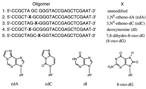
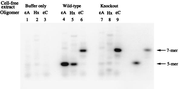
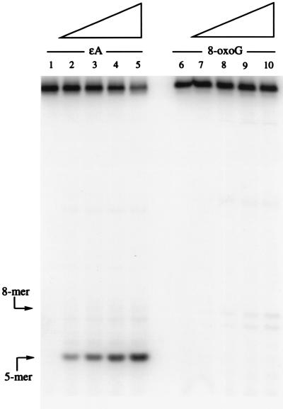
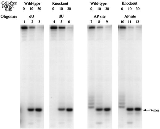
It has previously been reported that 1,N6-ethenoadenine (epsilonA), deaminated adenine (hypoxanthine, Hx), and 7,8-dihydro-8-oxoguanine (8-oxoG), but not 3,N4-ethenocytosine (epsilonC), are released from DNA in vitro by the DNA repair enzyme alkylpurine-DNA-N-glycosylase (APNG). To assess the potential contribution of APNG to the repair of each of these mutagenic lesions in vivo, we have used cell-free extracts of tissues from APNG-null mutant mice and wild-type controls. The ability of these extracts to cleave defined oligomers containing a single modified base was determined. The results showed that both testes and liver cells of these knockout mice completely lacked activity toward oligonucleotides containing epsilonA and Hx, but retained wild-type levels of activity for epsilonC and 8-oxoG. These findings indicate that (i) the previously identified epsilonA-DNA glycosylase and Hx-DNA glycosylase activities are functions of APNG; (ii) the two structurally closely related mutagenic adducts epsilonA and epsilonC are repaired by separate gene products; and (iii) APNG does not contribute detectably to the repair of 8-oxoG.


Figures

Figure 1
Figure 1
Structure of the modified nucleosides used and their positions in a defined 25-mer oligonucleotide. The unmodified oligomer is in sequence 1. The substitutions are shown as X (sequences 2–5).
Figure 2
Figure 2
Autoradiogram of denaturing 12% polyacrylamide gel comparing extent of nicking by mouse testes cell-free extracts of ɛA, Hx, and ɛC in control samples (lanes 1–3), wt mice (lanes 4–6) and ko mice (lanes 7–9). The positions of 5′ cleavage are shown on the far right, as indicated by arrows. The oligomers were allowed to react with 30 μg of protein for 1 h at 37°C in a standard nicking assay.
Figure 3
Figure 3
Quantitation of the 32P-labeled bands from PAGE of the same three oligomers in Fig. 2 as a function of protein concentration of testes extracts (0–30 μg), under the same reaction conditions as in Fig. 2. The percent of oligomer cleaved is expressed as % nicking efficiency. Note that in the ko mice (Right) there was no detectable ɛA or Hx cleavage above background.
Figure 4
Figure 4
(Left) PAGE of activities of wt and ko mouse testes extract toward 8-oxoG after 1-h incubation, which produces a 5′ 8-mer fragment as indicated by the arrow. Lane 1 is a control with buffer alone. Lanes 2–5 and 6–9 are with increasing amounts of testes protein (5, 10, 15, and 30 μg). (Right) Quantitation of the autoradiogram in Left. Note that there is no detectable difference between the wt and ko mouse extracts.
Figure 5
Figure 5
Autoradiogram of partially purified murine APNG-treated ɛA oligomer (Fig. 1, sequence 2) (Left) and 8-oxoG oligomer (Fig. 1, sequence 5) (Right) after treatment with increasing amounts of the recombinant protein, from 2.5 to 15 ng, shown by the slope of the triangles above. Lanes 1 and 6 are controls with no added protein. The reaction conditions were the same as in Fig. 2 except that HAP1 at 0.5 μg/ml was added to cleave the AP site on the 5′ side. The use of HAP1 alone under the same conditions does not cause any cleavage of either oligomer (data not shown). The positions of the size markers are indicated by arrows.
Figure 6
Figure 6
Activity of both mouse extracts against dU (lanes 1–6) and the AP site (lanes 7–12). The AP site was obtained by treating the dU-containing oligomer with purified UDG. The AP site was then created and incubated with testes cell-free extracts from both wt and ko mice. Lanes 7 and 10 illustrate the lability of the AP site under experimental conditions in the absence of any added enzyme. For this reason the purified HAP1 was added to the reaction mixture in Fig. 5.

References

    1. Sancar A. Annu Rev Biochem. 1996;65:43–81. - PubMed
    1. Friedberg E, Walker G C, Seide W. DNA Repair and Mutagenesis. Washington, DC: Am. Soc. Microbiol.; 1995.
    1. Singer B, Hang B. Chem Res Toxicol. 1997;10:713–732. - PubMed
    1. Mi L J, Mahl E, Chaung W, Boorstein R J. Mutat Res. 1997;374:287–295. - PubMed
    1. Engelward B P, Dreslin A, Christensen J, Huszar D, Kurahara C, Samson L. EMBO J. 1996;15:945–952. - PMC - PubMed

Publication types

MeSH terms