. 1997 Nov 25;94(24):12920-5.
doi: 10.1073/pnas.94.24.12920.

Cloning and expression of rat 25-hydroxyvitamin D3-1alpha-hydroxylase cDNA

Affiliations

Cloning and expression of rat 25-hydroxyvitamin D3-1alpha-hydroxylase cDNA

T Shinki et al. Proc Natl Acad Sci U S A. .

Abstract

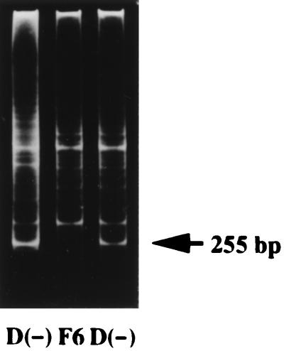
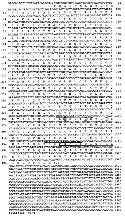
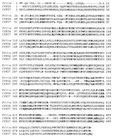
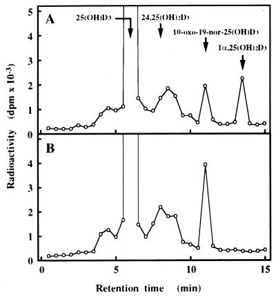
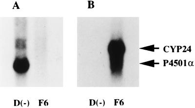
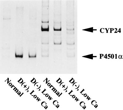
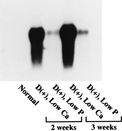
A full-length cDNA for the rat kidney mitochondrial cytochrome P450 mixed function oxidase, 25-hydroxyvitamin D3-1alpha-hydroxylase (P4501alpha), was cloned from a vitamin D-deficient rat kidney cDNA library and subcloned into the mammalian expression vector pcDNA 3.1(+). When P4501alpha cDNA was transfected into COS-7 transformed monkey kidney cells, they expressed 25-hydroxyvitamin D3-1alpha-hydroxylase activity. The sequence analysis showed that P4501alpha was of 2,469 bp long and contained an ORF encoding 501 amino acids. The deduced amino acid sequence showed a 53% similarity and 44% identity to the vitamin D3-25-hydroxylase (CYP27), whereas it has 42.6% similarity and 34% identity with the 25-hydroxyvitamin D3-24-hydroxylase (CYP24). Thus, it composes a new subfamily of the CYP27 family. Further, it is more closely related to the CYP27 than to the CYP24. The expression of P4501alpha mRNA was greatly increased in the kidney of vitamin D-deficient rats. In rats with the enhanced renal production of 1alpha,25-dihydroxyvitamin D3 (rats fed a low Ca diet), P4501alpha mRNA was greatly increased in the renal proximal convoluted tubules.


Figures

Figure 1
Figure 1
Electrophoretic bands (ethidium bromide fluorescence) produced by the method of mixed oligonucleotide-primed amplification of cDNA. Lanes 1 and 3, vitamin D-deficient rats D(−); lane 2, vitamin D-deficient rat treated for 48 h with 1 μg of 26,27-F6-1α,25(OH)2D3 (F6).
Figure 2
Figure 2
The nucleotide sequence of rat P4501α cDNA and its deduced amino acid sequence. Arrows indicate primer positions for isolation of P4501α fragment. The region of the heme-binding site is underlined. The conserved amino acids known to be important in ferredoxin-binding are circled. The start codon is shown in bold type.
Figure 3
Figure 3
Alignment of P4501α with closely related other CYPs. The closest relatives to P4501α were determined by using the blastp program provided by the National Center for Biotechnology Information, and the amino acid sequences were manually aligned to P4501α. CYP27, the predicted protein from the rat mitochondrial vitamin D3-25-hydroxylase (GenBank accession no. M38566); CYP24, the predicted protein from the rat mitochondrial 25(OH)D3-24-hydroxylase (GenBank accession no. X59506). Identical residues are shown in bold type, and dashes represent gaps for alignment purposes.
Figure 4
Figure 4
Metabolism of 25(OH)D3 in COS-7 cells transfected with the expression plasmid for P4501α. Metabolism of 25(OH)D3 by the cells transfected with 10 μg of pcDNA 1α (A) and the same amount of pcDNA empty vector (B). The retention times of 25(OH)D3, 24,25(OH)2D3, 10-oxo-19-nor-25(OH)D3, and 1α,25(OH)2D3 are indicated. 25(OH)D3 was metabolized into 1α,25(OH)2D3 only in the cells transfected with pcDNA 1α. The fraction containing 1α,25(OH)2D3 was further applied to HPLC under the following two conditions: column, Finepak SIL 4.6 × 250 mm; mobile phase, 8% 2-propanol in hexane; flow rate, 1 ml/min; column, J′-sphere ODS-AM 4.6 × 150 mm (YMC Co., Kyoto, Japan); mobile phase, 10% water in methanol; flow rate, 1.0 ml/min (data not shown). In all cases the putative 1α,25(OH)2D3 comigrated with authentic 1α,25(OH)2D3. Similar results were obtained in three independent sets of experiments.
Figure 5
Figure 5
Expression of CYP24 and P4501α mRNAs in rat kidney. Poly(A)+ RNAs (5 μg) were prepared from rats that had been treated with either 26,27-F6-1α,25(OH)2D3 or vehicle. 26,27-F6-1α,25(OH)2D3 (1 μg/rat) was intravenously administered to vitamin D-deficient low Ca rats. Rats were killed 48 h after administration of either 26,27-F6-1α,25(OH)2D3 or vehicle. The blots were probed with [32P]dCTP labeled P4501α (A) and CYP24 (B). Similar results were obtained in three independent sets of experiments.
Figure 6
Figure 6
Expression of CYP24 and P4501α mRNAs in the kidney of each rat model. Radioautography of RT-PCR products of CYP24 and P4501α mRNAs in microdissected PCT. Three-week-old rats were fed a vitamin D-replete or a vitamin D-deficient low Ca diet for 2 weeks. Microdissection and RT-PCR were performed as described (18, 19) with a slight modification. Final volume of the PCR reaction was 20 μl, and composition of the solution was 10 mM Tris⋅HCl (pH 8.3), 50 mM KCl, 2 mM MgCl2, gelatin 0.01%, 200 nM sense and antisense primers, 0.1 mM dNTPs, 74 kBq of [33P]dCTP, and 1 unit of Taq DNA polymerase. The program of PCR was as follows: 25 cycles of 94°C for 40 sec, 50°C for 1 min, and 72°C for 1 min. The expected size of RT-PCR products for P4501α and CYP24 were 253 and 482 bp, respectively.
Figure 7
Figure 7
Expression of CYP24 and P4501α mRNAs in 3-week-old rats maintained for 2–3 weeks on a vitamin D-repleted diet containing 1.2% Ca and 0.6% P (Normal), 0.03% Ca and 0.6% P (Low Ca), or 1.2% Ca and 0.1% P (Low P). Poly(A)+ RNA (5 μg) was electrophoresed and probed for P4501α. Similar results were obtained in three independent sets of experiments.

References

    1. DeLuca H F. FASEB J. 1988;2:224–236. - PubMed
    1. Suda T, Shinki T, Kurokawa K. Curr Opin Nephrol Hypertens. 1994;3:59–64. - PubMed
    1. Armbrecht H J, Okuda K, Wongsurawat N, Nemani R K, Chen M L, Boltz M A. J Steroid Biochem Mol Biol. 1992;43:1073–1081. - PubMed
    1. Okuda K, Usui E, Ohyama Y. J Lipid Res. 1995;36:1641–1652. - PubMed
    1. Nelson D R, Koymans L, Kamataki T, Stegeman J J, Feyereisen R, Waxman D J, Waterman M R, Gotoh O, Coon M J, Estabrook R W, Gunsalus I C, Nebert D W. Pharmacogenetics. 1996;6:1–42. - PubMed

MeSH terms

Associated data